Madhavi Katamaneni* | A.V.R. Mayuri
© 2022 IIETA. This article is published by IIETA and is licensed under the CC BY 4.0 license (http://creativecommons.org/licenses/by/4.0/).
OPEN ACCESS
The World Health Organization (WHO) made the announcement that the SARS virus-induced Coronavirus contamination-2019 (COVID-19) has been raised to the status of an international pandemic in March of 2020. If this virus is discovered at a relatively early stage in its life cycle, then it will be possible to contain it and treat it in an effective manner. Because of this fact, real-time polymerase chain reaction (RT-PCR) has emerged as the screening method of first desire for the rapid detection of COVID-19 in blood samples. This is a direct result of the fact Large-scale research have shown that the outcomes of an RT-PCR experiment can be misleadingly bad up to sixty two percent of the time. As a final result, the focus of these studies is on a thorough examination of COVID-19 detection and complexity through the utilisation of photos obtained from chest x-rays that are centred at the lung. In the beginning, the research looked into the many layers of computer-aided detection, such as deep learning and meta-heuristics techniques. After that, the research is concentrated on the processes of feature extraction, feature preference, and sophistication operations by utilising techniques such as gadget learning, deep learning, and bio-optimization. The study highlights the contemporary difficulties presented by artificial intelligence structures for the detection of COVID-19, which can help to put into action a hybrid system.
coronavirus disease-2019, bio-optimization algorithms, chest x-ray images, artificial intelligence approaches, deep learning and machine learning models
COVID-19 is an infectious illness that turned into reversed with the help of the SARS virus and declared a global pandemic with the aid of the WHO in March of 2020. The most significant COVID-19 outbreak to date has spread to 223 countries and territories around the world, resulting in over 129 million cases of infection and over 1.8 million fatalities worldwide [1]. As can be seen in Figure 1, the number of fatalities and inflammatory episodes that occur simultaneously is expanding at an alarming rate. The early discovery of COVID-19 is essential in order to impede the progression of the virus and to provide treatment in order to prevent its effects. The day-to-day rise in COVID-19 cases all over the world, in addition to the restrictions of contemporary diagnostic strategies, provide challenging problems in recognising and managing the pandemic.
Figure 1. (a) COVID-19 cases worldwide
Figure 1. (b). COVID-19 death rate statistics among various countries. (Sources: Center for Systems Science and Engineering (C.S.S.E.) at Johns Hopkins University, Baltimore, MD, USA)
Researchers [2] from every corner of the world are putting in a lot of effort to devise effective methods for identifying diseases and to speed up the discovery of vaccinations and treatments. Examination of blood samples, testing for viruses, and medical imaging are the three techniques that are probably utilised in diagnostic work the most commonly [3]. Blood testing has revealed the presence of antibodies against severe acute respiratory syndrome coronavirus 2, also known as SARS-CoV-2. This was discovered after the virus was given the name SARS. When it comes to recognising COVID-19, this test, on the other hand, has an accuracy of approximately 2 to 3 percentage points at best. Viral tests employ samples taken from the respiratory tract in order to recognise SARS-CoV-2 antigens.
The rapid diagnostic test (RDT) [4], sometimes known as the fast diagnostic check, is a type of antibody detection test that is quick and can yield results in as little as a half an hour. However, there is a restricted supply of RDT exam kits that may be purchased, and the efficacy of those kits is determined by the pattern rate further to the time frame in which contamination turned into first visible. Because the test is unable to differentiate between COVID-19 and other viral infections, it has the potential to produce results that can be mistakenly significant. As an end result, its usage is not recommended for the analysis of COVID-19. The technique known as reverse transcription-polymerase chain reaction (RT-PCR) [5] is the tool that is currently regarded as the gold standard for first-line screening. On the other hand, the findings of a comprehensive study revealed that the degrees of test result sensitivity varied between fifty and sixty percentage. It is now possible to obtain a preliminary RT-PCR stop result that is awful as a result of this. As a consequence of this, repeated RT-PCR tests are carried out all through the whole of a 14-day assertion length in order to guarantee the dependability of the test stop result for analysis [6].
To put this another way, an RT-PCR negative result for a suspected case of COVID-19 is only considered to be a real negative result if there are no lingering RT-PCR results acquired after multiple tests have been completed over the course of the 14-day be aware period. This can be stressful for the patients, and it can be expensive for the healthcare control, because certain nations do not always have RT-PCR test kits available [7]. Because of this, clinical imaging methods, such as chest CT and CXR tests, are used extensively in the process of identifying pneumonia brought on by COVID-19 [8-10]. This is a direct result of the aforementioned. CT has a greater sensitivity for early pneumonic alteration, contamination development, and opportunity analysis in this one-of-a-kind circumstance; however, intravenous evaluation medium administration is essential for the diagnosis of pulmonary thromboembolism. Despite recent developments in diagnostic technology [11], radiologic imaging by alone isn't always normally sufficient for determining whether or not or whether or not COVID-19 pneumonia is present. This is because radiologic imaging can't determine whether or not COVID-19 pneumonia is present. Imaging needs to be incorporated into the diagnostic processes that are carried out in hospitals and labs. In addition, the American College of Radiology and the Italian Society of Radiology (SIRM) no longer recommend using chest CT as a screening technique. The SIRM follows the lead of the ACR in this regard. Instead, they recommend it for individuals who are experiencing symptoms and who meet certain clinical criteria in order to have the examination performed. The most crucial characteristic of COVID-19 contamination is the bilateral distribution of floor glass opacities (GGO) [12], with or without consolidation in the posterior and peripheral lungs. Patients diagnosed with COVID-19 who are severely ill are likely to have a high risk of developing venous thromboembolism, which can include deep vein thrombosis and/or pulmonary embolism. This is a reasonable assumption to make given the severity of their illness. This is due to the fact that people with severe infections have an increased risk of developing such conditions. As a result of this condition, it'll provide up the question of whether or not or no longer evaluation medium changed into used all through the CT have a study [13, 14]. Because COVID-19 targets the respiratory system, chest radiography images are a key tool for the early identification and control of the disease.
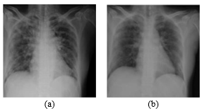
Figure 2. CXR findings: (a) GGO and (b) GGO with consolidation [8]
Because of this, CXRs have been utilised as a diagnostic tool of the first line in Italy as well as in a number of other countries all over the world [15]. The state of the lungs, as well as the degree of any illness or recovery, can be successfully determined with the assistance of radiological scans. These scans also help determine whether or not treatment is necessary [16]. The CXR scans of COVID-19 patients were examined by radiologists, and their findings revealed a significant number of anomalies. These findings have been reported [17]. In the CXR pictures, which are displayed in Figure 2, you can see some examples of the capabilities that COVID-19 possesses. The bilateral GGO and the bilateral and multifocal GGO with consolidation are both included in these cases. CXR is a device that is typically available in the majority of clinical settings; it is far more effective in terms of patient education and immediate diagnosis, hence saving a significant amount of time [18]. As a result of this, the CXR can be used for a variety of tasks, this includes, but isn't restricted to, patient man or woman triage, determining which treatments are maximum important for patients, and making the maximum green use of clinical assets.
Numerous investigations [19] made use of CXR photos for COVID-19 identification and classification, and the outcomes of those studies were fantastic, even though compared to CT photograph-based entirely COVID-19 detection [20]. In addition, CXR pictures were utilised in numerous other research.
The goal of this article is to provide an outline and update on artificial intelligence-based fully certainly approaches programmes in COVID-19 illness utilising radiological pix, which includes CXR and CT, with a concentrate on their capability in important applications, which include the following: analysis of COVID-19 in addition to forecasting the mortality hazard and severity of the disease.
The finishing touches of the object can be dissected into the following categories: The second phase includes the delivery of the datasets as well as their descriptions. In Section three, the most important procedures associated with the COVID-19 beauty standard are discussed. In the fourth step, we will talk about the artwork that is connected to a selection of different procedures. Section 5 details the challenging circumstances that have been identified as having occurred. The completion of the interest can be seen in segment 6.
2.1 CT scans
COVID-CT-Dataset [21] - The University of San Diego has begun compiling a records set for COVID-19, which will consist of 349 CT photographs conveying scientific discoveries. It asserts that it is the most significant example of its kind. An artificial intelligence model was trained until it reached an accuracy rate of 85 percent in order to demonstrate its potential. You may access the data set containing the statistics at https://github.com/UCSD-AI4H/COVID-CT. An image-primarily based model that operates with CT scans for COVID-19 diagnosis can be found at https://github.com/JordanMicahBennett/SMART-CT-SCAN BASED- COVID19 VIRUS DETECTOR/. This model can be downloaded and used.
2.2 Images of CX-rays
Radiography Database for COVID-19 (COVID-19) A group of researchers [22] from Qatar University in Doha and the University of Dhaka in Bangladesh, in addition to collaborators from Pakistan and Malaysia with medical doctors, have compiled a database of CXR images for COVID-19 extremely good instances along with regular and viral pneumonia photographs. There are 219 powerful COVID-19 pictures included in the most recent launch, along with 1341 regular pictures and 1345 pictures of viral pneumonia. The authors have indicated that they may continue with their plan to replace this database as soon as fresh x-ray images of patients suffering from COVID-19 pneumonia become available.
The Collection of Image Data for the COVID-19 [23], Joseph Paul Cohen was the one who initially made available a selection of open picture records from the COVID-19. The following URL, https://github.com/ieee8023/covid-chestxray-dataset, is where all of the images and information have been uploaded for public consumption.
This marks the launch of the brand-new COVIDx dataset, which includes 16,756 chest radiography images collected over the course of 13,645 patient times. The open-source chest radiography datasets that may be found at https://github.com/ieee8023/covid-chestxray-dataset and https://www.Kaggle.com/c/rsna- pneumonia-detection-project are used to generate the modern COVIDx dataset [24]. It is a combination of data provided by many different activities, including the Radiological Society of North America (RSNA), Dr. Joseph Paul Cohen, others involved in the RSNA Pneumonia Detection Challenge, and the group at MILA, which was involved in the COVID-19 picture facts series project for the purpose of making statistics available to the global community.
ChestX-ray8 [25]: The chest x-ray (CXR) is one of the most comprehensive radiological exams that can generally be obtained for the screening and diagnosis of a wide variety of lung conditions. Picture Archiving and Communication Systems (PACS) are utilised by the majority of contemporary healthcare facilities. These PACS can 2.be accessed at https://nihcc.App.Box.Com/v/ChestXray-NIHCC to see the numerous X-ray imaging studies and radiological opinions that are kept there.
2.3 Other images
Datasets for Thermal Images: There is no thermal image dataset available for screening patients with high fever. On the other hand, Kopaczka et al. [26] came up with the idea of a fully annotated thermal face database in addition to the software that would go along with it in order to recognise thermal facial functions. You can obtain information on similarly mind of related information that can be discovered through the utilisation of such systems by visiting the website https://www.Flir.Com.Au/discover/public-protection/thermal-imaging-for-detecting-elevated-bodytemperature. The website also contains information on how such systems can be used.
Basic steps involved in COVID-19 detection: This section provides a comprehensive breakdown of the many ranges that are available for COVID-19 detection and classification. Figure 3 depicts a basic block diagram of the COVID-19 detection and sorting system. This system is made up of many different kinds of currently available techniques at each level and may be seen as a whole in this picture.
Figure 3. Basic block diagram of SCDC with multiple techniques
Preprocessing: Computer-aided assessment for the reliable and rapid detection of COVID-19 has become a necessity in order to prevent the spread of the virus throughout the pandemic and to simplify the burden that is placed on the healthcare system. This evaluation can be done through the use of a computer. Imaging with CXR has a number of benefits that distinguish it from other imaging and detection methods [27]. On the topic of detecting COVID-19 from a limited number of the original X-ray images, a great number of articles have been cited. On the other hand, the effect of image enhancement and lung segmentation of a massive dataset on the detection of COVID-19 is not mentioned elsewhere in the literature. Image enhancement is a crucial step in the processing of photographs that draws attention to important details within an image and either reduces or eliminates positive secondary details in order to improve the incredible of the identification method [28]. Image enhancement is an important technique. The actual photographs will be used as a comparison to determine which of the target images will be most suitable for a given application. In order to investigate the impact that photo enhancement techniques [29] have on COVID-19 detection, five one-of-a-kind picture enhancement techniques were utilised. These techniques included histogram equalisation (HE), evaluation constrained adaptive histogram (CLAHE), photograph complement, gamma correction, and stability assessment enhancement technique (BCET).
Segmentation: Pattern recognition techniques are becoming increasingly important in the processing of photographs and various medical programmes. The correct determination of the location of the infection and an accurate evaluation of the severity of the condition are two essential objectives of infection localization. However, the results of prior study [30] demonstrate that the activation maps generated fundamentally through the underlying DL network can also fail to execute each purpose, that is, aside from the point locations in which biassed severity grading appears in many situations. Lung segmentation is essential to the identification and sophistication of COVID-19, which is necessary to overcome these challenges. In this way, the region of interest may be tracked all the way down to the sections of the lungs, which results in an improvement in the dependability. The scientific community has asserted that computational methods can segment CXR through the utilisation of the K-approach and fuzzy clustering procedures [31], and it has pushed for the development of hybrid algorithms for different COVID-19. As a result, automated and robust deep learning technologies with precise and quick detection are becoming increasingly popular.
Figure 4. Samples segmented outcomes [30]
Figure 4 depicts the segmented final sample results that were achieved by the application of CNN models. Figure 4(a) depicts the entrance photo, while Figure 4(b) illustrates the ground fact effects. Both of these figures can be found in the same figure. In addition, the segmented output can be shown in Figure 4(c), which was generated through the application of the CNN version and which localises the COVID-19 area. Within the CXR picture, the COVID-19 impacted area is indicated by the arrow in Figure 4(d).
Feature extraction: The statistical information that is included in the input image and is then utilised to keep track of the characteristics of the COVID-19 illness is referred to as features. In order to extract these skills, a number of different picture processing algorithms [32] are utilised. Gray stage dependency matrix (GLCM), quick Fourier remodel, discrete wavelet transforms (DWT), spatially grey stage dependency matrix (SGLD), and basic component evaluation (PCA) are some of the methods that can be used. In order to fulfil the hurdles presented by the actual worldwide COVID-19 detection, additional robust features will need to be retrieved. The number of characteristics gathered with the help of one technique changed into now not big ample to correctly identify COVID-19. The fusion method [33] of extracting functions through the application of one-of-a-kind procedures should, on the other hand, provide a large number of distinguishing characteristics for accurate identification. When viewed in this light, the concept of fusion can be understood to refer to the concatenation of the two separate vectors.
Feature selection: Feature selection is the process of selecting the optimal features from the available features. Usually, either machine learning or deep learning methods [34] are needed to train with the available features. So, if the total available features are increased, it will be difficult to perform the training procedure. Thus, feature selection is used to reduce the number of features and select the optimal features only. Machine learning and statistical pattern recognition both employ feature selection. This is critical in a variety of applications, including classification. In conventional methods [35], there are a lot of extracted features that are either worthless or don't provide much information. If these features aren't removed, the primary application's computational load will increase. Selecting a subset of features can improve the value of an evaluation function in a number of ways by using optimization approaches.
Classification: In the context of the COVID-19 epidemic, there will be a pressing demand for ready-to-use resources for facts collecting and artificial intelligence (AI) algorithms [36] to facilitate the search for efficient and secure treatments. The innovative incorporation of radiomics techniques and AI-based absolutely solutions into healthcare is already transforming established paradigms within the entire healthcare environment. This is made possible by the innovative digitalization of scientific information. To be more specific, diagnostic and selection manual systems for medical imaging are the most successful examples of innovation for health care that have been implemented.
AI-primarily based approaches [37] have resulted in the development of diagnostic applications that improve image capture, preprocessing, annotation, and interpretation. This provides an "augmentation" of the radiologists, as opposed to the impractical replacement of them. The application of AI in medical imaging has been particularly helpful for diagnosing neurodegenerative diseases and coronary heart conditions. This has a significant impact on the risk of developing breast and lung cancer.
Deep learning, also known as DL, and machine learning, often known as ML, are both subfields of AI that are focused on the development of systems that can learn from examples and embellish itself without being specifically taught [38]. ML stands for "machine learning" and refers to the testing of computer algorithms that can beautify themselves autonomously via the accumulation of experience and the beneficial resource of the utilisation of facts. Algorithms used in machine learning can make predictions or decisions without first being given explicit instructions to do so. This allows the algorithms to work more independently. In order to arrive at those decisions, they construct a model that is mostly predicated on sample records, sometimes known as "education statistics."
Machine learning algorithms [39] are utilised in an extensive kind of programmes, which include in medicine, e-mail filtering, speech recognition, and computer vision. These applications are utilised in regions in which it's far difficult or not possible to grow conventional algorithms to carry out the desired duties. Statistical learning is only a subset of device mastering; however, not all device reading is statistical learning. Computational statistics, which specialises in making predictions the usage of computer systems, is closely associated with a subset of device reading. The investigation of mathematical optimization opens up new avenues for you to explore in the areas of strategy, idea, and awareness within the field of machine learning. Data mining is a subfield of information analysis that focuses on conducting exploratory data assessment and analysis without first being instructed how to proceed. It is a connected problem of having an investigation carried out.
Deep learning [40] is a category of machine learning algorithms that uses numerous layers to routinely extract higher-level capabilities from raw data input. This is accomplished through a process known as deep learning. When it comes to image processing, for example, lower layers are able to recognise edges, while higher layers may also become aware of concepts associated with humans, such as digits, letters, or faces. For example, lower layers can recognise edges; higher layers may also become aware of such concepts. Deep-learning architectures [41] such as deep neural networks, deep perception networks, deep reinforcement gaining knowledge of, recurrent neural networks, and convolutional neural networks (CNN) had been completed to fields such as pc imaginative and prescient, speech recognition, natural language processing, device translation, bioinformatics, drug format, medical photo analysis, material inspection, and board undertaking applications, in which telecommunications and computer networks are utilised. Both DL and ML have been successfully implemented in a wide variety of domains [42], including clinical informatics and medical care. One of the most important research avenues makes use of both DL and ML in order to find COVID-19 and fight it. Numerous research projects had been kicked up in order to utilise and embellish COVID-19-related DL and ML techniques.
This section gives the detailed analysis of various methods for identifying the COVID-19 by using machine learning, deep learning and transfer learning models. This survey is concentrated on purely on artificial intelligence approaches for feature extraction, segmentation and classification operations. Further, this survey also focused on feature selection operations using evolutionary, swarm intelligence, natural selection, meta-heuristics- based bio-optimization methods.
3.1 Survey based on COVID-19 dataset
This section gives the detailed survey on COVID-19 detection and classification methods using two different datasets such CT and CXR. The challenges presented in artificial intelligence approaches with respect to these datasets are studied.
3.1.1 Survey on COVID-19 classification using CT images
A chest CT scan is a diagnostic procedure that does not involve the use of any invasive procedures in order to obtain a clear image of the chest region of a patient. It generates x-rays in a more ideal shape, which results in photographs of the chest that are more specific. It creates images that contain bones, fats, muscles, and organs, giving doctors a better look, which is vital when attempting to make accurate diagnosis [43]. There are several variations of the chest CT scan, the most common of which are the excessive-decision chest CT test and the spiral chest CT test. In a single revolution of the x-ray tube, the high-definition chest CT scan can provide more information than just a single slice, often known as a snapshot. The spiral chest CT test usefulness consists of a table that moves in a continuous motion along a passage that resembles a tunnel while an x-ray tube travels in a spiral pattern. The fact that a three-dimensional image of the lungs may be produced using the spiral CT is one of the test's primary benefits.
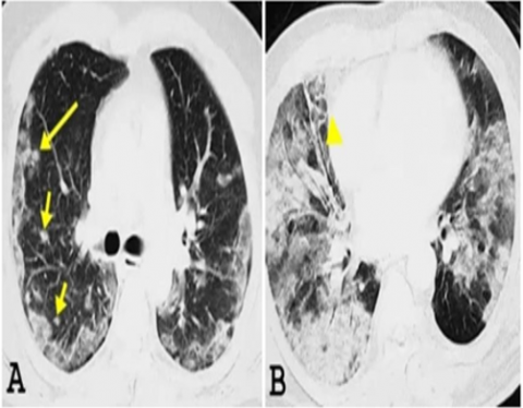
GGO, consolidation, reticulation/thickened interlobular septa, nodules, and lesion distribution are some important CT functions (left, proper or bilateral lungs). The most noticeable CT functions [44] seen in COVID-19 pneumonia are areas of floor-glass opacification that are bilateral and subpleural, as well as consolidation that affects the lower lobes. During the intermediate period, which lasts for four to fourteen days after the onset of symptoms, the crazy-paving pattern and a potentially visible Halo signal become important functions. The lung CT results of patients diagnosed with COVID-19 are depicted in Figure 5. Figure 5(a) is an axial CT photograph taken within the first week of treatment of a patient who is 37 years old. It demonstrates peripheral bilateral GGO as well as a few small-scattered pulmonary nodules (arrows). Figure 5(b) is an axial CT photograph taken of a patient who is 52 years old during the second week after their initial presentation. This scan demonstrates that the patient has diffuse GGO, consolidation, and bronchiectasis (arrowhead)
Figure 5. Samples COVID-19 affected CT images adapted from [44]
The first category of labour to be covered in this discussion focuses on diagnosis methods as a segmentation issue. A CT image dataset containing 46,096 photographs of both healthy and inflammatory patients that has been proposed by Chen et al. [45] is comprised of these images. These photographs have been categorised by qualified radiologists. It turned into amassed from 106 individuals hospitalised with 51 confirmed cases of COVID-19 pneumonia and 55 patients serving as controls. Only deep learning models were employed for the segmentation process in this work in order for it to successfully identify the diseased region in CT images of patients who were either healthy or infected.
It came to be mostly dependent on the UNet++ semantic segmentation version [46], which is utilised to extract valid regions contained inside the images. It employed 289 randomly selected CT images and compared them to another 600 CT images that were also randomly selected. The professional model that was developed as a result of this study was implemented at the Renmin Hospital of Wuhan University in Wuhan, Hubei province, China, in order to assist in the assessment of current COVID-19 cases. It was also made available for free download on the internet, making it possible to conduct a speedy investigation of current incidents in several other areas. An artificial intelligence framework that is cloud-based and entirely free to use has been developed in order to provide assistance with the detection of COVID-19 pneumonia around the world. The second type of artwork viewed COVID-19 to be a binary type problem. Li et al. [47] proposed the use of transfer reading on the RESNET50 in order to extract visual functions from volumetric chest CT scans. This was done using the COVNet algorithm. Using the U-Net model, lung segmentation has developed into being a pre-processing task that can be accomplished. The problem of making a diagnosis became additionally approached as a three-class category problem: separating healthy patients from persons with different kinds of pneumonia and people with COVID-19. The research conducted by Song et al. [40] employed data collected from 88 patients diagnosed with COVID-19, 101 patients diagnosed with pneumonia caused by bacteria, and 86 healthy individuals as controls. It developed a neural network known as DRE-Net (Relation Extraction neural network), which changed into basically built on ResNet50. On this network, the Feature Pyramid Network (FPN) [48] and the Attention module have been included so as to symbolise finer-grained components of the images. The various CT image-based COVID-19 detection methods are outlined in Table 1, together with segmentation, class styles, datasets, and performance evaluations. Sensitivity (SEN), specificity (SPE), accuracy (ACC), and area beneath curve are the overall performance parameters that are provided here (AUC).
Table 1. Representative works for CT based COVID-19 diagnosis
|
Ref. No. |
Segmentation Model |
Classification model |
Dataset |
Performance |
|
[45] |
UNet++ |
DLCNN |
46,096 CT images |
SEN=100%, SPE=93.55%, ACC=95.24% |
|
[49] |
M-Inception |
AlexNet |
453 of pathogenconfirmed COVID-19 |
ACC=82.9%, SPE=80.5%, SEN= 84% |
|
[50] |
VNET |
ResNet-18 feature extraction and classification |
A total of 618 CT samples were collected: 19, 224 CT samples Fluel175 CT samples from healthy people |
ACC= 86.7% |
|
[40] |
DRE-Net |
CNN |
777 CT images |
AUC=0.99, SEN= 0.93, ACC= 0.86, F-score 0.87 |
|
[51] |
CNN |
SVM |
46,096 CT images |
AUC=0.996, SEN=98.2%, SPE=92.2% |
|
[52] |
VB-Net |
V-shaped network. |
249 CT images |
ACC=91.6% |
|
[53] |
2D-CNN |
CAP Network |
970 CT volumes |
ACC= 94.98%, AUC= 97.91% |
|
[54] |
Capsule Network |
SVM |
150 CT images. |
ACC=99.68% |
|
[55] |
VGG-Net |
MobileNet |
249 CT images |
AUC=0.959, ACC=0.976 |
|
[56] |
3D U-Net++ |
CNN |
150 CT images. |
AUC=0.991, SEN=0.974, SPE= 0.922 |
3.1.2 Survey on COVID-19 classification using CXR images
The identification of the GGO area, which can be difficult with CT pictures since it is concentrated in a small space, is one of the challenges presented in the COVID-19 detection process. In addition, the traditional procedures did not slice and extract the features from CT photographs any more. In addition, there is a possibility that there is not availability of more than one well-known dataset for CT snapshots. The use of CT imaging comes with a number of drawbacks, one of which is the requirement for an excessively high impacted individual dose and superior value. When it comes to detecting chest pathology, digital CXR radiography is the imaging modality that has a cheaper cost and a wider availability. Because of this, automatic diagnosis of COVID-19 skills in CXR will make it a particularly valuable diagnostic tool [57] in the fight against the disease. Digital X-ray imaging computer-aided assessment is used for the diagnosis of first-rate disorders such as osteoporosis, the majority of malignancies, and cardiac illness.
Assessment enhancement, on the other hand, is utilised as a pre-processing step because it is extremely difficult to differentiate soft tissue with a terrible assessment in X-ray imagery. Lung segmentation of CXRs is a step that is necessary and vital for you to take in order to detect lung nodules, and the literature suggests a number of different segmentation procedures [58]. Consolidation has been identified in COVID-19 infected patients based on the results of CXR exams. In one study that was conducted in Hong Kong [59], three different patients received daily CXR examinations, and all of them demonstrated progress in the lung consolidation over the course of three to four days. Improvement can be shown on subsequent CXR exams as time passes after the first diagnosis. The patient with the 0.33 showed no significant changes during the course of 8 days. However, an identical test showed that the floor glass opacities within the right decrease lobe peripheral on the CT are not visible at the chest radiograph, which were taken one hour after the primary observe. This chest radiograph became taken after the first observe. Despite this, CXR is still recommended in addition to CT for more in-depth radiological investigation. Several different automatic strategies relating to CXR were been forward. In the following section, we will go over the most important work, and Table 2 will provide a more organised explanation of the many methods used. Both of these sections will take place simultaneously.
Figure 6. Samples COVID-19 affected CXR images adapted from the research [59]
Figure 6 depicts chest radiographs taken of an elderly male patient affected by the disease who was originally from Wuhan, China and later travelled to Hong Kong, China. These three chest radiographs were determined based on the daily chest radiographs that were obtained on the patient who is being discussed here. On day four, the consolidation that began on day zero in the proper lower region continued, and fresh consolidative modifications were made in the proper midzone outer border and perihilar place. This exchange in the midzone makes an improvement on the day 7 movie, which includes high GGO territory.
To this day, quite a few computer vision models for X-ray COVID-19 that are mostly based on deep learning have been proposed. The model COVID-Net [60], which was proposed using the resource of Darwin AI in Canada, is one of the most significant advances that has been made. This work provides a network structure for the detection of COVID-19 cases based on chest X-rays by combining human-driven principled network layout prototype with system-pushed design exploration.
The COVIDX-Net was proposed by Hemdan et al. [27] based fully largely on seven particular architectures of DCNNs; particularly VGG19, DenseNet201, InceptionV3, ResNetV2, InceptionResNetV2, Xception, etc. Brilliant version mixing led to F1- values of 0.89 and zero respectively. 91 for both typical and COVID-19 occurrences. In a similar fashion, Asmaa Abbas and colleagues [61] proposed a Decompose, Transfer, and Compose (DeTraC) approach for the category of COVID-19 chest X-ray photos. In order to carry out the diagnosis, the authors utilised CNN capabilities based on pre-professional models that were run on ImageNet and ResNet. Ghoshal et al. [32] introduced the Uncertainty-Aware COVID-19 Classification and Referral version with the recommended Drop weights in keeping with Bayesian Convolutional Neural Networks. This model became used to classify and refer to COVID-19 records (BCNN). In order to make the COVID-19 detection as large as possible, several kinds of prediction uncertainty in deep mastering were used into succeeding paintings. One of its far epistemic or model uncertainty bills for the model parameters uncertainty because it no longer takes into account all of the factors of the data or the lack of schooling data one of its far epistemic or model uncertainty bills for the model parameters in Table 2 outlines a few more CXR picture enabled COVID-19 detection and class methods, including segmentation, feature extraction, and sophistication styles. These methods are presented in chronological order. Table 2 presents a comparison of the general performance of a variety of techniques with regard to a few different datasets.
Table 2. Representative works for CXR based COVID-19 diagnosis
|
Ref. No |
Models used |
Dataset |
Performance |
|
[62] |
CLAHE-preprocessing, U-Net segmentation., adversarial+ based feature extraction. |
There are a total of 662 chest X-rays included in the 247 images that come from the Japanese Society of Radiological Technology (JSRT) Collection+ Shenzhen dataset. |
DSC=97.5% |
|
[61] |
CNN uses models that have already been trained on ImageNet and ResNet+ Decompose, Transfer, and Compose (DeTraC) for COVID-19 chest X-ray images. |
180 samples of normal CXRs (with a resolution of 4020 x 4892 pixels) from the Japanese Society of Radiological Technology (JSRT) and the Cohen JP. COVID-19 image data series. |
ACC=95.12%, SEN=97.91%, SPE=91.87%, PRE=93.36% |
|
[63] |
Pre-trained ResNet50 model with transfer learning |
The open-source GitHub repository shared by Dr. Joseph Cohen+Chest X-Ray Images (Pneumonia) |
ACC=97% |
|
[24] |
Lightweight residual projection expansion projection- extension (PEPX) layout sample provided by COVID-Net. |
The COVIDx collection includes 16,756 chest radiography images from 13,645 patient cases retrieved from open access data repositories. |
ACC=92.4% |
|
[27] |
COVIDX-Net: based on seven different architectures of DCNNs; namely VGG19, DenseNet201, InceptionV3, ResNetV2, Inception ResNetV2, Xception, and MobileNetV2 |
COVID-19 cases provided by Dr. Joseph Cohen and Dr. Adrian Rosebrock |
F1-scores=0.91 |
|
[64] |
Fined tuned versions of (VGG16, VGG19, DenseNet201, Inception- ResNet-V2, Inception-V3, Resnet50, MobileNet-V2 and Xception). |
5856 images (4273 pneumonia and 1583 normal). |
ACC=96% |
|
|
Deep features from Resnet50 + SVM classification |
Data available in the repository of GitHub, Kaggle and Open-i as per their validated X-ray images. |
ACC=95.38%, FPR=95.52%, F1- score=91.41%, Kappa=90.76% |
|
|
Various fine-tuned models: VGG19, MobileNet, Inception, Inception- Resnet V2, Xception |
1427 X-Ray images. 224 images with confirmed Covid-19, 700 common pneumonia, and 504 images of normal conditions are included |
ACC=95.57, SPE=99.99 |
|
[32] |
Drop weights-based BCNN. |
68 Posterior-Anterior (PA) X-ray photographs of lungs with COVID-19 cases from updated the dataset with Kaggle's Chest X- Ray Images from healthy sufferers, resulting in a total of 5941 PA chest radiography spanning 4 directions (Normal: 1583, Bacterial Pneumonia: 2786, non-COVID-19 Viral Pneumonia: 1504, and COVID-19). |
ACC=88.39% |
|
[60] |
3-step technique to fine-tune a pre- trained ResNet-50 architecture to improve model performance |
|
ACC=96.23% |
3.2 Survey on CXR segmentation
This phase provides a targeted evaluation of several CXR picture segmentation approaches for localising the COVID-19 area, which enables the COVID-19 to be classified into the appropriate category. U-Net-primarily based CNN model for lung picture segmentation was carried out, and the type was completed with the aid of the worthwhile resource of method by making use of three different CNN architectures (ResNet, Inception, and VGG). However, this approach has difficulties dealing with the computational complexity. In addition, DEFU-Net has been established for the purpose of CXR photo segmentation. DEFU-Net is a modified version of U-Net that was developed through the use of the utilisation of the utilisation of the utilisation of twin encoder fusion (DEF)-based U-Net. In this instance, DEF is given an advantageous beneficial resource by virtue of the blending of Inception-CNN and Densely-CNN. The network is made deeper with the purpose of making it easier to retrieve contextual characteristics thanks to the densely connected recurrent pathway. Locating the inception blocks with dilatation allows for an increase in the diameter of the network as well as an improvement in the depiction of abilities. The inception blocks have the ability to gather information about the surrounding space both globally and locally from many receptive fields. At the same time, the two pathways are combined through the use of summing capabilities, which helps to maintain the contextual and geographic statistics necessary for element interpretation. This multi-learning-scale version is currently profiting from the excellent developers of the Chest X-ray dataset. However, this method struggles with having a poor level of type accuracy. After that, the FractalCovNet model became an advanced method for segmenting CT and CXR images; nowadays, this is a frequent practise thanks to the utilisation of U-Net and fractal blocks. The same FractalCovNet structure can also be utilised for the enhancement of CT and CXR pictures.
DeepSDM, which utilised rich details from the Signed Distance Map, was introduced by the authors in. (SDM). Through the utilisation of the multi-project methodology, the binary segmentation masks and SDM are determined upon simultaneously. In addition, the SDM regression assignment uses a boundary-primarily based simply definitely weighting technique, which compels the model to focus additional attention on pneumothorax and its contour. In addition, for joint segmentation and class operations, the COVID Smart Data based absolutely truly Network (COVID-SDNet) is proposed. However, using this strategy resulted in a lower F1 score, which in turn produced a reduction in the tool's sensitivity. The authors of proposed a technique known as the conditional generative adverse network (C-GAN) for the purpose of getting lung picture segmentation. After that, binary sturdy invariant scalable key-factors, also known as BRISK, are utilised so that the functions can be extracted from the segmented final effects. In conclusion, a DNN that is completely based on the VGG-19 model is employed for sophistication. Using a channel and spatial hobby module, the authors of suggested a three-terminal interest (TTA) model in order to robotically spotlight the objective area and improve the general overall performance of lung segmentation. At the same time, the 3-terminal hobby mechanism makes use of the advanced semantics of high-scale capabilities in order to enhance the area and recognition capabilities of the eye mechanism, reduce the noise caused by the ancient beyond, and emphasise the capabilities associated with intention. In addition, the self-hobby deep neural community is an advanced version of TTA. It has been developed with the execution of the proposed interest modules to U-Net for segmentation of lung regions and finished experiments even as converting the locations of the eye modules within the baseline network. Our Inception Residual Recurrent CNN version was proposed by the authors of for the purpose of COVID-19 identification and accurate segmentation was carried out using it. In addition, hybrid slime mould contained whale optimization set of regulations. Optimization is applied in order to decrease the losses caused by segmentation styles.
3.3 Survey on feature extraction methods
This section gives the detailed analysis of low level, statistical, texture feature extraction methods by using deep learning and machine learning models. Further, this section also deals with the high-level feature extraction methods. In authors used hybrid features by combining CNN features, Scale Invariant Feature Transform (SIFT), and GIST features. Further, Long Short-Term Memory (LSTM) was used to perform the classification operation. This method suffering with the high complexities. In authors used the wiener filtering for preprocessing the CXR image. Then, features are extracted by using local binary patterns (LBP), gray level run length matrix (GLRM), gray level co-occurrence matrix (GLCM) descriptors. Further, classification operation is performed by using ANN, with the salp swarm algorithm (SSA) based optimal feature selection. But this method suffers with the low efficiency. Further, deep learning models such as VGG19, VGG16, ResNet101, ResNet50, ResNet18 models are used for deep feature extraction methods are used. Further, the classification operation is performed by using SVM model with multiple kernels like Gaussian, Cubic, Quadratic and Linear. But these kernels resulted in low classification performance.
In authors used the Deep Neural Network (DNN) based Bi-LSTM network for deep feature extraction and classification. But this method resulted in considerable low performance with high computational complexity. Further, ResNET50v2 model is used to extract the deep features from the CXR images. Then, features are fused by using analysis of variance and features are selected by mutual information feature selection (MIFS) method. Then, KNN and SVM based machine learning models are used to classify the three classes. But, the MIFS selected the low seismic features with reduced probabilities because it extracted the DWT based low features from CXR images.
In the histogram-oriented gradient (HOG), CNN based joint feature extraction. Initially, modified anisotropic diffusion filtering is used to preprocess the CXR images. Then, the HOG based texture features and CNN based high level features are extracted from the dataset. Finally, VGGNet classifier is used, but this method is suffering with the higher training complexities.
In attribute filters, Gabor filter banks, and morphological operators are utilized for feature extraction. Contextual features reduced by convolutional filters (CFRCF) are used to optimize the deep features, which extracts the hidden relationship between features through nonlinear sub-statistical, global features. Finally, SVM classifier is used for classification. In authors utilized the Bag of Deep Visual Words (BoDVW) for feature extraction process. Further, shallow learner-based CNN is used for classification. Further, U-Net based capsule network is used for deep feature extraction. Then, DenseCapsNet, DenseNet121, and ResNet50 models are used for classification.
3.4 Survey on optimization techniques
This section gives the detailed analysis of various feature selection methods by using evolutionary and natural selection-based feature selection methods. These methods resulted in better selection of features as compared to deep learning features selections. In authors used different feature selection methods such as Advanced Squirrel Search Optimization Algorithm (ASSOA), which selected the CNN extracted features. Then, Multi-layer Perceptron based ResNet-50 is used for classification. But, the ASSOA is high computational complexities. Further, quasi-reflected slime mold algorithm (QRSMA) based meta-heuristic optimization utilized for feature selection, which is and improved version of conventional SMA. In authors developed the modified whale optimization algorithm with population reduction (mWOAPR) approach for efficient segmentation of COVID-19 region, which is an enhanced version of standard WOA.
In used the Nystrom sampling and coalition game theory for feature reduction process and selects the best features, which selected InceptionV3, Xception, and VGG16 extracted features. Further, fractional-order and marine predators’ algorithm (FO-MPA) is utilized for most similar feature selection, which grabbed Inception extracted features. In developed the MobileNetV3 is used for feature extraction, swarm based aquila optimizer is used for feature selection. This method effectively reduces the number of features, but it suffers with the loss of important features during the feature selection.
In authors used the grey wolf optimizer (GWO) for feature selection, and developed the optimized CNN (OptCoNet) for CXR based COVID-19 classification. But this method is capable of extracting only low-level features. In authors implemented a hybrid network, where deep features are extracted by using ResNet50, Inception-v3, and VGG19 models. Then, particle swarm optimization (PSO) with extreme gradient boosting is used to select the optimal features [65]. But this method suffers with the high computational complexity. Further, genetic algorithm (GA) based approach is used for selection of best features. Then, MLP based CNN is used for the classification process. In authors used three optimization methods such as Guided Whale Optimization Algorithm (GWOA), PSO and Stochastic Fractal Search (SFS). Initially, features are extracted by AlexNet models, and best features are selected by using SFS algorithm. Then, SVM model is used for classification process, and the network loss values are optimized using GWOA algorithm. Further, overall performance is improved by using PSO approach.
Authors implemented Fuzzy C-Mean (FCM) clustering for segmentation of COVID-19 region. Further, optimal features are selected by using Information Gain (IG) optimization and classification is performed using back propagation-based ANN. In authors used WOA for optimization of GAN based hyperparameters. In authors used ensemble of ResNet50-Error correcting model for feature extraction and GWO for optimization for optimal feature selection. Further, MLP based CNN is optimized by using WOA and BAT algorithms. This method suffering with the high computational complexity. Table 3 presents summary of few artificial intelligence approaches, with respect to the feature extraction, feature selection and classification models. Further, Table 3 also compares the performance of various approaches on different datasets.
Table 3. Representative works for CXR based COVID-19 diagnosis
|
Feature extraction |
Feature selection |
Classification |
Accuracy |
|
DenseNet121 |
gravitational search algorithm |
social ski driver with DenseNet121 |
98.38% |
|
Binary PSO |
Binary PSO |
distance biased Naïve Bayes |
97.34% |
|
Shapely Adaptive explanations |
Bayesian Adaptive Synthetic optimization |
Extreme Gradient Boosting |
98.38% |
|
CNN |
fractional-order cuckoo search |
SVM |
96.49% |
|
Otsu’s segmentation method |
manta ray foraging optimization |
Opposition based learning |
97.39% |
|
DCNN |
Chimp Optimization Algorithm |
Extreme Learning Machines |
99.11 % |
|
CNN |
Clustering-based Golden Ratio Optimizer |
|
99.31%, |
|
CNN |
Dragonfly algorithm |
SVM |
98.39% |
|
|
Hybrid Social Group Optimization algorithm. |
SVM |
99.65% |
|
11 CNN models |
multi-objective spotted hyena optimizer |
CNN |
95.20% |
|
- |
barnacle mating optimization |
CRNN |
97.31% |
|
CNN |
Multi-objective Adaptive Differential Evolution |
CNN |
99.02% |
|
Fractional Multichannel Exponent Moments |
Manta-Ray Foraging Optimization |
ANN |
98.09% |
In the field of medical image processing, even though variety of technics are developing every year, they are suffering with the new type of challenges. The conventional computer aided detection and classification methods were suffering with the many challenges, which resulted in reduced performance and misprediction. Further, the variants of COVID-19 also evolving, thus the artificial intelligence models must detect & classify all the updated variants.
Selection of dataset: The identification of GGO area is one of the hurdles faced in COVID-19 detection utilizing CT imaging. Furthermore, traditional approaches for segmenting and extracting features from CT scans have failed due to the small region. Dataset plays the major role in artificial intelligence models, but standard COVID-19 datasets for CT scans are not available. Moreover, the use of CT imaging has the disadvantage of requiring a high patient dosage and increasing the expense. It establishes digital CXR radiography as the imaging modality of choice for diagnosing chest pathology due to its cheaper cost and broader availability. There are multiple COVID-19 based CXR image datasets are available, which helps to classify the COVID-19 effectively.
During Pre-processing: Multiple global problems such as noises, artifacts, improper extraction of disease dependent features, and complex backgrounds are affecting the performance of COVID-19 classification. During the pre-processing of CXR images, one of the critical difficulties is the non- uniformity distribution of illumination and contrast variation between foreground and background. The conventional preprocessing methods are failed to synchronize the background, foreground issues and they are eliminating the wanted regions during the removal of artifacts.
During segmentation: Segmenting the disease affected region from CXR images is difficult task, as they comprise different symptoms like shape, gray colors, texture regions, and most of the background areas are similar. The background often contains unwanted elements, which can make it difficult to correctly segment the region of interest as the symptoms are manifest. Further, different variants of COVID19 based CXR images have different region of interest. Thus, the conventional segmentation methods are failed to segment the similar background areas from CXR images. There is no standard segmentation dataset is available, the ground truth labels used in the conventional methods are created by external tools. Further, the improper localization of COVID-19 resulted in loss of huge disease specific features, which impacted the performance of classification operation. Therefore, it is observed that the conventional methods without segmentation resulted in superior performance compared to the with segmentation models.
During Feature Extraction: The number of images presented in the CXR dataset is expanding every year, which resulted in increment of features. Therefore, it is difficult to train the deep learning methods with huge features or multiple number of images. Further, multiple number of diseases in CXR images may contain the similar features and sometimes features of one disease also depending on the features of other disease. Hence, the features must be extracted carefully based on the relationship with other diseases, but the conventional feature extraction methods are failed to satisfy these properties.
During Feature Selection: Feature selection must be done carefully, but the conventional methods missing the important features during the selection process. Further, selection of accurate bio- optimization based method also impact the performance of the feature selection, which should be done carefully with low computational complexity, respectively.
During Classification: As the size of the dataset is increasing every year, the computational complexity of the conventional deep learning methods also increasing. Thus, the deep learning methods need to train effectively to improve the classification & predication performance. Further, the number of layers and type of layers in the considered deep learning models must be take accurately to result best performance.
This article was mainly focused on the detailed survey on computer aided COVID-19 detection & classification methods using deep learning and bio-optimization approaches. Initially, the process of basic COVID-19 classification mechanism is discussed with various stages of operations. The problems presented in CT image based COVID-19 classification methods are addressed. Then, the survey on CXR image based artificial intelligence approaches was conducted with respect to segmentation, feature extraction, feature selection and classification stages. The detailed survey helps to identify the various challenges presented in each stage of computer aided systems. In addition, the problems presented in the conventional machine learning models, image processing methods, deep learning models, artificial intelligence approaches and meta-heuristic-based bio- optimization approaches are also addressed for the various hybrid combinations. From the survey on segmentation methods, it is observed that there is no standard segmentation dataset available, the ground truth labels utilised in traditional approaches are generated by external tools. Furthermore, incorrect localisation of COVID-19 resulted in the loss of a large number of disease-specific characteristics, which reduced classification operation performance. Therefore, it was discovered that traditional approaches without segmentation produced better results when compared to models with segmentation. Finally, this survey is effectively used to identify the best suitable combination or method in each stage, which can significantly improve the performance of the system.
[1] Monshi, M.M.A., Poon, J., Chung, V., Monshi, F.M. (2021). CovidXrayNet: Optimizing data augmentation and CNN hyperparameters for improved COVID-19 detection from CXR. Computers in Biology and Medicine, 133: 104375. https://doi.org/10.1016/j.compbiomed.2021.104375
[2] Shi, F., Wang, J., Shi, J., et al. (2020). Review of artificial intelligence techniques in imaging data acquisition, segmentation, and diagnosis for COVID-19. IEEE Reviews in Biomedical Engineering, 14: 4-15. https://doi.org/10.1109/RBME.2020.2987975
[3] Štifanić, D., Musulin, J., Jurilj, Z., et al. (2021). Semantic segmentation of chest X-ray images based on the severity of COVID-19 infected patients. EAI Endorsed Transactions on Bioengineering and Bioinformatics, 1: e3. http://dx.doi.org/10.4108/eai.7-7-2021.170287
[4] Vidal, P.L., de Moura, J., Novo, J., Ortega, M. (2021). Multi-stage transfer learning for lung segmentation using portable X-ray devices for patients with COVID-19. Expert Systems with Applications, 173: 114677. https://doi.org/10.1016/j.eswa.2021.114677
[5] Pirouz, B., Shaffiee Haghshenas, S., Shaffiee Haghshenas, S., Piro, P. (2020). Investigating a serious challenge in the sustainable development process: analysis of confirmed cases of COVID-19 (new type of coronavirus) through a binary classification using artificial intelligence and regression analysis. Sustainability, 12(6): 2427. https://doi.org/10.3390/su12062427
[6] Ibrahim, A.U., Ozsoz, M., Serte, S., Al-Turjman, F., Yakoi, P.S. (2021). Pneumonia classification using deep learning from chest X-ray images during COVID-19. Cognitive Computation. https://doi.org/10.1007/s12559-020-09787-5
[7] Shankar, K., Perumal, E. (2021). A novel hand-crafted with deep learning features based fusion model for COVID-19 diagnosis and classification using chest X-ray images. Complex & Intelligent Systems, 7(3): 1277-1293. https://doi.org/10.1007/s40747-020-00216-6
[8] Hussain, E., Hasan, M., Rahman, M.A., Lee, I., Tamanna, T., Parvez, M.Z. (2021). CoroDet: A deep learning based classification for COVID-19 detection using chest X-ray images. Chaos, Solitons & Fractals, 142: 110495. https://doi.org/10.1016/j.chaos.2020.110495
[9] Shelke, A., Inamdar, M., Shah, V., Tiwari, A., Hussain, A., Chafekar, T., Mehendale, N. (2021). Chest X-ray classification using deep learning for automated COVID-19 screening. SN Computer Science, 2(4): 300. https://doi.org/10.1007/s42979-021-00695-5
[10] Karhan, Z., Fuat, A.K.A.L. (2020). Covid-19 classification using deep learning in chest X-ray images. In 2020 Medical Technologies Congress (TIPTEKNO), Antalya, Turkey, pp. 1-4. https://doi.org/10.1109/TIPTEKNO50054.2020.9299315
[11] Pham, T.D. (2021). Classification of COVID-19 chest X-rays with deep learning: new models or fine tuning? Health Information Science and Systems, 9(1): 9. https://doi.org/10.1007/s13755-020-00135-3
[12] Majid, A., Khan, M.A., Nam, Y., Tariq, U., Roy, S., Mostafa, R.R., Sakr, R.H. (2021). COVID19 classification using CT images via ensembles of deep learning models. Computers, Materials and Continua, 69(1): 319-337. http://dx.doi.org/10.32604/cmc.2021.016816
[13] Ismael, A.M., Şengür, A. (2021). Deep learning approaches for COVID-19 detection based on chest X-ray images. Expert Systems with Applications, 164: 114054. https://doi.org/10.1016/j.eswa.2020.114054
[14] Suppakitjanusant, P., Sungkanuparph, S., Wongsinin, T., Virapongsiri, S., Kasemkosin, N., Chailurkit, L., Ongphiphadhanakul, B. (2021). Identifying individuals with recent COVID-19 through voice classification using deep learning. Scientific Reports, 11(1): 19149. https://doi.org/10.1038/s41598-021-98742-x
[15] Amyar, A., Modzelewski, R., Li, H., Ruan, S. (2020). Multi-task deep learning based CT imaging analysis for COVID-19 pneumonia: Classification and segmentation. Computers in Biology and Medicine, 126: 104037. https://doi.org/10.1016/j.compbiomed.2020.104037
[16] Ravi, V., Narasimhan, H., Chakraborty, C., Pham, T.D. (2022). Deep learning-based meta-classifier approach for COVID-19 classification using CT scan and chest X-ray images. Multimedia Systems, 28(4): 1401-1415. https://doi.org/10.1007/s00530-021-00826-1
[17] Serte, S., Demirel, H. (2021). Deep learning for diagnosis of COVID-19 using 3D CT scans. Computers in Biology and Medicine, 132: 104306. https://doi.org/10.1016/j.compbiomed.2021.104306
[18] Horry, M.J., Chakraborty, S., Paul, M., Ulhaq, A., Pradhan, B., Saha, M., Shukla, N. (2020). COVID-19 detection through transfer learning using multimodal imaging data. IEEE Access, 8: 149808-149824. https://doi.org/10.1109/ACCESS.2020.3016780
[19] Waheed, A., Goyal, M., Gupta, D., Khanna, A., Al-Turjman, F., Pinheiro, P.R. (2020). Covidgan: data augmentation using auxiliary classifier GAN for improved COVID-19 detection. IEEE Access, 8: 91916-91923. https://doi.org/10.1109/ACCESS.2020.2994762
[20] Ibrahim, D.M., Elshennawy, N.M., Sarhan, A.M. (2021). Deep-chest: Multi-classification deep learning model for diagnosing COVID-19, pneumonia, and lung cancer chest diseases. Computers in Biology and Medicine, 132: 104348. https://doi.org/10.1016/j.compbiomed.2021.104348
[21] Zhao, J., Zhang, Y., He, X., Xie, P. (2020). Covid-CT-dataset: A CT scan dataset about COVID-19. arXiv preprint arXiv:2003.13865,490.
[22] Chowdhury, M.E., Rahman, T., Khandakar, A., et al. (2020). Can AI help in screening viral and COVID-19 pneumonia? IEEE Access, 8: 132665-132676. https://doi.org/10.1109/ACCESS.2020.3010287
[23] Cohen, J.P., Morrison, P., Dao, L. (2020). COVID-19 image data collection. arXiv preprint arXiv:2003.11597.
[24] Wang, L., Lin, Z.Q., Wong, A. (2020). Covid-net: A tailored deep convolutional neural network design for detection of covid-19 cases from chest x-ray images. Scientific Reports, 10(1): 19549. https://doi.org/10.1038/s41598-020-76550-z
[25] Wang, X., Peng, Y., Lu, L., Lu, Z., Bagheri, M., Summers, R.M. (2017). Chestx-ray8: Hospital-scale chest x-ray database and benchmarks on weakly-supervised classification and localization of common thorax diseases. In Proceedings of the IEEE Conference on Computer Vision and Pattern Recognition, Honolulu, HI, USA, pp. 2097-2106. https://doi.org/10.1109/CVPR.2017.369
[26] Kopaczka, M., Kolk, R., Merhof, D. (2018). A fully annotated thermal face database and its application for thermal facial expression recognition. In 2018 IEEE international instrumentation and measurement technology conference (I2MTC), Houston, TX, USA, pp. 1-6. https://doi.org/10.1109/I2MTC.2018.8409768
[27] Hemdan, E.E.D., Shouman, M.A., Karar, M.E. (2020). Covidx-net: A framework of deep learning classifiers to diagnose covid-19 in x-ray images. arXiv preprint arXiv:2003.11055.
[28] Hu, S., Gao, Y., Niu, Z., et al. (2020). Weakly supervised deep learning for COVID-19 infection detection and classification from CT images. IEEE Access, 8: 118869-118883. https://doi.org/10.1109/ACCESS.2020.3005510
[29] Dansana, D., Kumar, R., Bhattacharjee, A., Hemanth, D.J., Gupta, D., Khanna, A., Castillo, O. (2020). Early diagnosis of COVID-19-affected patients based on X-ray and computed tomography images using deep learning algorithm. Soft Computing. https://doi.org/10.1007/s00500-020-05275-y
[30] Basu, S., Mitra, S., Saha, N. (2020). Deep learning for screening COVID-19 using chest x-ray images. In 2020 IEEE Symposium Series on Computational Intelligence (SSCI), Canberra, ACT, Australia, pp. 2521-2527. https://doi.org/10.1109/SSCI47803.2020.9308571
[31] Toğaçar, M., Ergen, B., Cömert, Z. (2020). COVID-19 detection using deep learning models to exploit Social Mimic Optimization and structured chest X-ray images using fuzzy color and stacking approaches. Computers in Biology and Medicine, 121: 103805. https://doi.org/10.1016/j.compbiomed.2020.103805
[32] Ghoshal, B., Tucker, A. (2020). Estimating uncertainty and interpretability in deep learning for coronavirus (COVID-19) detection. arXiv preprint arXiv:2003.10769.
[33] Kc, K., Yin, Z., Wu, M., Wu, Z. (2021). Evaluation of deep learning-based approaches for COVID-19 classification based on chest X-ray images. Signal, Image and Video Processing, 15(5): 959-966.
[34] Jain, R., Gupta, M., Taneja, S., Hemanth, D.J. (2021). Deep learning based detection and analysis of COVID-19 on chest X-ray images. Applied Intelligence, 51(3): 1690-1700. https://doi.org/10.1007/s10489-020-01902-1
[35] Jain, G., Mittal, D., Thakur, D., Mittal, M.K. (2020). A deep learning approach to detect COVID-19 coronavirus with X-ray images. Biocybernetics and Biomedical Engineering, 40(4): 1391-1405. https://doi.org/10.1016/j.bbe.2020.08.008
[36] Gupta, A., Gupta, S., Katarya, R. (2021). InstaCovNet-19: A deep learning classification model for the detection of COVID-19 patients using Chest X-ray. Applied Soft Computing, 99: 106859. https://doi.org/10.1016/j.asoc.2020.106859
[37] Abed, M., Mohammed, K.H., Abdulkareem, G.Z., et al. (2021). A comprehensive investigation of machine learning feature extraction and classification methods for automated diagnosis of COVID-19 based on X-ray images. Computers, Materials, & Continua, 66(3): 3289-3310. https://doi.org/10.2196/preprints.18947
[38] Apostolopoulos, I.D., Aznaouridis, S.I., Tzani, M.A. (2020). Extracting possibly representative COVID-19 biomarkers from X-ray images with deep learning approach and image data related to pulmonary diseases. Journal of Medical and Biological Engineering, 40(3): 462-469. https://doi.org/10.1007/s40846-020-00529-4
[39] Sakib, S., Tazrin, T., Fouda, M.M., Fadlullah, Z.M., Guizani, M. (2020). DL-CRC: deep learning-based chest radiograph classification for COVID-19 detection: A novel approach. IEEE Access, 8: 171575-171589. https://doi.org/10.1109/ACCESS.2020.3025010
[40] Song, Y., Zheng, S., Li, L., et al. (2021). Deep learning enables accurate diagnosis of novel coronavirus (COVID-19) with CT images. IEEE/ACM transactions on computational biology and bioinformatics, 18(6): 2775-2780. https://doi.org/10.1109/TCBB.2021.3065361
[41] Ko, H., Chung, H., Kang, W.S., et al. (2020). COVID-19 pneumonia diagnosis using a simple 2D deep learning framework with a single chest CT image: model development and validation. Journal of Medical Internet Research, 22(6): e19569. https://doi.org/10.2196/19569
[42] Ozdemir, M.A., Ozdemir, G.D., Guren, O. (2021). Classification of COVID-19 electrocardiograms by using hexaxial feature mapping and deep learning. BMC Medical Informatics and Decision Making, 21(1): 170. https://doi.org/10.1186/s12911-021-01521-x
[43] Chen, J.I.Z. (2021). Design of accurate classification of COVID-19 disease in X-ray images using deep learning approach. Journal of ISMAC, 3(2): 132-148. https://doi.org/10.36548/jismac.2021.2.006
[44] Nayak, S.R., Nayak, D.R., Sinha, U., Arora, V., Pachori, R.B. (2021). Application of deep learning techniques for detection of COVID-19 cases using chest X-ray images: A comprehensive study. Biomedical Signal Processing and Control, 64: 102365. https://doi.org/10.1016/j.bspc.2020.102365
[45] Chen, J., Wu, L., Zhang, J. et al. (2020). Deep learning-based model for detecting 2019 novel coronavirus pneumonia on high-resolution computed tomography. Scientific Reports, 10: 19196. https://doi.org/10.1038/s41598-020-76282-0
[46] Zhou, Z., Rahman Siddiquee, M.M., Tajbakhsh, N., Liang, J. (2018). Unet++: A nested u-net architecture for medical image segmentation. In Deep Learning in Medical Image Analysis and Multimodal Learning for Clinical Decision Support, Granada, Spain, pp. 3-11. https://doi.org/10.1007/978-3-030-00889-5_1
[47] Li, L., Qin, L., Xu, Z., et al. (2020). Artificial intelligence distinguishes COVID-19 from community acquired pneumonia on chest CT. Radiology, 200905. https://doi.org/10.1148/radiol.2020200905
[48] Lin, T.Y., Dollár, P., Girshick, R., He, K., Hariharan, B., Belongie, S. (2017). Feature pyramid networks for object detection. In 2017 IEEE Conference on Computer Vision and Pattern Recognition (CVPR), Honolulu, HI, USA, pp. 2117-2125. https://doi.org/10.1109/CVPR.2017.106
[49] Wang, S., Kang, B., Ma, J., et al. (2021). A deep learning algorithm using CT images to screen for Corona Virus Disease (COVID-19). European Radiology, 31(8): 6096-6104. https://doi.org/10.1007/s00330-021-07715-1
[50] Xu, X., Jiang, X., Ma, C., et al. (2020). A deep learning system to screen novel coronavirus disease 2019 pneumonia. Engineering, 6(10): 1122-1129. https://doi.org/10.1016/j.eng.2020.04.010
[51] Gozes, O., Frid-Adar, M., Greenspan, H., et al. (2020). Rapid ai development cycle for the coronavirus (COVID-19) pandemic: Initial results for automated detection & patient monitoring using deep learning CT image analysis. arXiv preprint arXiv:2003.05037.
[52] Shan, F., Gao, Y., Wang, J., et al. (2020). Lung infection quantification of COVID-19 in CT images with deep learning. arXiv preprint arXiv:2003.04655.
[53] Jin, C., Chen, W., Cao, Y., et al. (2020). Development and evaluation of an artificial intelligence system for COVID-19 diagnosis. Nature communications, 11(1): 5088. https://doi.org/10.1038/s41467-020-18685-1
[54] Barstugan, M., Ozkaya, U., Ozturk, S. (2020). Coronavirus (covid-19) classification using CT images by machine learning methods. arXiv preprint arXiv:2003.09424.
[55] Shi, H., Han, X., Jiang, N., et al. (2020). Radiological findings from 81 patients with COVID-19 pneumonia in Wuhan, China: a descriptive study. The Lancet Infectious Diseases, 20(4): 425-434. https://doi.org/10.1016/S1473-3099(20)30086-4
[56] Jin, S., Wang, B., Xu, H., et al. (2020). AI-assisted CT imaging analysis for COVID-19 screening: Building and deploying a medical AI system in four weeks. MedRxiv.
[57] Rajaraman, S., Siegelman, J., Alderson, P.O., Folio, L.S., Folio, L.R., Antani, S.K. (2020). Iteratively pruned deep learning ensembles for COVID-19 detection in chest X-rays. IEEE Access, 8: 115041-115050. https://doi.org/10.1109/ACCESS.2020.3003810
[58] Zebin, T., Rezvy, S. (2021). COVID-19 detection and disease progression visualization: Deep learning on chest X-rays for classification and coarse localization. Applied Intelligence, 51(2): 1010-1021. https://doi.org/10.1007/s10489-020-01867-1
[59] Ng, M.Y., Lee, E.Y., Yang, J., et al. (2020). Imaging profile of the COVID-19 infection: Radiologic findings and literature review. Radiology: Cardiothoracic Imaging, 2(1): e200034. https://doi.org/10.1148/ryct.2020200034
[60] Farooq, M., Hafeez, A. (2020). Covid-ResNet: A deep learning framework for screening of Covid19 from radiographs. arXiv preprint arXiv:2003.14395.
[61] Abbas, A., Abdelsamea, M.M., Gaber, M.M. (2021). Classification of COVID-19 in chest X-ray images using DeTraC deep convolutional neural network. Applied Intelligence, 51(2): 854-864. https://doi.org/10.1007/s10489-020-01829-7
[62] Gaál, G., Maga, B., Lukács, A. (2020). Attention U-net based adversarial architectures for chest x-ray lung segmentation. arXiv preprint arXiv:2003.10304.
[63] Narin, A., Kaya, C., Pamuk, Z. (2021). Automatic detection of coronavirus disease (COVID-19) using X-ray images and deep convolutional neural networks. Pattern Analysis and Applications, 24(3): 1207-1220. https://doi.org/10.1007/s10044-021-00984-y
[64] El Asnaoui, K., Chawki, Y., Idri, A. (2021). Automated methods for detection and classification pneumonia based on x-ray images using deep learning. In Artificial Intelligence and Blockchain for Future Cybersecurity Applications, pp. 257-284. https://doi.org/10.1007/978-3-030-74575-2_14
[65] Lu, M.S., Liu, H.Y., Yuan, X.P. (2021). Thermal fault diagnosis of electrical equipment in substations based on image fusion. Traitement du Signal, 38(4): 1095-1102. https://doi.org/10.18280/ts.380420