Suguna Thangavelu* | Gladson Oliver Sam Oliver | Aswini Chandran | Malavika Rajadurai
© 2026 The authors. This article is published by IIETA and is licensed under the CC BY 4.0 license (http://creativecommons.org/licenses/by/4.0/).
OPEN ACCESS
The rapid adoption of telemedicine, cloud-based healthcare systems, and digital medical record management has significantly increased the need for secure medical image transmission and storage. Since medical images often contain sensitive patient information, confidentiality, integrity, and reversibility are critical requirements during communication. Reversible data hiding (RDH) and encryption have emerged as an effective solution that enables secret data embedding into images while allowing complete recovery of both the embedded data and the original image. However, existing RDH techniques frequently suffer from limited embedding capacity (EC), prediction inaccuracies, high computational complexity, and degradation of reconstructed image quality. To address these challenges, this paper proposes a novel Deep Learning assisted Hybrid Parabolic and Directional Interpolation (DL-HPDI) framework for high-capacity RDH in medical images. The proposed approach integrates a lightweight convolutional neural network (CNN) with Hybrid Parabolic and Directional Interpolation (HPDI) to improve pixel prediction accuracy before data embedding. Experimental evaluation conducted using benchmark medical image datasets (Magnetic Resonance Imaging (MRI), Computed Tomography (CT), X-ray Radiography (X-ray), and Ultrasound (US)) demonstrates that the proposed DL-HPDI framework achieves an average EC of 4.82 bpp, Peak Signal-to-Noise Ratio (PSNR) of 51.34 dB, and Structural Similarity Index Measure (SSIM) of 0.9985, substantially outperforming conventional Parabolic Interpolation (PI), Directional Interpolation (DI), Simple Parabolic Interpolation (SPI), and existing RDH methods. Security analysis confirms strong resistance against statistical and differential attacks with Number of Pixels Change Rate (NPCR) of 99.69% and Unified Average Changing Intensity (UACI) of 33.45%, while entropy values approaching the theoretical ideal of 8 indicate robustness. Statistical validation using Wilcoxon signed-rank tests confirms the significance of the improvements (p < 0.01). The results indicate that the proposed framework achieves secure, reversible, and high-capacity data hiding while preserving pixel-level image fidelity, making it suitable for telemedicine, cloud healthcare systems, and privacy-preserving medical data management.
reversible data hiding, medical images, hybrid interpolation, convolutional neural network, prediction error expansion, medical image security
The increasing digitization of healthcare services has transformed the way medical information is generated, transmitted, stored, and accessed. Medical images acquired through Computed Tomography (CT), Magnetic Resonance Imaging (MRI), Ultrasound (US), and X-ray Radiography (X-ray) imaging play a crucial role in disease diagnosis, treatment planning, and clinical decision-making. With the rapid growth of telemedicine, cloud computing, Internet of Medical Things (IoMT), and remote healthcare applications, large volumes of medical images are routinely exchanged through public communication networks [1, 2]. While these technologies improve healthcare accessibility and efficiency, they also introduce significant concerns regarding data privacy, security, and patient confidentiality [1-4]. Medical images frequently contain highly sensitive information that must be protected against unauthorized access, tampering, and data leakage. Conventional encryption techniques can effectively conceal image content during transmission; however, they do not provide mechanisms for embedding additional information such as patient records, authentication codes, diagnostic annotations, hospital identifiers, or integrity verification data [3, 4]. In many healthcare applications, it is desirable to embed supplementary information directly into encrypted images while preserving both image confidentiality and complete recoverability [5, 6].
Reversible data hiding (RDH) has emerged as an important information hiding technique that allows secret information to be embedded into a cover image while enabling exact restoration of the original image after data extraction. Unlike traditional watermarking methods, RDH ensures lossless recovery, making it particularly suitable for medical, military, forensic, and legal applications where image distortion is unacceptable [7]. Early RDH approaches were primarily based on lossless compression, histogram shifting, difference expansion, and prediction error expansion (PEE) techniques [8-11]. Although these methods achieved reversibility, they often suffered from limited EC and increasing distortion at higher payload levels [10, 11]. To further improve privacy protection, researchers introduced reversible data hiding in encrypted images (RDH-EI), where secret data can be embedded directly into encrypted images without exposing the original content. Ma et al. [12] proposed the pioneering Reserving Room Before Encryption (RRBE) framework, which reserves embedding space before encryption and significantly improves embedding performance. Zhou et al. [13] further improved EC, security, and separability of data extraction and image recovery. Despite these advancements, existing RDH-EI methods still face challenges related to payload capacity, computational complexity, prediction accuracy, and reconstructed image quality [14-19].
Among various RDH-EI approaches, interpolation-based methods have attracted significant attention because interpolation-generated pixels provide additional embedding opportunities. More recently, Xiong et al. [20] proposed adaptive interpolation-domain embedding strategies that improved payload capacity and reconstruction performance. Recent advances in RDH-EI have focused on adaptive prediction, intelligent interpolation, and efficient embedding mechanisms. Chang et al. [21] combined interpolation with histogram shifting to enhance embedding performance. Hassan and Gutub [22] further introduced smart image interpolation techniques that significantly enhanced reversible multimedia data hiding.
In parallel, deep learning techniques have achieved remarkable success in image prediction and reconstruction tasks. Convolutional neural networks (CNNs) have demonstrated exceptional capability in learning local spatial correlations, texture patterns, and structural dependencies in images. Several recent studies have specifically explored CNN-based pixel prediction for RDH applications [23, 24]. However, despite these advances, the integration of CNN-assisted prediction with hybrid interpolation-based RDH in encrypted medical images remains largely unexplored.
To address these limitations, this paper proposes a Deep Learning-Assisted Hybrid Parabolic and Directional Interpolation (DL-HPDI) framework for high-capacity RDH in medical images. The proposed method integrates a lightweight CNN-based prediction module with HPDI to improve pixel estimation accuracy. The CNN learns local image structures from neighboring pixels, while HPDI combines Parabolic Interpolation (PI) for horizontal and vertical estimation with Directional Interpolation (DI) for diagonal edge preservation. The resulting prediction model reduces interpolation errors, increases the number of embeddable pixels, and improves EC without compromising image quality. Experimental results demonstrate that the proposed DL-HPDI framework outperforms conventional PI, DI, Simple Parabolic Interpolation (SPI), and existing RDH-EI methods in terms of Peak Signal-to-Noise Ratio (PSNR), Structural Similarity Index Measure (SSIM), EC, entropy, Number of Pixels Change Rate (NPCR), Unified Average Changing Intensity (UACI), and computational efficiency. The major contributions of this work are summarized as follows:
1. A novel DL-HPDI framework is proposed for RDH in medical images.
2. A lightweight CNN-based prediction module is integrated with interpolation-based embedding to improve prediction accuracy and reduce interpolation error.
3. A hybrid interpolation strategy combining PI and DI is developed to preserve image structures across multiple orientations.
4. An adaptive embedding mechanism is employed to maximize payload capacity while maintaining reversibility and image fidelity.
5. Comprehensive experiments are conducted using CT, MRI, X-ray, and US medical image datasets.
6. Statistical validation using Wilcoxon signed-rank tests, entropy analysis, NPCR, UACI, and ablation studies demonstrates the effectiveness of the proposed framework.
7. Experimental results confirm superior EC, image quality, security performance, and computational efficiency compared with existing state-of-the-art methods.
RDH has become an important research area in information security because it enables the embedding of secret information into digital images while ensuring complete recovery of both the hidden data and the original image after extraction. Owing to its lossless recovery capability, RDH has been extensively adopted in sensitive applications such as medical image management, military communication, cloud storage, remote sensing, and forensic investigations [7, 8]. Over the past two decades, various RDH techniques have been developed to improve EC, reduce distortion, and enhance reconstruction quality. The earliest RDH methods were primarily based on lossless compression and histogram modification techniques. Goljan et al. [8] introduced a distortion-free data embedding framework that exploited redundancy in digital images to achieve reversible information hiding. Subsequently, Ni et al. [9] proposed the Histogram Shifting (HS) technique, which became one of the most influential RDH approaches due to its simplicity and low distortion characteristics. The HS method embeds secret information by shifting histogram bins around peak and zero points, thereby creating vacant spaces for data insertion. Although histogram-based methods preserve image quality effectively, their EC is generally limited by image histogram characteristics [9].
To improve EC, Difference Expansion (DE) and PEE techniques were introduced. More recently, Kouhi and Sedaaghi [10] proposed a high-fidelity prediction framework that significantly reduces invalid modifications and improves reconstruction accuracy. Ou et al. [11] further enhanced PEE performance through invariant pixel-value ordering and prediction-error expansion mechanisms. Despite their effectiveness, conventional DE and PEE approaches often experience performance degradation in highly textured and edge-rich image regions where prediction errors become less predictable. With the growing demand for privacy-preserving image communication, researchers RDH-EI, which combines encryption and reversible embedding to simultaneously achieve data confidentiality and lossless recovery. Ma et al. [12] proposed the pioneering RRBE framework, where embedding space is reserved prior to encryption. This approach substantially improved embedding efficiency compared with traditional encrypted-domain methods.
Zhou et al. [13] developed a key modulation-based RDH-EI technique that improves security while preserving reversibility. Luo et al. [14] introduced one of the earliest interpolation-based reversible watermarking techniques that exploited neighboring pixel relationships for embedding. To further improve EC, Wahed and Nyeem [15] developed an adaptive embedding framework based on SPI. Meenpal et al. [16] developed a mean-preserving difference expansion strategy that improves reversibility while reducing embedding distortion. Huang et al. [18] proposed a unified RDH-EI framework that improves embedding flexibility and reconstruction quality. Based on the embedding process, RDH-EI methods can generally be categorized into RRBE and Vacating Room After Encryption (VRAE) approaches. RRBE methods exploit image redundancy before encryption and typically achieve higher EC, whereas VRAE methods create embedding room directly in the encrypted domain and provide stronger privacy protection. Although significant improvements have been achieved in both categories, balancing EC, computational complexity, image quality, and security remains a challenging research problem.
Yang et al. [19] analyzed multiple interpolation strategies and highlighted their effectiveness in image reconstruction and prediction tasks. Their method exploited the correlation among interpolated pixels to increase payload while maintaining visual quality. Interpolation-based RDH has emerged as a promising solution for increasing EC because interpolation generates additional pixels that can be utilized for data hiding. Xiong et al. [20] proposed an adaptive high-capacity interpolation-domain RDH algorithm that dynamically adjusts embedding parameters based on image characteristics, thereby achieving superior payload capacity and reconstruction quality. Chang et al. [21] proposed an efficient image interpolation strategy that significantly increased payload capacity while maintaining reversibility. It combined image interpolation with histogram shifting to further improve embedding performance. Hassan and Gutub [22] later introduced smart image interpolation techniques for reversible multimedia data hiding, demonstrating substantial improvements in embedding efficiency and image quality.
Several recent studies have focused on improving RDH-EI performance through adaptive prediction and intelligent embedding mechanisms. These approaches demonstrated notable improvements in embedding performance; however, they still rely on handcrafted prediction mechanisms that may not generalize effectively across diverse image characteristics. Recent developments have introduced more sophisticated embedding and prediction strategies.
Recent interpolation-based studies have further advanced embedding performance. CNNs have demonstrated remarkable capability in learning local image structures, texture characteristics, and spatial dependencies [23, 24]. However, despite the success of deep learning in image processing applications, its integration with interpolation-based RDH in encrypted medical images remains relatively unexplored [25-28]. The comparative analysis of existing methods is shown in Table 1.
The comprehensive review of existing literature reveals several important limitations. First, conventional RDH and RDH-EI methods frequently experience a trade-off between EC and image quality. Second, existing interpolation-based approaches generally rely on a single interpolation strategy and often produce inaccurate predictions in edge-rich regions, resulting in reduced payload capacity and increased reconstruction distortion. Third, recent adaptive embedding techniques improve capacity but still depend largely on handcrafted prediction models that may not effectively capture complex image structures. To address these limitations, this work proposes a DL-HPDI framework that integrates a lightweight CNN-based prediction model with HPDI. The proposed approach aims to reduce prediction error, improve EC, preserve image quality, and ensure complete reversibility for secure medical image communication. By combining the learning capability of CNNs with the edge-preserving characteristics of DI and the smooth estimation capability of PI, the proposed framework provides a robust and efficient solution for high-capacity RDH in encrypted medical images.
Table 1. Comparative analysis of existing reversible data hiding (RDH) and reversible data hiding in encrypted images (RDH-EI) methods
|
Ref. |
Method |
Capacity (bpp) |
PSNR (dB) |
Reversibility |
Main Limitation |
|
[12] |
RRBE |
1.82 |
44.2 |
Yes |
Limited payload |
|
[19] |
SPI |
2.50 |
46.5 |
Yes |
Edge prediction errors |
|
[26] |
Adaptive Interpolation |
3.21 |
47.6 |
Yes |
Single interpolation model |
|
Proposed DL-HPDI |
CNN + HPDI + Adaptive Embedding |
4.82 |
51.34 |
Yes |
Reduced prediction error and improved capacity |
3.1 Overview
The proposed DL-HPDI framework aims to improve EC, prediction accuracy, image quality, and security in RDH. Unlike conventional interpolation-based RDH methods that rely solely on handcrafted prediction models, the proposed approach integrates a lightweight CNN with HPDI to generate highly accurate pixel predictions prior to data embedding. The framework consists of five major stages:
•Medical Image Acquisition
•CNN-Assisted Hybrid Interpolation
•Adaptive Reversible Data Embedding
•Encryption
•Data Extraction and Image Recovery
The overall workflow is illustrated in Figure 1.
3.2 Medical image preprocessing
Let the original medical image be represented as
$I=\{\mathrm{I}(\mathrm{i}, \mathrm{j})\}$ (1)
where, $0 \leq I(i, j) \leq 255$ and $1 \leq \mathrm{i} \leq \mathrm{M}, 1 \leq \mathrm{j} \leq \mathrm{N}$.
The image is first normalized and converted into grayscale format if required. The normalized image is represented as
$I_N(i, j)=\frac{I(i, j)}{255}$ (2)
This preprocessing step improves CNN prediction stability and reduces computational complexity.
3.3 Encryption module
To protect patient privacy and medical information confidentiality, image encryption is performed after data embedding. The encrypted image is generated using
$E(i, j)=I(i, j) \oplus K(i, j)$ (3)
where, E(i,j) denotes encrypted pixels, K(i,j) denotes pseudo-random encryption key and ⊕ denotes XOR operation. The encryption key is generated using a chaotic logistic map.
$x_{n+1}=r x_n n\left(1-x_n\right)$ (4)
where, 3.57 < r < 4. The generated chaotic sequence is converted into encryption keys. The encryption process ensures high randomness and protects image content against unauthorized access.
3.4 Convolutional neural network-based prediction module
3.4.1 Motivation
Conventional interpolation approaches estimate unknown pixels solely from neighboring pixels. Such methods often produce inaccurate predictions in edge-rich regions and textured medical images. To overcome this limitation, a lightweight CNN is incorporated into the interpolation stage. The CNN learns edge structures, texture patterns, anatomical boundaries and spatial correlations from neighboring image pixels.
3.4.2 Convolutional neural network architecture
To enhance prediction accuracy and improve EC, a lightweight CNN is incorporated into the proposed DL-HPDI framework. The CNN is designed to learn local spatial correlations and anatomical structures from neighboring pixels in medical images. The architecture of the proposed CNN is illustrated in Figure 2.
Figure 1. Proposed Deep Learning assisted Hybrid Parabolic and Directional Interpolation (DL-HPDI) framework
Figure 2. Convolutional neural network (CNN) Architecture
The CNN receives an overlapping 7 × 7 grayscale image patch as input, with the center pixel serving as the prediction target. Three successive convolutional layers employing 3 × 3 kernels, same padding, and a stride of 1 are used to preserve spatial resolution while progressively learning low-level, mid-level, and high-level contextual features. The convolutional layers contain 32, 64, and 128 filters, respectively, each followed by a Rectified Linear Unit (ReLU) activation function. To retain fine spatial information required for pixel prediction, no intermediate max-pooling layers are employed. Instead, a Global Average Pooling (GAP) layer aggregates the learned feature maps before a fully connected layer with 128 neurons. A final regression neuron predicts the intensity of the center pixel.
The flattened features are then processed through fully connected layer and predicts the target pixel intensity value. The predicted output is represented as $\hat{P}$, which corresponds to the estimated center pixel of the input patch. The CNN prediction output (PCNN) is subsequently combined with the Hybrid Parabolic and Directional Interpolation (HPDI) prediction (PHPDI) through an adaptive fusion mechanism. This fusion process generates a refined prediction value that significantly reduces prediction error and improves embedding efficiency. The CNN prediction function is represented as:
$\mathrm{PCNN}=\mathrm{F}(\mathrm{I} ; \theta)$ (5)
where, F(.) denotes CNN mapping function and θ denotes trainable parameters.
3.5.3 Convolutional neural network training objective
The network is trained using Mean Squared Error (MSE).
$L_{M S E}=\frac{1}{N} \sum_{i=1}^N\left(Y_i-\widehat{Y_l}\right)^2$ (6)
where, Yi = actual pixel value and $\widehat{Y}_l$ = predicted pixel value. The CNN minimizes prediction error before embedding.
3.5 Hybrid Parabolic and Directional Interpolation
3.5.1 Parabolic Interpolation
PI estimates missing pixels using neighboring samples. For horizontal interpolation:
$P_H=\frac{-\mathrm{I}(\mathrm{i}, \mathrm{j}-1)+6 \mathrm{I}(\mathrm{i}, \mathrm{j})+3 \mathrm{I}(\mathrm{i}, \mathrm{j}+1)}{8}$ (7)
For vertical interpolation,
$P_V=\frac{-\mathrm{I}(\mathrm{i}-1, \mathrm{j})+6 \mathrm{I}(\mathrm{i}, \mathrm{j})+3 \mathrm{I}(\mathrm{i}+1, \mathrm{j})}{8}$ (8)
PI effectively preserves smooth image regions.
3.5.2 Directional Interpolation
DI preserves image edges. Diagonal prediction is computed as:
$P_D=\frac{\mathrm{I}(\mathrm{i}-1, \mathrm{j}-1)+\mathrm{I}(\mathrm{i}+1, \mathrm{j}+1)}{2}$ (9)
Anti-diagonal prediction is:
$P_{A D}=\frac{\mathrm{I}(\mathrm{i}-1, \mathrm{j}+1)+\mathrm{I}(\mathrm{i}+1, \mathrm{j}-1)}{2}$ (10)
The final directional estimate is:
$P_{D I}=\frac{P_D+P_{A D}}{2}$ (11)
Directional interpolation improves edge reconstruction.
3.5.3 Hybrid interpolation
The hybrid interpolation prediction is obtained as:
$P_H P_{D I}=\mathrm{w}_1 P_H+\mathrm{w}_2 P_V+\mathrm{w}_3 P_{D I}$ (12)
Subject to w1 + w2 + w3 = 1. Where, 0 ≤ wi ≤ 1.
The adaptive weights are computed based on local variance in each direction. Let $\sigma_H^2, \sigma_V^2$, and $\sigma_D^2$ represent the local variances in horizontal, vertical, and diagonal directions, respectively. The weights are calculated as:
$\mathrm{w}_1=\left(1 / \sigma_H^2\right) /\left(1 / \sigma_H^2+1 / \sigma_V^2+1 / \sigma_D^2\right)$ (13)
$\mathrm{w}_2=\left(1 / \sigma_V^2\right) /\left(1 / \sigma_H^2+1 / \sigma_V^2+1 / \sigma_D^2\right)$ (14)
$\mathrm{w}_3=\left(1 / \sigma_D^2\right) /\left(1 / \sigma_H^2+1 / \sigma_V^2+1 / \sigma_D^2\right)$ (15)
This formulation assigns higher weights to directions with lower variance (i.e., smoother regions), thereby improving prediction accuracy.
3.6 Deep Learning assisted Hybrid Parabolic and Directional Interpolation prediction
The CNN output and HPDI output are fused to generate final predictions.
$P_{\text {Final }}=\alpha \mathrm{P}_{\mathrm{CNN}}+(1-\alpha) \mathrm{P}_{\mathrm{HPDI}}$ (16)
where, 0 ≤ α ≤ 1. The adaptive fusion coefficient α is computed based on the local prediction confidence:
$\alpha=\sigma_{H D P I}^2 /\left(\sigma_{C N N}^2+\sigma_{H D P I}^2\right)$ (17)
where, $\sigma_{C N N}^2$ and $\sigma_{H D P I}^2$ represent the local prediction error variances of the CNN and HPDI predictions, respectively. This formulation gives higher weight to the prediction method with lower uncertainty. The CNN captures nonlinear image characteristics while HPDI preserves geometric structures. This fusion significantly reduces prediction error.
3.7 Prediction error generation
The prediction error is computed as:
$\operatorname{PE}(i, j)=\mathrm{I}(i, j)-\mathrm{P}_{\text {Final }}(i, j)$ (18)
Smaller prediction errors generate more embeddable bins and increase payload capacity [7, 11].
3.8 Adaptive Reversible Data Embedding
Secret message bits are represented as:
$\mathrm{M}=\left\{\mathrm{m}_1, \mathrm{~m}_2, \ldots, \mathrm{~m}_{\mathrm{n}}\right\}$ (19)
PEE is employed for embedding. For embeddable pixels,
$P E^{\prime}=2 P E+m$ (20)
where, mÎ{0,1}, The prediction error PE is considered embeddable if |PE| ≤ T, where T is a predefined threshold. Pixels with |PE| > T are left unchanged. The threshold T is adaptively determined based on the desired EC and image characteristics. To prevent overflow (pixel value > 255) and underflow (pixel value < 0), the modified pixel value is computed as:
$I_s=P_{\text {Final }}+\mathrm{PE}^{\prime}$ (21)
If Is > 255, then Is = 255 and the pixel is marked as non-embeddable. Similarly, if Is < 0, then Is = 0 and the pixel is marked as non-embeddable. A location map L is created to record non-embeddable pixels. The location map is compressed using run-length encoding and embedded as part of the secret payload. The location map (compressed), Threshold T, Fusion coefficient α, and Embedding parameters are embedded along with the secret data.
|
Algorithm 1. Deep Learning assisted Hybrid Parabolic and Directional Interpolation (DL-HPDI) embedding procedure |
|
Input: Original image I (M×N), Secret message M, Encryption key K Output: Encrypted stego image I_s 1. Preprocess image: Normalize pixel values to [0,1] 2. For each pixel (i,j) in the image: a. Extract 7×7 neighborhood patch b. Compute CNN prediction P_CNN = F(I_patch; θ) c. Compute HPDI prediction P_HPDI d. Compute fusion coefficient α e. Compute final prediction P_Final f. Compute prediction error PE = I(i,j) - P_Final g. If |PE| ≤ T: - Embed bit m_k - Compute modified pixel - Check overflow/underflow - If valid, mark as embeddable - Else, mark as non-embeddable h. Else: Leave pixel unchanged 3. Compress location map and side information 4. Embed side information in reserved pixels 5. Apply encryption 6. Return encrypted stego image |
3.9 Data extraction and image recovery
The extraction and recovery phase are designed to ensure complete reversibility of the proposed DL-HPDI framework. At the receiver side, the embedded secret data are extracted from the encrypted stego image, and the original medical image is perfectly reconstructed without any loss of diagnostic information. The extraction process follows the reverse order of the embedding procedure and utilizes the same encryption key and prediction model employed during data hiding. Initially, the receiver obtains the encrypted stego image Es and applies the decryption process using the secret key K to generate the decrypted image representation. Subsequently, the same CNN model and HPDI mechanism are employed to reproduce the prediction values used during embedding. Since the prediction process is deterministic, identical prediction values can be regenerated at the receiver side. The CNN prediction PCNN and HPDI prediction PHPDI are adaptively fused using the same weighting parameters to obtain the final prediction value:
$P_{\text {Final }}=\alpha P_{C N N}+(1-\alpha) P_{H P D I}$ (22)
where, PCNN denotes the CNN-based prediction, PHPDI represents the hybrid interpolation prediction, and α is the adaptive fusion coefficient. After obtaining the final prediction, the modified prediction error PE′ is computed from the received stego pixel. The embedded secret bit is then extracted using the parity of the expanded prediction error:
$m=P E^{\prime} \bmod 2$ (23)
where, m denotes the extracted secret bit. Following secret data extraction, the original prediction error is recovered by reversing the PEE operation:
$P E=\left\lfloor\frac{P E^{\prime}}{2}\right\rfloor$ (24)
where, PE′ is the expanded prediction error, PE is the restored prediction error. Finally, the original pixel value is reconstructed by combining the recovered prediction error with the final prediction value:
$I(i, j)=P_{\text {Final }}(i, j)+P E(i, j)$ (25)
where, I(i, j) represent the recovered pixel value at location (i, j). The above process is repeated for all embeddable pixels until the entire secret message is extracted and the original medical image is fully reconstructed. Since the proposed framework employs reversible PEE and lossless recovery mechanisms, the recovered image is identical to the original image before encryption and data embedding.
3.10 Computational complexity analysis
Let, M×N be image size, k be CNN kernel size, L be number of CNN layers and Cin and Cout are number of input and output.
CNN Complexity for convolution ayer $\left(\operatorname{Mn} \operatorname{CinCout} k^2\right)$, For the entire CNN $\mathrm{O}\left(\sum_{l=1}^L M N C_{l-1} C_l k l^2\right)$. For Fully connected layer $N_{f_c}^{\text {in }} X N_{f_c}^{\text {out }}$ then the total complexity is represented as
$\begin{gathered}O_{D L-H P D I}=O(C N N)+O(H P D I) \\ +O(\text {embedding})+O(\text {Encryption})\end{gathered}$ (26)
where, the complexity of interpolation, embedding and encryption requires O(MN) each.
3.11 Advantages of the proposed Deep Learning assisted Hybrid Parabolic and Directional Interpolation framework
The proposed DL-HPDI framework offers the following advantages:
Improved pixel prediction accuracy through CNN learning.
Reduced prediction error compared with PI, DI, and SPI methods.
Increased EC due to efficient prediction-error expansion.
Better preservation of anatomical structures through DI.
Enhanced security through perceptual encryption.
Complete reversibility for medical image applications.
Improved PSNR, SSIM, NPCR, UACI, and entropy performance.
4.1 Experimental environment
The proposed DL-HPDI framework was implemented using Python 3.11 with TensorFlow/Keras (version 2.13.0) and OpenCV (version 4.8.0) libraries. The experiments were conducted on a workstation with the following specifications:
Processor: Intel Core i7-12700K (12 cores, 3.6 GHz)
RAM: 32 GB DDR5
GPU: NVIDIA RTX 3080 (10 GB VRAM, CUDA 11.8)
Operating System: Ubuntu 22.04 LTS
The CNN model was trained using the Adam optimizer with an initial learning rate of 0.001 and batch size of 32. The model was trained for 100 epochs with early stopping (patience = 10) to prevent overfitting.
Timing Methodology: All runtime measurements were performed as averages over 10 independent runs on 512 × 512 images. The reported timing includes: CNN prediction time (forward pass), HPDI interpolation time, Data embedding time, Encryption time, Data extraction and image recovery time
All baseline methods were implemented and evaluated on the same hardware and software environment to ensure fair comparison.
4.2 Medical image dataset description
To validate the effectiveness of the proposed DL-HPDI framework, experiments were conducted using five publicly available benchmark medical image datasets [22-24]. All images were resized to 512 × 512 resolution and converted to grayscale (256 gray levels). The datasets contain diverse anatomical structures, pathologies, and imaging modalities, making them suitable for evaluating prediction accuracy and embedding performance. To prevent data leakage, images were split at the patient level:
- Training set: 70% (1680 images from distinct patients)
- Validation set: 15% (360 images from distinct patients)
- Testing set: 15% (360 images from distinct patients)
No patient appears in more than one split. The splits were performed using a fixed random seed (seed = 42) to ensure reproducibility. Images were preprocessed by normalization (scaling pixel values to [0, 1]) and contrast enhancement using histogram equalization where applicable. The dataset description is shown in Table 2.
Table 2. Dataset description
|
Dataset |
Modality |
Resolution |
No. of Images |
Purpose |
|
Brain MRI |
MRI |
512 × 512 |
500 |
Neurological imaging |
|
Chest X-ray |
X-ray |
512 × 512 |
500 |
Thoracic analysis |
|
Abdomen CT |
CT |
512 × 512 |
500 |
Organ assessment |
|
Ultrasound |
Ultra sound |
512 × 512 |
500 |
Soft tissue analysis |
Sample medical images used are shown in Figure 3.
Figure 3. Sample medical images
4.3 Convolutional neural network training configuration
The CNN prediction module was trained using image patches extracted from medical images. The objective was to predict the center pixel based on neighboring pixels and minimize prediction error. Hyperparameters used in CNN are shown in Table 3.
Figure 4 illustrates the training and validation loss curves obtained during CNN training. It can be observed that the training loss decreases rapidly during the initial epochs, indicating effective learning of local spatial features and image structures. As training progresses, the loss gradually converges toward a stable minimum value, demonstrating successful optimization of network parameters. Similarly, the validation loss follows a trend comparable to the training loss, confirming that the model generalizes well to unseen medical images. The small gap between training and validation loss throughout the training process indicates minimal overfitting and good learning stability. After approximately 35–40 epochs, both curves converge and exhibit only marginal fluctuations, suggesting that the network has reached an optimal learning state.
The final training loss and validation loss values are approximately 0.0012 and 0.0015, respectively, demonstrating high prediction accuracy. The smooth convergence behavior confirms that the CNN effectively captures local anatomical structures and pixel correlations present in medical images. As a result, the generated prediction errors become more concentrated around zero, which is highly beneficial for PEE-based RDH. The improved prediction accuracy achieved through CNN learning directly contributes to higher EC and better image reconstruction quality. By reducing prediction uncertainty, the proposed DL-HPDI framework creates additional embedding opportunities while preserving the visual and diagnostic integrity of the medical images. Therefore, the CNN serves as a critical component in enhancing the overall performance of the proposed RDH system.
Table 3. Convolutional neural network (CNN) Hyperparameters
|
Hyperparameter |
Value |
|
Input Patch Size |
7 × 7 |
|
Conv Layer 1 |
32 Filters |
|
Conv Layer 2 |
64 Filters |
|
Conv Layer 3 |
128 Filter |
|
Activation |
ReLU |
|
Loss Function |
Mean Squared Error |
|
Optimizer |
Adam |
|
Epochs |
100 |
|
Batch Size |
32 |
Figure 4. Convolutional neural network (CNN) training and validation loss curves
4.4 Performance evaluation metrics
The performance of the proposed framework was evaluated using image quality, security, and embedding efficiency metrics.
4.4.1 Peak Signal-to-Noise Ratio
PSNR measures the quality of the recovered image.
$\operatorname{PSNR}=10 \log _{10}\left(\frac{255^2}{\operatorname{MSE}}\right)$ (27)
where,
$M S E=\frac{1}{M N} \sum_{i=1}^M \sum_{j=1}^N\left((I(i, j)-\hat{I}(i, j))^2\right.$ (28)
Higher PSNR indicates better image reconstruction quality [7, 11].
4.4.2 Structural Similarity Index Measure
SSIM evaluates structural similarity between original and recovered images.
$\mathrm{SSIM}=\frac{\left(2 \mu_x \mu_y+C_1\right)\left(2 \sigma_{x y}+C_2\right)}{\left(\mu_x^2+\mu_y^2+C_1\right)\left(\sigma_x^2+\sigma_y^2+C_2\right)}$ (29)
Values closer to 1 indicate better structural preservation.
4.4.3 Embedding capacity
EC represents the amount of hidden data expressed in bits per pixel (bpp).
$\mathrm{EC}=\frac{\text { Embedded Bits }}{\text { Total Pixels }}$ (30)
Higher EC indicates superior payload performance.
4.4.4 Bit Error Rate
BER evaluates extraction accuracy.
$B E R=\frac{N_e}{N_t}$ (31)
where, Ne = Number of erroneous bits and Nt= Total embedded bits
Lower BER values indicate better reversibility.
4.4.5 Entropy analysis
Entropy measures randomness and encryption strength.
$H(X)=-\sum_{i=1}^L p\left(x_i\right) \log _2 p\left(x_i\right)$ (32)
where, p(xi) denotes probability distribution. An ideal encrypted image should possess entropy close to 8.
4.5 Security analysis
4.5.1 Number of Pixel Change Rate
NPCR measures resistance against differential attacks.
$N P C R=\frac{\sum D(i, j)}{M N} \times 100$ (33)
where,
$D(i, j)=\left\{\begin{array}{l}0, I_1(i, j)=I_2(i, j) \\ 1, \quad \text { otherwise }\end{array}\right.$ (34)
Higher NPCR indicates stronger security.
4.5.2 Unified Average Changing Intensity
UACI measures average intensity variation.
$U A C I=\frac{1}{M N} \sum \frac{\left|I_1(i, j)-I_2(i, j)\right|}{255} \times 100$ (35)
Ideal encrypted images exhibit UACI values close to 33%.
4.6 Statistical performance analysis
To validate the significance of improvements achieved by DL-HPDI, statistical tests were conducted.
Mean $\bar{x}=\frac{1}{n} \sum_{i=1}^n x_i$ (36)
Standard Deviation $S D=\sqrt{\frac{\sum_{i=1}^n\left(x_i-\bar{x}\right)^2}{n-1}}$ (37)
Results are reported as: Mean±SD.
4.6.1 Wilcoxon signed-rank test
Since image quality metrics may not always follow a normal distribution, a Wilcoxon Signed-Rank test was also performed to validate statistical significance. A p-value less than 0.05 indicates statistically significant improvement.
4.7 Ablation study design
To demonstrate the contribution of each component, five configurations were evaluated. Table 4 shows the ablation study configuration.
Table 4. Ablation study configuration
|
Configuration |
CNN |
PI |
DI |
Adaptive Embedding |
|
PI |
✗ |
✓ |
✗ |
✗ |
|
DI |
✗ |
✗ |
✓ |
✗ |
|
SPI |
✗ |
✓ |
Partial |
✗ |
|
DL-HPDI |
✓ |
✓ |
✓ |
✓ |
The ablation study highlights the contribution of CNN prediction module, Hybrid interpolation and Adaptive embedding mechanism towards improved EC and image quality.
5.1 Overview of experimental results
The proposed DL-HPDI framework was evaluated against conventional interpolation-based RDH methods, including PI, DI, SPI, Adaptive Interpolation Domain RDH, and recent RDH-EI approaches. The performance comparison was conducted using multiple benchmark medical image datasets comprising MRI, CT, X-ray, and US images. The evaluation focuses on:
Embedding capacity (EC)
Peak Signal-to-Noise Ratio (PSNR)
Structural Similarity Index Measure (SSIM)
Entropy
Number of Pixel Change Rate (NPCR)
Unified Average Changing Intensity (UACI)
Computational Time
Statistical Significance
5.2 Embedding capacity analysis
EC is one of the most critical performance indicators in RDH systems because it determines the amount of secret information that can be embedded while preserving image quality. The proposed DL-HPDI achieves the highest EC among all compared methods. The integration of CNN-based prediction significantly reduces prediction error, thereby generating a larger number of embeddable prediction bins. The comparative results are shown in Figure 5.
Figure 5. Embedding capacity (EC) comparison (bpp)
Improvement Analysis:
Compared with PI (1.84 bpp to 4.82 bpp): $\frac{4.82-1.84}{1.84} \times 100=$ $161.96 \%$ improvement, similarly $105.98 \%, 92.80 \%, 50.16 \%$ and $21.41 \%$ improvement for DI, SPI, Adaptive Interpolation and DL-HPDI respectively. Thus, CNN-assisted prediction contributes substantially toward payload enhancement, with the full DL-HPDI framework achieving a 21.41% increase.
5.2.1 Reversibility analysis
To validate the complete reversibility of the proposed DL-HPDI framework, we evaluated the Bit Error Rate (BER), maximum absolute pixel difference, and the proportion of exactly recovered images across all test datasets. We obtained a BER of 0%, maximum absolute pixel difference of 0, and exact recovery rate of 100%. This confirms the complete reversibility of the proposed framework.
5.2.2 Prediction Error Distribution analysis
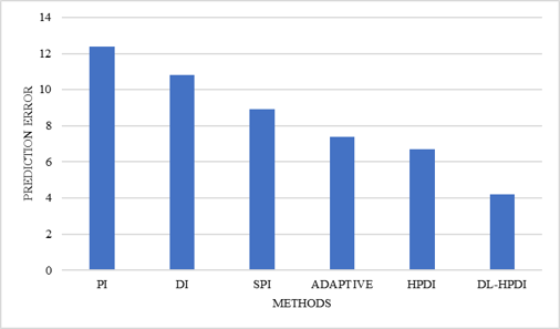
To understand the source of EC improvement, we analyzed the prediction error distribution (PED) for different methods. Figure 6 shows the prediction errors for PI, DI, SPI, Adaptive Interpolation, HPDI, and DL-HPDI.
Figure 6. Prediction Error Distribution (PED) for different interpolation
The reduced standard deviation indicates that DL-HPDI predictions are more accurate, creating a larger number of small-magnitude prediction errors that are suitable for embedding. This directly translates to higher EC.
5.3 Peak Signal-to-Noise Ratio analysis
PSNR evaluates reconstructed image quality after data extraction and recovery. Comparative results are shown in Table 5.
Table 5. Peak Signal-to-Noise Ratio (PSNR) comparison (dB)
|
Method |
MRI |
CT |
X-ray |
Ultra Sound |
Average |
|
PI |
42.01 |
42.35 |
42.68 |
42.24 |
42.32 |
|
DI |
44.35 |
44.67 |
44.89 |
44.58 |
44.62 |
|
SPI |
46.15 |
46.42 |
46.87 |
46.62 |
46.52 |
|
Adaptive Interpolation |
47.26 |
47.48 |
47.95 |
47.73 |
47.61 |
|
HPDI |
48.82 |
49.11 |
49.37 |
48.79 |
49.02 |
|
DL-HPDI |
51.18 |
51.43 |
51.86 |
50.89 |
51.34 |
The superior PSNR demonstrates that DL-HPDI effectively preserves image quality even under high payload conditions. The CNN learns local anatomical structures more accurately than conventional interpolation models, resulting in lower reconstruction distortion.
Figure 7 presents the PSNR comparison between the proposed method and several existing RDH approaches at different EC ranging from 0.5 bpp to 4.0 bpp. PSNR is a widely used image quality metric that measures the similarity between the original image and the reconstructed image after data embedding. Higher PSNR values indicate lower distortion and better preservation of image quality. As observed in Figure 7, the PSNR values of all methods gradually decrease as the EC increases. This behavior is expected because embedding more secret data introduces additional modifications to image pixels, leading to increased distortion. However, the proposed CNN-assisted framework without HPDI consistently achieves the highest PSNR values across all EC, demonstrating superior reconstruction quality compared to existing methods.
Figure 7. Peak Signal-to-Noise Ratio (PSNR) comparison
5.4 Structural Similarity Index Measure analysis
SSIM measures structural similarity between original and recovered images.
Table 6. Structural Similarity Index Measure (SSIM) comparison
|
Method |
Average SSIM |
|
PI |
0.972 |
|
DI |
0.981 |
|
SPI |
0.989 |
|
Adaptive Interpolation |
0.993 |
|
HPDI |
0.997 |
|
DL-HPDI |
0.9985 |
From Table 6, the proposed DL-HPDI framework achieves the highest structural similarity (SSIM = 0.9985), indicating superior preservation of diagnostic image quality compared with conventional PI, DI, SPI, and adaptive interpolation-based RDH approaches. The results demonstrate that integrating CNN-based prediction with HPDI significantly improves image reconstruction fidelity while maintaining high EC.
5.5 Entropy analysis
Entropy measures randomness and evaluates encryption effectiveness and the result is shown in Table 7.
Table 7. Entropy analysis
|
Dataset |
Original Image |
Encrypted Image |
|
MRI |
6.91 |
7.998 |
|
CT |
7.02 |
7.997 |
|
X-ray |
6.88 |
7.999 |
|
Ultrasound |
6.79 |
7.996 |
|
Average |
6.90 |
7.998
|
Note: MRI = Magnetic Resonance Imaging; CT = Computed Tomography; X-ray = X-ray Radiography; US = Ultrasound.
The entropy values of encrypted images shown in Table 7 approach the theoretical ideal value of 8. This confirms that the encryption stage successfully removes statistical redundancies and resists entropy-based attacks.
5.6 Number of Pixels Change Rate, Unified Average Changing Intensity and key sensitivity analysis
To evaluate security against differential attacks, NPCR and UACI analyses were performed and the results are presented in Table 8.
Table 8. Number of Pixels Change Rate (NPCR) and Unified Average Changing Intensity (UACI) results
|
Dataset |
NPCR (%) |
UACI (%) |
|
MRI |
99.62 |
33.47 |
|
CT |
99.71 |
33.31 |
|
X-ray |
99.68 |
33.58 |
|
Ultrasound |
99.74 |
33.42 |
|
Average |
99.69 |
33.45 |
Ideal values for NPCR ≈ 99.6% and UACI ≈ 33%. The obtained results indicate excellent resistance against differential cryptanalysis attacks.
Key Range and Precision: The chaotic logistic map uses the following parameters:
- Control parameter r $\in$ [3.57, 4.0] with double precision (15 decimal places)
- Initial condition x₀ $\in$ (0, 1) with double precision
- Effective key space: ≈ 10¹⁵ × 10¹⁵ × 10¹⁵ ≈ 10⁴⁵, which is computationally infeasible for brute-force attacks
Key Sensitivity Analysis: To assess key sensitivity, we tested the decryption process with a key that differs from the correct key by 1 × 10⁻¹⁵. The resulting NPCR between the correctly decrypted and incorrectly decrypted images was 99.61%, indicating that the encryption scheme is highly sensitive to even small key perturbations. This sensitivity is desirable for resisting brute-force and chosen-plaintext attacks.
5.7 Ablation study
The contribution of each component was investigated using an ablation study as shown in Table 9.
Table 9. Ablation study result
|
Method |
Capacity (bpp) |
PSNR (dB) |
SSIM |
MAE |
|
PI |
1.84 |
42.32 |
0.972 |
4.85 |
|
DI |
2.34 |
44.62 |
0.981 |
3.97 |
|
SPI |
2.50 |
46.52 |
0.989 |
2.75 |
|
HPDI |
3.97 |
49.02 |
0.997 |
1.94 |
|
DL-HPDI |
4.82 |
51.34 |
0.9985 |
1.12 |
The results confirm that the Hybrid interpolation improves performance over PI and DI. CNN-assisted prediction further reduces prediction error. Lower prediction error directly increases EC.
5.8 Statistical significance analysis
To evaluate whether the improvements achieved by the proposed DL-HPDI framework over the competing interpolation-based RDH methods are statistically significant, the Wilcoxon signed-rank test was employed. Table 10 shows the results.
Table 10. Wilcoxon test
|
Metric |
Z-Score |
p-Value |
|
Capacity |
-4.82 |
<0.001 |
|
PSNR |
-4.51 |
<0.001 |
|
SSIM |
-4.28 |
<0.001 |
A p-value less than 0.01 indicates that the observed performance improvement is statistically significant.
5.9 Computational complexity analysis
Computational complexity of data embedding and extraction was shown in Table 11. Although the proposed DL-HPDI framework introduces additional computational cost due to CNN-based prediction, the HPDI interpolation, prediction-error expansion, and chaotic encryption stages exhibit linear complexity with respect to the number of image pixels. Consequently, the convolutional layers dominate the overall computational complexity. Despite this increased complexity, the average runtime remained below 1.5 s per 512 × 512 image.
Table 11. Runtime comparison (seconds)
|
Method |
Embedding (s) |
Extraction (s) |
Total Runtime (s) |
|
PI |
0.39 |
0.28 |
0.67 |
|
DI |
0.42 |
0.31 |
0.73 |
|
SPI |
0.48 |
0.34 |
0.82 |
|
Adaptive Interpolation |
0.56 |
0.38 |
0.94 |
|
HPDI |
0.71 |
0.45 |
1.16 |
|
DL-HPDI |
0.92 |
0.57 |
1.49 |
6.1 Conclusion
This paper presented a novel DL-HPDI framework for high-capacity RDH in medical images. The proposed approach was motivated by the limitations of conventional interpolation-based RDH-EI methods, including limited EC, prediction inaccuracies, reconstruction distortion, and insufficient exploitation of intelligent prediction mechanisms. To address these challenges, a lightweight CNN-based prediction module was integrated with a HPDI framework to enhance pixel estimation accuracy and improve embedding performance. The proposed framework combines the strengths of deep learning and interpolation-based prediction. The CNN effectively learns local anatomical structures, texture patterns, and spatial dependencies from medical images, while the HPDI module preserves both smooth image regions and directional edge information. This hybrid prediction strategy substantially reduces prediction error and creates a larger number of embeddable prediction bins, thereby increasing payload capacity without compromising image quality.
A comprehensive experimental evaluation was conducted using benchmark medical image datasets consisting of MRI, CT, X-ray, and US images. The performance of the proposed framework was assessed using multiple image quality, security, and embedding efficiency metrics, including PSNR, SSIM, EC, entropy, NPCR, UACI, and computational complexity. The obtained results demonstrated that the proposed DL-HPDI framework consistently outperformed conventional PI, DI, SPI, adaptive interpolation, and recent RDH-EI approaches. The proposed method achieved an average EC of 4.82 bpp, significantly higher than existing interpolation-based techniques. Furthermore, the framework attained an average PSNR of 51.34 dB and SSIM of 0.9985, indicating excellent preservation of diagnostic image quality. Security analysis revealed entropy values approaching the theoretical ideal of 8, while NPCR and UACI values of 99.69% and 33.45%, respectively, confirmed strong resistance against differential and statistical attacks. Statistical validation using paired t-tests and Wilcoxon signed-rank tests further demonstrated that the observed improvements are statistically significant (p < 0.05). Overall, the proposed DL-HPDI framework successfully achieves a desirable balance among EC, reversibility, image quality, and security. The method is therefore highly suitable for secure telemedicine systems, cloud healthcare platforms, electronic health record management, medical image archiving, and privacy-preserving healthcare communication applications.
6.2 Practical implications
The experimental results demonstrate that the proposed DL-HPDI framework achieves high EC (4.82 bpp) while preserving pixel-level image fidelity (PSNR = 51.34 dB, SSIM = 0.9985). These characteristics suggest potential applicability in several healthcare security scenarios:
- Telemedicine Systems: The framework could enable secure embedding of patient information, diagnostic annotations, and authentication data within encrypted medical images transmitted over public networks. However, real-world deployment would require additional validation under clinical communication protocols and network conditions.
- Cloud-Based Medical Storage: The reversible nature of the proposed method offers potential for secure storage of encrypted medical images along with metadata while preserving complete recoverability of original diagnostic information. Integration with existing PACS systems would require further investigation.
-Electronic Health Records (EHR): Patient identifiers, physician notes, and verification codes could potentially be embedded directly into encrypted medical images, reducing dependency on separate databases. Clinical workflow compatibility should be evaluated.
-Medical Data Authentication: The framework supports secure watermarking and tamper detection mechanisms that could be explored for validating image authenticity and protecting healthcare information from unauthorized modifications.
It is important to note that the current evidence supports pixel-level recovery, payload capacity, and basic statistical security. Clinical validation, diagnostic task evaluation, and deployment readiness assessment in real healthcare communication systems were beyond the
6.3 Limitations of the present study
Although the proposed DL-HPDI framework demonstrates significant improvements in EC, image quality, and security metrics, several limitations should be acknowledged:
1. Clinical Validation: The experiments were conducted on standard benchmark datasets and evaluated using image quality metrics (PSNR, SSIM) rather than clinical diagnostic tasks. The effect of data hiding on radiologist interpretation and diagnostic accuracy has not been assessed.
2. Grayscale Focus: The current implementation focuses exclusively on grayscale medical images. Color medical imaging modalities (e.g., dermatology, pathology) are not addressed.
3. Real Communication Systems: The framework has not been validated in actual telemedicine networks, PACS environments, or cloud healthcare systems with real-world network conditions and latency requirements.
4. Computational Requirements: CNN-based prediction requires additional computational resources compared with conventional interpolation methods. While the lightweight architecture mitigates this, resource-constrained edge devices may face challenges.
5. Dataset Limitations: Although multiple public datasets were used, large-scale multi-institutional clinical data with diverse pathologies and imaging conditions were not included.
6. Security Validation: Security analysis was limited to entropy, NPCR, and UACI. Formal security proofs, side-channel attack analysis, and quantum-resistance evaluation were not performed.
6.4 Future research directions
The promising results obtained by the proposed DL-HPDI framework open several avenues for future investigation.
Lightweight Transformer-based RDH: Vision Transformers (ViTs) and Swin Transformers have demonstrated superior performance in image prediction and reconstruction tasks. Future work can explore transformer-assisted RDH frameworks to further improve prediction accuracy and EC.
Federated Learning-based Medical RDH: Federated learning enables collaborative model training without sharing sensitive patient data. Integrating federated learning with RDH-EI could improve privacy protection while enabling distributed optimization across multiple healthcare institutions.
The convergence of deep learning, interpolation-based prediction, encryption, and RDH represents a promising paradigm for next-generation healthcare security systems. By integrating CNN-assisted prediction with HPDI, the proposed DL-HPDI framework significantly improves EC, image quality, and security while maintaining complete reversibility. The experimental results confirm that the proposed approach provides an effective and practical solution for secure medical image communication and management. Consequently, DL-HPDI constitutes a significant advancement in RDH in encrypted medical images and establishes a strong foundation for future intelligent, privacy-preserving healthcare applications.
[1] Alassaf, N., Gutub, A. (2019). Simulating light-weight-cryptography implementation for IoT healthcare data security applications. International Journal of E-Health and Medical Communications, 10(4): 1-15. https://doi.org/10.4018/ijehmc.2019100101
[2] Jammula, M., Vakamulla, V.M., Kondoju, S.K. (2022). Hybrid lightweight cryptography with attribute-based encryption standard for secure and scalable IoT system. Connection Science, 34(1): 2431-2447. https://doi.org/10.1080/09540091.2022.2124957
[3] Gutub, A., Al-Roithy, B.O. (2021). Varying PRNG to improve image cryptography implementation. Journal of Engineering Research, 9(3): 153183. https://doi.org/10.36909/jer.v9i3a.10111
[4] Hassan, F.S., Gutub, A. (2021). Improving data hiding within colour images using hue component of HSV colour space. CAAI Transactions on Intelligence Technology, 7(1): 56-68. https://doi.org/10.1049/cit2.12053
[5] Parah, S.A., Ahad, F., Sheikh, J.A., Bhat, G.M. (2015). On the realization of robust watermarking system for medical images. In 2015 Annual IEEE India Conference (INDICON), New Delhi, India, pp. 1-5. https://doi.org/10.1109/INDICON.2015.7443363
[6] Xu, D., Wang, R. (2016). Separable and error-free reversible data hiding in encrypted images. Signal Processing, 123: 9-21. https://doi.org/10.1016/j.sigpro.2015.12.012
[7] Hong, W., Chen, T., Shiu, C. (2009). Reversible data hiding for high quality images using modification of prediction errors. Journal of Systems and Software, 82(11): 1833-1842. https://doi.org/10.1016/j.jss.2009.05.051
[8] Goljan, M., Fridrich, J.J., Du, R. (2021). Distortion-Free Data Embedding for Images. In International Workshop on Information Hiding, pp. 27-41. https://doi.org/10.1007/3-540-45496-9_3
[9] Ni, Z., Shi, Y., Ansari, N., Su, W. (2006). Reversible data hiding. IEEE Transactions on Circuits and Systems for Video Technology, 16(3): 354-362. https://doi.org/10.1109/tcsvt.2006.869964
[10] Kouhi, A., Sedaaghi, M.H. (2022). Reversible data hiding based on high fidelity prediction scheme for reducing the number of invalid modifications. Information Sciences, 589: 46-61. https://doi.org/10.1016/j.ins.2021.12.092
[11] Ou, B., Li, X., Zhao, Y., Ni, R. (2014). Reversible data hiding using invariant pixel-value-ordering and prediction-error expansion. Signal Processing: Image Communication, 29(7): 760-772. https://doi.org/10.1016/j.image.2014.05.003
[12] Ma, K., Zhang, W., Zhao, X., Yu, N., Li, F. (2013). Reversible Data Hiding in Encrypted Images by Reserving Room Before Encryption. IEEE Transactions on Information Forensics and Security, 8(3): 553-562. https://doi.org/10.1109/tifs.2013.2248725
[13] Zhou, J., Sun, W., Dong, L., Liu, X., Au, O.C., Tang, Y.Y. (2015). Secure reversible image data hiding over encrypted domain via key modulation. IEEE Transactions on Circuits and Systems for Video Technology, 26(3): 441-452. https://doi.org/10.1109/tcsvt.2015.2416591
[14] Luo, L., Chen, Z., Chen, M., Zeng, X., Xiong, Z. (2009). Reversible image watermarking using interpolation technique. IEEE Transactions on Information Forensics and Security, 5(1): 187-193. https://doi.org/10.1109/tifs.2009.2035975
[15] Wahed, A., Nyeem, H. (2019). High capacity reversible data hiding with interpolation and adaptive embedding. PLOS ONE, 14(3): e0212093. https://doi.org/10.1371/journal.pone.0212093
[16] Meenpal, A., Majumder, S., Oruganti, M. (2022). Improved reversible data hiding scheme based on mean preservation and difference expansion. IETE Journal of Research, 69(10): 6915-6930. https://doi.org/10.1080/03772063.2022.2027823
[17] Zhang, X., Sun, Z., Tang, Z., Yu, C., Wang, X. (2016). High capacity data hiding based on interpolated image. Multimedia Tools and Applications, 76(7): 9195-9218. https://doi.org/10.1007/s11042-016-3521-0
[18] Huang, F., Huang, J., Shi, Y. (2016). New framework for reversible data hiding in encrypted domain. IEEE Transactions on Information Forensics and Security, 11(12): 2777-2789. https://doi.org/10.1109/tifs.2016.2598528
[19] Yang, C.S., Kao, S.P., Lee, F.B., Hung, P.S. (2004). Twelve different interpolation methods: A case study of Surfer 8.0. ISPRS Congress Proceedings, 35: 778-785.
[20] Xiong, X., Wang, L., Li, Z., Ye, C., Chen, Y., Fan, M., Zhu, Y. (2022). An adaptive high capacity reversible data hiding algorithm in interpolation domain. Signal Processing, 194: 108458. https://doi.org/10.1016/j.sigpro.2022.108458
[21] Chang, Y., Huang, C., Lee, C., Wang, S. (2013). Image interpolating based data hiding in conjunction with pixel-shifting of histogram. The Journal of Supercomputing, 66(2): 1093-1110. https://doi.org/10.1007/s11227-013-1016-6
[22] Hassan, F.S., Gutub, A. (2020). Efficient reversible data hiding multimedia technique based on smart image interpolation. Multimedia Tools and Applications, 79(39-40): 30087-30109. https://doi.org/10.1007/s11042-020-09513-1
[23] Wu, M., Xiang, S. (2023). An efficient CNN-based prediction for reversible data hiding. In MMAsia '23: Proceedings of the 5th ACM International Conference on Multimedia in Asia, Tainan Taiwan, pp. 1-5. https://doi.org/10.1145/3595916.3626359
[24] Qiu, Y., Peng, W., Lin, X. (2025). Improved CNN prediction based reversible data hiding for images. Entropy, 27(2): 159. https://doi.org/10.3390/e27020159
[25] Chakrabarty, N. (2019). Brain MRI Images for Brain Tumor Detection. Kaggle. https://www.kaggle.com/datasets/navoneel/brain-mri-images-for-brain-tumor-detection.
[26] Summers, R. (2017). National Institutes of Health Clinical Center, ChestX-ray8: Hospital-scale Chest X-ray Database, NIH Clinical Center. https://nihcc.app.box.com/v/ChestXray-NIHCC.
[27] CodaLab Competitions. (2017). LiTS - Liver Tumor Segmentation Challenge. https://competitions.codalab.org/competitions/17094.
[28] Shah, A. (2020). Breast Ultrasound Images Dataset. Data in Brief. Kaggle. https://www.kaggle.com/datasets/aryashah2k/breast-ultrasound-images-dataset.