İsa Avcı*![]() | Ahmed Alzabaq
| Ahmed Alzabaq![]()
© 2023 IIETA. This article is published by IIETA and is licensed under the CC BY 4.0 license (http://creativecommons.org/licenses/by/4.0/).
OPEN ACCESS
Convolutional Neural Network (CNN)-based deep learning techniques have recently demonstrated increased potential and effectiveness in image recognition applications, such as those involving medical images. Deep-learning models can recognize targets with performance comparable to radiologists when used with CXR. The primary goal of this research is to examine a deep learning technique used on the radiography dataset to detect COVID-19 in X-ray medical images. The proposed system consists of several stages, from pre-processing, passing through the feature reduction using more than one technique, to the classification stage based on a proposed model. The test was applied to the COVID-19 Radiography dataset of normal and three lung infections (COVID-19, Viral Pneumonia, and Lung Opacity). The proposed CNN model has shown its ability to classify COVID, normal, and other lung infections with perfect accuracy of 99.94%. Consequently, the AI-based early-stage detection algorithms will be enhanced, increasing the accuracy of the X-ray-based modality for the screening of various lung diseases.
COVID-19, convolutional neural network (CNN), linear discriminant analysis (LDA), gray level co-occurrence matrix (GLCM), radiography
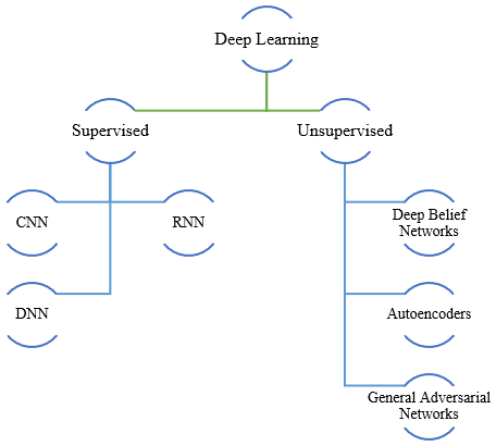
Artificial intelligence is a new subject that focuses on building models from data, and in recent years, it has become more widely used in the creation of tools to support specialists in the interpretation of medical images [1]. Pneumonia is a lung inflammation that mostly affects the alveoli, which are tiny air sacs, and is typically brought on by a bacterial or viral infection. Chest pain, breathing difficulties, fever, and coughing up phlegm are common symptoms of pneumonia, however, their severity might vary. On chest radiographs, pneumonia typically appears as an area of opacity (CXRs). The diagnosis of pneumonia is challenging, though, because increased opacity on CXRs may be caused by several different lung diseases, including pulmonary edema, hemorrhage, and volume loss [2]. The unique COVID-19 virus, which started from animals and quickly spread over the world, was discovered in Wuhan Province, China, in December 2019. Physical contact, such as shaking hands with an infected person, as well as airborne transmission of COVID-19 are the most straightforward methods [3]. Through the respiratory system, the virus enters lung cells, replicates there, and kills those cells. Due to its RNA composition and mutational features, COVID-19 is exceedingly challenging to diagnose and treat [4]. The most typical COVID-19 signs and symptoms are fever, coughing, shortness of breath, lightheadedness, headaches, and muscle aches [5]. The virus is extremely dangerous and can cause those with compromised immune systems to pass away [6]. While chest CT can be an early screening technique for COVID-19, there are considerable imaging similarities between this and other types of viral and inflammatory lung disease. As a result, distinguishing COVID-19 from other viral pneumonia is difficult [7]. Radiologists can also take their time identifying the features. Furthermore, manual reading of CT images is a time-consuming and exhausting activity that leads to human mistakes [8]. Thus, technology based on artificial intelligence (AI)-based automated analysis can aid radiologists in the analysis of COVID-19 from CT scans. Deep learning (DL) is a significant milestone in AI [9]. One of the most used DL designs is the convolutional neural network (CNN) [10]. CNN has been frequently used in the health sector due to its powerful properties [11]. CNN approaches, in conjunction with radiological imaging, can aid in the accurate detection and categorization of COVID-19 [12]. A CT-Classification technique was used to classify recent CNN pictures of COVID-19 and Non-COVID-19 [13]. Figure 1 is depicted various DL approaches for COVID-19 detection [14].
Figure 1. Respiratory disease detection methods based on DL [14]
The remainder of the paper is organized as follows: Section 2 included a brief review of the literature. Section 3 describes the proposed approach as well as the experimental setup, model training, and evaluation. Section 4 presents the analysis and discussion of the analysis and discussion of the results.
Many researchers have recently conducted studies in the medical field, particularly in medical image processing techniques. They used Machine Learning (ML) and Deep Learning (DL) techniques, among other things. ML technologies are widely recognized as important tools for improving disease prediction and diagnosis. However, efficient extraction approaches are required to create improved ML models. DL models are commonly used in medical imaging systems because they can automatically extract features or use pre-trained networks.
Asif et al. [15] tried to use digitized chest x-ray images to automatically identify COVID-19 pneumonia patients while improving detection accuracy with deep convolutional neural networks (CNN) in 2020. The collection consists of 864 COVID-19 images, 1345 images of viral pneumonia, and 1341 images of regular chest x-rays. This work suggests the CNN-based model Inception V3 with transfer learning for the identification of patients with coronavirus pneumonia utilizing chest X-ray radiographs, and it obtains a classification accuracy of more than 98%.
Hussain and Shiren [16] used chest X-rays to test Convolutional Neural Networks (CNNs) for COVID-19 classification. In this study, the effectiveness of CNN with one, three, and four convolution layers are compared. 13,808 CXR images make up the dataset used in this investigation. Their initial test findings show that the CNN model with three convolution layers can reliably recognize objects on X-ray pictures with 96% accuracy using three divisions of the dataset.
Tang et al. [17] observed that deep CNNs could accurately and effectively differentiate between normal and abnormal chest radiographs in this investigation, where they also obtained good diagnostic accuracy. The CNN model, trained on adult patient datasets and refined on pediatric patient datasets, achieved an accuracy of 94.64%, sensitivity of 96.5%, and specificity of 92.86% for classifying normal from pneumonia.
Pandit et al. [18] showed a model with 1428 chest radiographs of healthy individuals and patients with common bacterial pneumonia who tested positive for COVID-19 (no infection). Using the pre-trained VGG16 model, the network was successfully trained on comparatively tiny chest radiographs for classification tasks in this study. The accuracy rates for the study were 96% and 92.5% for the two classes (COVID and non-COVID) and three output classes (COVID, non-COVID pneumonia, and normal).
Jadon et al. [19] tested well-known data scarcity techniques in deep learning to detect COVID-19 in 2021. Data augmentation, transfer learning, few-shot learning, and unsupervised learning are examples of these. They also proposed a bespoke few-shot learning strategy employing Siamese networks to detect COVID-19. Their experimental results demonstrated that by using few-shot learning methodologies, they could develop an efficient and accurate deep-learning model for COVID-19 identification with less data. The accuracy attains 96.4% utilizing the proposed strategy, up from 83% using baseline models.
Uddin et al. [20] presented an evaluation of a Convolutional Neural Network (CNN) to detect COVID-19 from chest X-ray (CXR) images, which makes the test faster and more reliable. The designed model focuses on enhancing accuracy and employs a transfer learning approach as well as a custom model. Deep feature extraction has been accomplished using pre-trained deep CNN models such as VGG16, InceptionV3, MobileNetV2, and ResNet50. Among the CNN models used, InceptionV3 has the highest accuracy of 98%.
Mahesh et al. [21] developed and presented research on how Convolutional Neural Networks (CNNs) perform very well in the identification of COVID images. The CNNs have the potential to diagnose respiratory disorders with the highest accuracy, despite the requirement for a large number of pictures, with an accuracy of 95% train accuracy and 98% validation accuracy.
Yang et al. [22] employed transfer learning techniques to overcome the shortage of data and decrease the training time. The modified VGG16 deep transfer learning architecture was utilized to perform binary and multi-class classification of X-ray image issues. Enhanced VGG16 detected COVID-19 and pneumonia X-ray pictures with 99% accuracy. The algorithms’ accuracy and validity were assessed using well-known public datasets from X-ray and CT-scan.
Arias-Garzón et al. [23] processed the images and classified them as positive or negative for COVID-19 using current deep learning models (VGG19 and U-Net). The proposed system consists of three main components: a classification model trained using a transfer learning scheme; a preprocessing stage with lung segmentation, which eliminates the environs that do not include relevant information for the activity and that may produce biased results; and finally, results in analysis and interpretation via heat maps visualization. The COVID-19 detection accuracy of the best models was about 97%.
In 2022, Hossain et al. [24] showed a refined ResNet50 model that accurately identified COVID-19 from chest X-ray images using transfer learning. To upgrade the ResNet50 model, they added two more completely connected layers to the original ResNet50 model. They employed ten distinct pre-trained weights that were developed using supervised learning, self-supervised learning, and other techniques on a range of large-scale datasets. The suggested work obtained 99.17% validation accuracy and 99.95% train accuracy for COVID instances in the two-class classification (COVID and Normal).
Boulila et al. [25] intended to maximize the utility of chest X-ray images without jeopardizing the privacy of the data contained in them. The recommended approach consists of two steps: training/testing the DL algorithm on the encrypted images, then encrypting the dataset with partially homomorphic encryption. In tests utilizing the COVID-19 Radiography dataset, the MobileNetV2 model achieves an accuracy of 94.2% over plain data and 93.3% over encrypted data.
Abdul Gafoor et al. [26] used Deep Learning Multi-layered networks to classify the chest images as COVID positive or negative. The suggested model is based on a dataset of Coronavirus-infected individuals in whom the radiologist noted multilobar involvements in chest X-rays. The research analyzed 6500 photos in total. The Convolutional Neural Network (CNN) model was trained, and a validation accuracy of 94% was attained.
Asif et al. [27] presented a robust deep-learning strategy for identifying COVID-19 patients from chest X-rays, with high accuracy and a low false negative rate. The suggested approach is a lightweight shallow CNN with optimum parameters for identifying COVID-19 instances from chest X-ray pictures. The study makes use of open-source chest X-rays from healthy persons and COVID-19 patients. In the classification of healthy and COVID-19-infected individuals, their model achieves 99.68% accuracy.
Sarki et al. [28] presented a DL-focused technique for classifying and detecting COVID-19 instances in x-ray pictures. Their model is completely automated and can categorize binary classes with 100% accuracy using VGG16 and multi-class classes with 93.75% accuracy using a constructed CNN.
Hashmi et al. [29] suggested a model that is effective for detecting pneumonia and is trained on digital chest X-ray pictures. This model might help radiologists in their decision-making. The weighted predictions from cutting-edge deep learning models like ResNet18, Xception, InceptionV3, DenseNet121, and MobileNetV3 are combined in an ideal manner via a revolutionary method based on a weighted classifier. This method is a form of supervised learning in which the network forecasts the outcome based on the caliber of the training dataset. The deep learning models are tuned using transfer learning to achieve an accuracy of 98.43%.
Karar et al. [30] suggested a deep learning framework that includes two significant improvements. First, a set of binary classifiers have been used to streamline the challenging multi-label classification of X-ray pictures for each tested case of health condition. To diagnose probable ailments for a patient, that simulates a clinical setting. Second, the COVID-19 and pneumonia classifiers’ cascaded architecture allows for the flexible deployment of numerous finely calibrated deep learning models at once, resulting in the best performance in terms of confirming infected patients. Eleven pre-trained convolutional neural network models are used in this investigation. The proposed classifiers’ findings demonstrated the best detection accuracy of 99.9%.
Brunese et al. [31] presented a three-phased strategy, the first of which is to determine whether pneumonia can be seen on a chest X-ray. Differentiating between COVID-19 and pneumonia is the second. The final phase aims to identify the regions in the X-ray indicative of COVID-19 presence. The efficiency of the suggested approach was proved through experimental analysis of 6523 chest X-rays from various institutions. The average time for COVID-19 identification was 2.5 seconds, and the average accuracy was 97%.
Apostolopoulos et al. [32] examined the significance of the retrieved features for the classification job, the cutting-edge convolutional neural network known as Mobile Net is used and trained from scratch. For training MobileNet v2, which has demonstrated great performance in similar tasks, a sizable dataset of 3905 X-ray pictures pertaining to 6 diseases is used. A classification accuracy of 87.66% is attained among the seven classes.
Mabrouk et al. [33] presented Ensemble Learning (EL), a computer-aided classification of pneumonia that aims to make chest X-ray image diagnosis simpler. Instead of constructing CNN models from scratch, their idea is based on existing Convolutional Neural Network (CNN) models, which have recently been used to improve the performance of numerous medical tasks. The chest X-ray data set is used to train the suggested model. The proposed EL method beats other cutting-edge techniques currently in use and achieves an accuracy of 93.91%.
Ullah et al. [34], suggested a brand-new CovidDetNet classification strategy to accurately and quickly detect COVID-19 from chest radiograph images. The generated images from image resizing are then fed into a CovidDetNet model created to detect COVID-19 for this purpose. COVID-19 detection has a 98.40% accuracy rate. All results are compared in Table 1.
Table 1. Summary of existing studies related to COVID-19 detection based on CNN models
|
Ref. no. |
Dataset |
Technique |
Best Accuracy |
|
[15] |
COVID-19 Radiography |
Convolutional Neural Networks (CNN) |
Up to 98% |
|
[16] |
COVID-19 Radiography |
Convolutional Neural Networks (CNN) |
96% |
|
[17] |
COVID-19 Radiography |
Convolutional Neural Networks (CNN) |
94.64% |
|
[18] |
COVID-19 Radiography |
CNN model (VGG16) |
96% |
|
[19] |
COVID-19 Radiography |
Convolutional Neural Networks (CNN) |
96.4% |
|
[20] |
COVID-19 Radiography |
CNN model (InceptionV3) |
98% |
|
[21] |
COVID-19 Radiography |
Convolutional Neural Networks (CNN) |
98% |
|
[22] |
COVID-19 Radiography |
CNN model (VGG16) |
99% |
|
[23] |
COVID-19 Radiography |
CNN models (VGG19 and U-Net) |
97% |
|
[24] |
COVID-19 Radiography |
CNN model (ResNet50) |
99.17% |
|
[25] |
COVID-19 Radiography |
CNN model (MobileNetV2) |
94.2% |
|
[26] |
COVID-19 Radiography |
Convolutional Neural Networks (CNN) |
94% |
|
[27] |
COVID-19 Radiography |
Convolutional Neural Networks (CNN) |
99.68% |
|
[28] |
COVID-19 Radiography |
CNN model (VGG16) |
93.75% |
|
[29] |
COVID-19 Radiography |
ResNet18, Xception, InceptionV3, DenseNet121, and MobileNetV3 |
98.43% |
|
[30] |
COVID-19 Radiography |
Convolutional Neural Networks (CNN) |
99.9% |
|
[31] |
COVID-19 Radiography |
Convolutional Neural Networks (CNN) |
97% |
|
[32] |
COVID-19 Radiography |
Convolutional Neural Networks (CNN) |
87.66% |
|
[33] |
COVID-19 Radiography |
Convolutional Neural Networks (CNN) |
93.91% |
|
[34] |
COVID-19 Radiography |
Convolutional Neural Networks (CNN) |
98.40% |
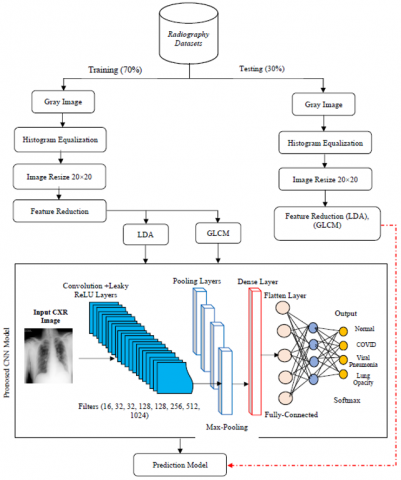
The proposed system which is shown in Figure 2 aims to detect infection with the Coronavirus through X-ray images using a COVID-19 Radiography dataset. The proposed system goes through several stages, where the images are processed in several ways, and then the features are reduced in more than one way such as LDA and GLCM. Then, the proposed Convolutional Neural Network (CNN) is utilized to classify cases as patients with (COVID-19, Viral Pneumonia, or Lung Opacity) from healthy people as the final stage in the system.
Figure 2. The proposed detection model is based on CNN
3.1 Dataset description
Table 2. COVID-19 radiography dataset categories
|
|
Category |
No. of Chest X-ray (CXR) Images |
|
First release |
COVID-19 |
219 |
|
Normal |
1341 |
|
|
Viral pneumonia |
1345 |
|
|
Final release |
COVID-19 |
3616 |
|
Normal |
10.192 |
|
|
Viral Pneumonia |
1345 |
|
|
Lung Opacity (Non-COVID lung infection) |
6012 |
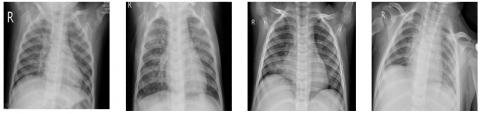
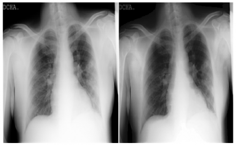
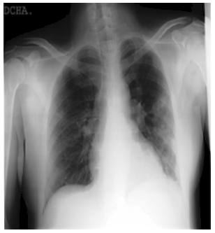
Together with collaborators from Pakistan and Malaysia, a research team from Qatar University in Doha, Qatar, and the University of Dhaka in Bangladesh has created a database of chest X-ray images for COVID-19-positive patients as well as photos of typical and viral pneumonia. This dataset of COVID-19, normal, and other lung infections is being provided in phases. Table 2 shows the details of the COVID-19 Radiography Dataset. Some COVID and Normal patients’ CXR images of this dataset are shown in Figure 3 to Figure 6.
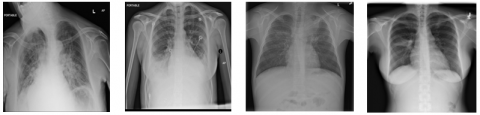
The normal chest X-ray shows clean lungs with no spots of aberrant opacification. Lung opacity is characterized by a localized lobar consolidation, in this case in the right upper lobe (white arrows), whereas viral pneumonia is characterized by a more widespread “interstitial” pattern in both lungs. Pictures that do not meet the aforementioned characteristics are classified as having COVID-19 disease.
Figure 3. Normal CXR image example from the COVID-19 Radiography Database
Figure 4. COVID CXR image example from the COVID-19 Radiography Database
Figure 5. Viral Pneumonia CXR image example from the COVID-19 Radiography Database
Figure 6. Lung Opacity CXR image example from the COVID-19 Radiography Database
3.2 Splitting COVID-19 dataset
Data splitting is a standard strategy for model validation that divides a given dataset into training and testing sets. Following that, the training data is utilized to fit and assess statistics and machine learning models. It can test and evaluate the accuracy of various models’ predictions without worrying about potential overfitting of the training set if a separate set of data is kept for validation [35]. It can use the aforementioned data splitting algorithms after defining a splitting ratio. The 80:20 rule states that 80% of the data is utilized for training and 20% for testing [36]. Alternative ratios, such as 70:30, 60:40, and even 50:50, are employed in practice. There appears to be no clear information on what ratio is excellent or best for a certain dataset. The 80:20 split is based on the well-known Pareto principle; however, it is only a guideline based on experience. There is currently no consensus on the appropriate data-splitting ratio based on theoretical or numerical research [37].
3.3 Data preprocessing
Data may be seen as the model algorithm quickly analyzing the aspects of the data. Data pre-processing is the most crucial and crucial step for a deep learning algorithm to function well in terms of generalization [38]. This stage includes a group of sub-processes as follows:
3.3.1 Histogram equalization
Histogram equalization is a spatial domain contrast enhancement strategy in image processing that uses the image’s histogram. Histogram equalization frequently increases the processed image’s global contrast. This technique works nicely with both bright and dark images [39]. The histogram was done by applying Eq. (1) as follows [40]:
$h[i]=\sum_{x=1}^N \sum_{y=1}^M\left\{\begin{array}{cc}0 & \text { if } f[x \cdot y]=i \\ 1 & \text { otherwise }\end{array}\right\}$ (1)
The cumulative distribution is then calculated by Eqns. (2) and (3) as follows:
$\operatorname{cdf}\left(X_i\right)=\sum_{i=0}^k p\left(X_i\right)$ (2)
$g[x . y]=\frac{C D F\left[f[x . y]-C D F_{\min }\right]}{(N \times M)-C D F_{\min }} \times(L-1)$ (3)
For gray-level values from 0 to 7, the minimum value which corresponds to zero calculates the number of 0, 1, 2, 3, 4, 5, 6, and 7 in an image and then finds the total number of pixels. Then, the running sum of each gray-level value was computed. Normalize by dividing by the total number of pixels, the resulting values ranged between 0.1 to 1. It is applied grayscale image and histogram equalization on the XRY image in Figure 7.
Figure 7. (a) Grayscale image (b) Equalize the CXR image
3.3.2 Image resize
Image resizing technology is quickly becoming a research hotspot due to the advent of varied display device sizes in recent years, which unavoidably results in images being reduced in size or enlarged (a process known as resizing). This method considers the content of the image in addition to the geometry size limits when resizing the images because each content requires various approaches. To accomplish the goal of resizing, high degrees of key parts are kept as nearly unchanged as feasible while less important areas are modified a few sizes larger. Image retargeting is another name for the practice of image resizing [41]. In this proposed system, the image was resized by 20 * 20 as shown in Figure 8.
Figure 8. 20×20 image resize
3.4 Feature reduction
Features are the specific information needed to address a particular application and represent important aspects of images. The training set requirements and classification accuracy are significantly impacted by the choice of input characteristics. The procedure of extracting features from photographs reduces the number of resources needed by capturing the visual content of the images [42]. The Linear Discriminant Analysis (LDA), and Gray Level Co-occurrence Matrix (GLCM) were used as feature reduction techniques.
3.4.1 Linear discriminant analysis (LDA)
A statistical, machine learning, and pattern recognition method for discovering linear combinations of characteristics that separate or describe at least two classes of events or objects, LDA is an extension of Fisher’s linear discriminant. The final result could be applied as a linear classifier or, more crucially, for dimensionality reduction before further classification. Projecting the original data matrix into a lower-dimensional space is the main objective of the LDA approach [43].
3.4.2 Gray level co-occurrence matrix (GLCM)
A simple method called GLCM is used to extract from images the textural properties including Energy, Contrast, Correlation, and Homogeneity. When used in calculations, the histogram solely conveys data about density distribution; it contains no information about the spatial relationships between pixels. One statistical method is the use of co-occurrence matrices to extract features that indicate the distribution of intensities and relative locations of nearby pixels in a picture [44]. If image H of dimension M×M, the co-occurrence matrix C can be written as [45]:
$\begin{aligned} & c(i . k) =\sum_{y=1}^M \sum_{z=1}^M\left\{\begin{array}{ll}1 & \text { if } H(y \cdot z)=i \text { and } H(y+\Delta y \cdot z+\Delta z)=j \\ 0 & \text { otherwise }\end{array}\right\}\end{aligned}$ (4)
where, (∆y, ∆z) denotes the separation between the relevant pixel and its neighbor.
3.5 Classify positive and negative cases using the proposed convolutional neural network (CNN) model
The proposed CNN model consists of 28 layers as follows:
·Convolutional Neural Network (CNN) (8) layers.
·Leaky ReLU (8) layers.
·Max Pooling (8) layers.
·Flatten (1) layer.
·Dense (3) layer.
Explains these layers in some detail in Table 3.
Table 3. The proposed model CNN layers
|
No. |
Layer Type |
Filters |
Size/Stride |
Activation Function |
|
1 |
Convolutional |
16 |
3/1 |
ــ |
|
2 |
Leaky ReLU |
ــ |
ــ |
ــ |
|
3 |
Max Pooling |
ــ |
1/1 |
ــ |
|
4 |
Convolutional |
32 |
3/1 |
ــ |
|
5 |
Leaky ReLU |
ــ |
ــ |
ــ |
|
6 |
Max Pooling |
ــ |
1/1 |
ــ |
|
7 |
Convolutional |
32 |
3/1 |
ــ |
|
8 |
Leaky ReLU |
ــ |
ــ |
ــ |
|
9 |
Max Pooling |
ــ |
1/1 |
ــ |
|
10 |
Convolutional |
128 |
3/1 |
ــ |
|
11 |
Leaky ReLU |
ــ |
ــ |
ــ |
|
12 |
Leaky ReLU |
ــ |
ــ |
ــ |
|
13 |
Max Pooling |
ــ |
1/1 |
ــ |
|
14 |
Dense |
ــ |
ــ |
Linear |
|
15 |
Convolutional |
256 |
3/1 |
ــ |
|
16 |
Leaky ReLU |
ــ |
ــ |
ــ |
|
17 |
Max Pooling |
ــ |
1/1 |
ــ |
|
18 |
Convolutional |
512 |
3/1 |
|
|
19 |
Leaky ReLU |
ــ |
ــ |
ــ |
|
20 |
Max Pooling |
ــ |
1/1 |
ــ |
|
21 |
Convolutional |
1024 |
3/1 |
|
|
22 |
Leaky ReLU |
ــ |
ــ |
ــ |
|
23 |
Max Pooling |
ــ |
1/1 |
ــ |
|
24 |
Dense |
ــ |
ــ |
Linear |
|
25 |
Convolutional |
50 |
3/1 |
Linear |
|
26 |
Max Pooling |
ــ |
1/1 |
ــ |
|
27 |
Flatten |
ــ |
ــ |
ــ |
|
28 |
Dense |
ــ |
ــ |
Softmax |
The common evaluation techniques for the classification model are Confusion Matrix, Precision, Recall, F1 Score, and Accuracy. The Confusion Matrix, as its name suggests, produces a matrix and describes the entire performance of the model. True Positive (TP), False Positive (FP), False Negative (FN), and True Negative are among its four terminologies (TN) [46]. The following formulas are used to determine the models:
Accuracy $=\frac{T P+T N}{T P+T N+F P+F N}$ (5)
Precision $=\frac{T P}{T P+F P}$ (6)
Recall $=\frac{T P}{T P+F N}$ (7)
$F-$ score $=2 * \frac{\text { precision } * \text { recall }}{\text { precision }+ \text { recall }}$ (8)
In this study, CXR images are examined. In this research project, 30% of the data are used for testing and 70% are used for training. Using the corresponding suggested CNN model classify normal and COVID-19 images. The performance outcomes of our suggested CNN are explained in Table 4.
Table 4. Results of the proposed CNN model
|
Accuracy |
Precision |
Recall |
F1-Score |
|
99.94% |
100% |
100% |
100% |
The proposed network has been designed with high accuracy which contributed to obtaining the highest accuracy of disease identification compared to other studies depending on deep learning, specifically the convolutional neural network. In addition to the high impact of preprocessing and feature reduction techniques that are the basis for obtaining this high rate of detection. The accuracy increased when processing pre-processing, extracting features and building the new model. Therefore, if the processing operations are canceled, it will not be possible to extract the best features, and therefore, it is impossible to build a new model. Therefore we will rely on the traditional processing conducted by the convolutional neural network, and we will obtain results similar to the rest of the studies.
4.1 Comparison results
Numerous investigations have been carried out in the last two years to identify and categorize COVID-19. For this problem, researchers are searching for a more practical answer. In this section, given in Table 5, we looked at and contrasted our proposed model with previously published, comparable studies.
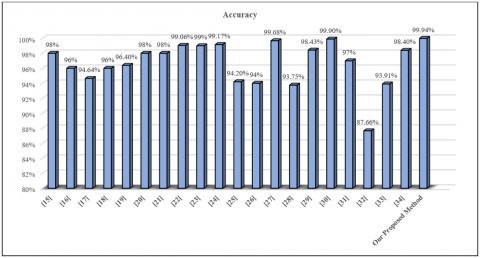
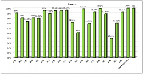
From Table 5 it is clear that the proposed CNN model outperformed the rest of the models to work on the same COVID-19 Radiography dataset to detect respiratory system infection. The proposed system helps with early and accurate detection, contributing to the possibility of increasing treatment and increasing the number of recoveries from the disease. The reason may be due to the existence of some errors in predicting the disease for not using pre-processing methods for the data, in addition to using more than one method to extract features in the proposed method, which allows for obtaining the best features which increase the accuracy and speed of prediction. Figure 9 to Figure 12 respectively explain the accuracy, precision, recall, and f-score comparison with related studies that are mentioned in section 2.
Figure 9. Accuracy comparisons
Table 5. Comparison results with related studies
|
Ref. No. |
Technique |
Dataset |
Accuracy |
Precision |
Recall |
F-score |
|
[15] |
CNN |
COVID-19 Radiography |
98% |
98.2% |
97% |
98% |
|
[16] |
CNN |
COVID-19 Radiography |
96% |
96% |
94% |
96% |
|
[17] |
CNN |
COVID-19 Radiography |
94.64% |
96.52% |
96.50% |
94.63% |
|
[18] |
CNN |
COVID-19 Radiography |
96% |
97.27% |
92.64% |
96% |
|
[19] |
CNN |
COVID-19 Radiography |
96.4% |
96.5% |
96.2% |
95.9% |
|
[20] |
CNN |
COVID-19 Radiography |
98% |
99% |
100% |
99% |
|
[21] |
CNN |
COVID-19 Radiography |
98% |
96.4% |
100% |
97.9% |
|
[22] |
CNN |
COVID-19 Radiography |
99.06% |
99.07% |
99.06% |
99.06% |
|
[23] |
CNN |
COVID-19 Radiography |
99% |
99% |
99% |
99% |
|
[24] |
CNN |
COVID-19 Radiography |
99.17% |
99.31% |
99.03% |
99.17% |
|
[25] |
CNN |
COVID-19 Radiography |
94.2% |
94.4% |
94.2% |
94.2% |
|
[26] |
CNN |
COVID-19 Radiography |
94% |
90% |
90% |
90% |
|
[27] |
CNN |
COVID-19 Radiography |
99.68% |
99.66% |
99.66% |
99.65% |
|
[28] |
CNN |
COVID-19 Radiography |
93.75% |
95.45% |
100% |
93.70% |
|
[29] |
CNN |
COVID-19 Radiography |
98.43% |
98.26% |
99% |
98.43% |
|
[30] |
CNN |
COVID-19 Radiography |
99.9% |
99.89% |
99.9% |
99.9% |
|
[31] |
CNN |
COVID-19 Radiography |
97% |
97.2% |
98% |
97.67% |
|
[32] |
CNN |
COVID-19 Radiography |
87.66% |
88.21% |
87.34% |
87.65% |
|
[33] |
CNN |
COVID-19 Radiography |
93.91% |
93.89% |
93.99% |
93.92% |
|
[34] |
CNN |
COVID-19 Radiography |
98.40% |
98.34% |
98.56% |
98.41% |
|
Our Proposed Met-hod |
Propos-ed CNN |
COVID-19 Radiography |
99.94% |
100% |
100% |
100% |
Figure 10. Precision comparisons
Figure 11. Recall comparisons
Figure 12. F-score comparisons
Lung disease is one of the diseases that can be healed if caught early enough. Early detection can lead to a cure. However, most patients fail to discover their ailment before it becomes chronic, raising the global death toll. Image Classification is obtaining information from an image, which is important in medical image classification. The convolutional Neural Network (CNN) model is a deep learning architecture used to classify lung disease correctly. Lung opacity extends throughout the human body's lung area. This infection is diagnosed via a chest x-ray. Doctors use this x-ray picture to make diagnoses or track the progress of lung opacity treatments. In the current study, we discussed a deep-based approach for COVID-19 subject prediction using X-ray images and the deep learning approach. With a perfect accuracy of 99.94%, the suggested CNN model has demonstrated its capacity to identify the COVID, normal, and other lung infections classes. As a result, the early-stage detection algorithms based on AI will be improved, making the X-ray-based modality more reliable in screening different lung illnesses. We will test the suggested approach on various pulmonary diseases for future work.
[1] Sethi, R., Mehrotra, M., Sethi, D. (2020). Deep learning based diagnosis recommendation for COVID-19 using chest X-rays images. In 2020 Second International Conference on Inventive Research in Computing Applications (ICIRCA), Coimbatore, India, pp. 1-4. https://doi.org/10.1109/ICIRCA48905.2020.9183278
[2] Wang, L., Lin, Z.Q., Wong, A. (2020). Covid-net: A tailored deep convolutional neural network design for detection of covid-19 cases from chest x-ray images. Scientific Reports, 10(1): 1-12. https://doi.org/10.1038/s41598-020-76550-z
[3] Asif, S., Wenhui, Y., Jin, H., Jinhai, S. (2020). Classification of COVID-19 from chest X-ray images using deep convolutional neural network. In 2020 IEEE 6th international conference on computer and communications (ICCC), Chengdu, China, pp. 426-433. https://doi.org/10.1109/ICCC51575.2020.9344870
[4] Rajaraman, S., Siegelman, J., Alderson, P.O., Folio, L.S., Folio, L.R., Antani, S.K. (2020). Iteratively pruned deep learning ensembles for COVID-19 detection in chest X-rays. IEEE Access, 8: 115041-115050. https://doi.org/10.1109/ACCESS.2020.3003810
[5] Song, Y., Zheng, S., Li, L., Zhang, X., Zhang, X., Huang, Z., Chen, J.W., Wang, R.X., Zhao, H.Y., Chong, Y.T., Shen, J., Zha, Y.F., Yang, Y. (2021). Deep learning enables accurate diagnosis of novel coronavirus (COVID-19) with CT images. IEEE/ACM Transactions on Computational Biology and Bioinformatics, 18(6): 2775-2780. https://doi.org/10.1109/TCBB.2021.3065361
[6] Narin, A., Kaya, C., Pamuk, Z. (2021). Automatic detection of coronavirus disease (COVID-19) using X-ray images and deep convolutional neural networks. Pattern Anal Applic, 24: 1207-1220. https://doi.org/10.1007/s10044-021-00984-y
[7] Panwar, H., Gupta, P.K., Siddiqui, M.K., Morales-Menendez, R., Singh, V. (2020). Application of deep learning for fast detection of COVID-19 in X-Rays using nCOVnet. Chaos, Solitons & Fractals, 138: 109944. https://doi.org/10.1016/j.chaos.2020.109944
[8] Heidari, M., Mirniaharikandehei, S., Khuzani, A.Z., Danala, G., Qiu, Y., Zheng, B. (2020). Improving the performance of CNN to predict the likelihood of COVID-19 using chest X-ray images with preprocessing algorithms. International Journal of Medical Informatics, 144: 104284. https://doi.org/10.1016/j.ijmedinf.2020.104284
[9] Minaee, S., Kafieh, R., Sonka, M., Yazdani, S., Soufi, G.J. (2020). Deep-COVID: Predicting COVID-19 from chest X-ray images using deep transfer learning. Medical Image Analysis, 65: 101794. https://doi.org/10.1016/j.media.2020.101794
[10] Gavini, V., Lakshmi, G.R.J. (2022). CT image denoising model using image segmentation for image quality enhancement for liver tumor detection using CNN. Traitement du Signal, 39(5): 1807-1814. https://doi.org/10.18280/ts.390540
[11] Sitaula, C., Hossain, M.B. (2021). Attention-based VGG-16 model for COVID-19 chest X-ray image classification. Applied Intelligence, 51(5): 2850-2863. https://doi.org/10.1007/s10489-020-02055-x
[12] Shelke, A., Inamdar, M., Shah, V., Tiwari, A., Hussain, A., Chafekar, T., Mehendale, N. (2021). Chest X-ray classification using deep learning for automated COVID-19 screening. SN Computer Science, 2(434): 1-9. https://doi.org/10.1007/s42979-021-00823-1
[13] Gianchandani, N., Jaiswal, A., Singh, D., Kumar, V., Kaur, M. (2020). Rapid COVID-19 diagnosis using ensemble deep transfer learning models from chest radiographic images. Journal of Ambient Intelligence and Humanized Computing, 1-13. https://doi.org/10.1007/s12652-020-02669-6
[14] Shoeibi, A., Khodatars, M., Alizadehsani, R., Ghassemi, N., Jafari, M., Moridian, P., Khadem, A., Sadeghi, D., Hussain, S., Zare, A., Sani, Z.A., Bazeli, J., Khozeimeh, F., Khosravi, A., Nahavandi, S., Acharya, U.R., Gorriz, J.M. (2020). Automated detection and forecasting of COVID-19 using deep learning techniques: A review. https://doi.org/10.48550/arXiv.2007.10785
[15] Asif, S., Yi, W.H., Hou, J., Yi, T., Si, J.H. (2020). Classification of COVID-19 from chest X-ray images using deep convolutional neural networks. Journal of Medrxiv. https://doi.org/10.1101/2020.05.01.20088211.
[16] Hussain, M.G., Shiren, Y. (2021). Recognition of covid-19 disease utilizing x-ray imaging of the chest using CNN. In 2021 International Conference on Computing, Electronics & Communications Engineering (ICCECE), Southend, United Kingdom, pp. 71-76. https://doi.org/10.1109/iCCECE52344.2021.9534839
[17] Tang, Y.X., Tang, Y.B., Peng, Y., Yan, K., Bagheri, M., Redd, B.A., Summers, R.M. (2020). Automated abnormality classification of chest radiographs using deep convolutional neural networks. NPJ Digital Medicine, 3(1): 1-8. https://doi.org/10.1038/s41746-020-0273-z
[18] Pandit, M.K., Banday, S.A., Naaz, R., Chishti, M.A. (2021). Automatic detection of COVID-19 from chest radiographs using deep learning. Radiography, 27(2): 483-489. https://doi.org/10.1016/j.radi.2020.10.018
[19] Jadon, S. (2021). COVID-19 detection from scarce chest x-ray image data using few-shot deep learning approach. In Medical Imaging 2021: Imaging Informatics for Healthcare, Research, and Applications SPIE, 11601: 161-170. https://doi.org/10.1117/12.2581496
[20] Uddin, A., Talukder, B., Monirujjaman Khan, M., Zaguia, A. (2021). Study on convolutional neural network to detect COVID-19 from chest X-rays. Mathematical Problems in Engineering, 1-11. https://doi.org/10.1155/2021/3366057
[21] Mahesh, P., Prathyusha, Y.G., Sahithi, B., Nagendram, S. (2021). Covid-19 detection from chest x-ray using convolution neural networks. In Journal of Physics: Conference Series, IOP Publishing, 1804(1): 012197. https://dx.doi.org/10.1088/1742-6596/1804/1/012197
[22] Yang, D., Martinez, C., Visuña, L., Khandhar, H., Bhatt, C., Carretero, J. (2021). Detection and analysis of COVID-19 in medical images using deep learning techniques. Scientific Reports, 11(1): 1-13. https://doi.org/10.1038/s41598-021-99015-3
[23] Arias-Garzón, D., Alzate-Grisales, J.A., Orozco-Arias, S., Arteaga-Arteaga, H.B., Bravo-Ortiz, M.A., Mora-Rubio, A., Saborit-Torres, J.M., Serrano, J.A.M., de la Iglesia Vayá, M., Cardona-Morales, O., Tabares-Soto, R. (2021). COVID-19 detection in X-ray images using convolutional neural networks. Machine Learning with Applications, 6: 100138. https://doi.org/10.1016/j.mlwa.2021.100138
[24] Hossain, M.B., Iqbal, S.H.S., Islam, M.M., Akhtar, M.N., Sarker, I.H. (2022). Transfer learning with fine-tuned deep CNN ResNet50 model for classifying COVID-19 from chest X-ray images. Informatics in Medicine Unlocked, 30: 100916. https://doi.org/10.1016/j.imu.2022.100916
[25] Boulila, W., Ammar, A., Benjdira, B., Koubaa, A. (2022). Securing the classification of COVID-19 in chest x-ray images: A privacy-preserving deep learning approach. 2022 2nd International Conference of Smart Systems and Emerging Technologies (SMARTTECH), Riyadh, Saudi Arabia, pp. 220-225. https://doi.org/10.1109/SMARTTECH54121.2022.00055
[26] Abdul Gafoor, S., Sampathila, N., KS, S. (2022). Deep learning model for detection of COVID-19 utilizing the chest X-ray images. Cogent Engineering, 9(1): 2079221. https://doi.org/10.1080/23311916.2022.2079221
[27] Asif, S., Zhao, M., Tang, F., Zhu, Y. (2022). A deep learning-based framework for detecting COVID-19 patients using chest X-rays. Multimedia Systems, 28: 1495-1513. https://doi.org/10.1007/s00530-022-00917-7.
[28] Sarki, R., Ahmed, K., Wang, H., Zhang, Y., Wang, K. (2022). Automated detection of COVID-19 through convolutional neural network using chest x-ray images. Plos One, 17(1): e0262052. https://doi.org/10.1371/journal.pone.0262052
[29] Hashmi, M.F., Katiyar, S., Keskar, A.G., Bokde, N.D., Geem, Z.W. (2020). Efficient pneumonia detection in chest Xray images using deep transfer learning. Diagnostics, 10(6): 417. https://doi.org/10.3390/diagnostics10060417
[30] Karar, M.E., Hemdan, E.E.D., Shouman, M.A. (2021). Cascaded deep learning classifiers for computer-aided diagnosis of COVID-19 and pneumonia diseases in X-ray scans. Complex & Intelligent Systems, 7(1): 235-247. https://doi.org/10.1007/s40747-020-00199-4
[31] Brunese, L., Mercaldo, F., Reginelli, A., Santone, A. (2020). Explainable deep learning for pulmonary disease and coronavirus COVID-19 detection from X-rays. Computer Methods and Programs in Biomedicine, 196: 105608. https://doi.org/10.1016/j.cmpb.2020.105608
[32] Apostolopoulos, I.D., Aznaouridis, S.I., Tzani, M.A. (2020). Extracting possibly representative COVID-19 biomarkers from X-ray images with deep learning approach and image data related to pulmonary diseases. Journal of Medical and Biological Engineering, 40(3): 462-469. https://doi.org/10.1007/s40846-020-00529-4
[33] Mabrouk, A., Díaz Redondo, R.P., Dahou, A., Abd Elaziz, M., Kayed, M. (2022). Pneumonia detection on chest x-ray images using ensemble of deep convolutional neural networks. Applied Sciences, 12(13): 6448. https://doi.org/10.3390/app12136448
[34] Ullah, N., Khan, J.A., Almakdi, S., Khan, M.S., Alshehri, M., Alboaneen, D., Raza, A. (2022). A novel CovidDetNet deep learning model for effective COVID-19 infection detection using chest radiograph images. Applied Sciences, 12(12): 6269. https://doi.org/10.3390/app12126269
[35] Nurhopipah, A., Hasanah, U. (2020). Dataset splitting techniques comparison for face classification on CCTV images. IJCCS (Indonesian Journal of Computing and Cybernetics Systems), 14(4): 341-352. https://doi.org/10.22146/ijccs.58092
[36] Nguyen, Q.H., Ly, H.B., Ho, L.S., et al. (2021). Influence of data splitting on performance of machine learning models in prediction of shear strength of soil. Mathematical Problems in Engineering. https://doi.org/10.1155/2021/4832864
[37] Awwalu, J., Nonyelum, O.F. (2019). On holdout and cross validation: A comparison between neural network and support vector machine. International Journal of Trend in Research and Development, 6(2): 235-239.
[38] Schintler, L.A., McNeely, C.L. (2019). Encyclopedia of big data. Springer, Cham. https://doi.org/10.1007/978-3-319-32010-6_300054
[39] Xie, Y., Ning, L., Wang, M., Li, C. (2019). Image enhancement based on histogram equalization. In Journal of Physics: Conference Series, IOP Publishing, 1314(1): 012161. https://dx.doi.org/10.1088/1742-6596/1314/1/012161
[40] Peng, Y.T., Chen, Y.R., Chen, Z., Wang, J.H., Huang, S.C. (2022). Underwater image enhancement based on histogram-equalization approximation using physics-based dichromatic modeling. Sensors, 22(6): 2168. https://doi.org/10.3390/s22062168
[41] Zhang, Z. (2015). The comparison of classic image resizing methods. Atlantis Press, 101-104. https://doi.org/10.2991/icicci-15.2015.22
[42] Kumar, D. (2020). Feature extraction and selection of kidney ultrasound images using GLCM and PCA. Procedia Computer Science, 167: 1722-1731. https://doi.org/10.1016/j.procs.2020.03.382
[43] Ricciardi, C., Valente, A.S., Edmund, K., Cantoni, V., Green, R., Fiorillo, A., Picone, I., Santini, S., Cesarelli, M. (2020). Linear discriminant analysis and principal component analysis to predict coronary artery disease. Health Informatics Journal, 26(3): 2181-2192. https://doi.org/10.1177/1460458219899210
[44] Yamuna, S., Abirami, S. (2015). Feature extraction of face value through Gray-Level Co-occurence matrix. International Journal of Open Information Technologies, 3(6): 32-35.
[45] Al-Abaji, M.A., Salih, M.M. (2018). The using of PCA, wavelet and GLCM in face recognition system, a comparative study. Journal of University of Babylon for Pure and Applied Sciences, 26(10): 131-139. https://doi.org/10.29196/jubpas.v26i10.1848
[46] Markoulidakis, I., Rallis, I., Georgoulas, I., Kopsiaftis, G., Doulamis, A., Doulamis, N. (2021). Multiclass confusion matrix reduction method and its application on net promoter score classification problem. In the 14th PErvasive Technologies Related to Assistive Environments Conference (PETRA 2021), Association for Computing Machinery, New York, NY, USA, pp. 412-419. https://doi.org/10.1145/3453892.3461323