Ömer Aldanma![]() | Habibe Beyza Atardağ
| Habibe Beyza Atardağ![]() | Esra Yüzgeç Özdemir*
| Esra Yüzgeç Özdemir*![]() | Fatih Özyurt
| Fatih Özyurt![]()
© 2024 The authors. This article is published by IIETA and is licensed under the CC BY 4.0 license (http://creativecommons.org/licenses/by/4.0/).
OPEN ACCESS
This study is an artificial intelligence (AI)-supported system that aims to help dentists and students by analyzing dental X-rays and detecting certain diseases in teeth. This system aims to help students in the learning process by quickly detecting procedures such as dentin decay, root canal treatment, implants, crowns, fillings in dental X-rays. The datasets obtained through Roboflow were subjected to labeling process. The dataset consists of approximately 2500 dental X-ray images containing dental diseases and procedures performed on teeth, consisting of 5 different classes. The classes identified in these images were labeled. After this labeling process, a deep learning model was developed using YOLOv8 architecture. Eigen-CAM was added to the model and its performance was tested. Eigen-CAM helped to finalize the results of the model by visualizing them. After all these processes, the model was integrated into a web interface and made available for use. The results of this study show that the proposed method is very fast and effective in analyzing dental X-rays. The results of the study have made significant contributions to dentists and dental students in terms of early diagnosis and learning process and have the potential to positively affect clinical decision support processes.
artificial intelligence, Eigen-CAM, X-ray analysis, YOLOv8
1.1 Background
Oral and dental health is very important for the general health and quality of life of individuals. The mouth is considered not only as the starting point of the digestive system, but also as an important component of social interactions and aesthetic perception [1]. Oral and dental health problems are a common problem in the field of health that negatively affects the quality of life of patients. With the neglect of regular oral care, bacterial plaque accumulates on tooth surfaces, damaging tooth enamel over time and leading to dental caries. This process not only causes pain or tooth loss, but also has the potential to lead to periodontal diseases, jawbone problems and systemic diseases. In addition to all these, the scientific links between oral and dental health and general health increase the importance of studies in this field [2].
Advances in medical technology and digital systems have significantly changed the organization of healthcare, and this change is also having an impact in dentistry. Recent research emphasizes that oral health can be used as a diagnostic window for diabetes, heart disease and other systemic diseases. These results emphasize that oral health is not an isolated concern, but a crucial element of comprehensive health care and the need for modern diagnostic tools and methods.
Dentistry is an interdisciplinary field that aims to protect and improve the oral and dental health of individuals and requires accurate diagnoses and effective treatments. Diagnosis with traditional dentistry methods consists of an intraoral examination process. Radiologic imaging techniques are also widely used to support these examinations [3]. The use of intra-oral and extra-oral radiologic imaging methods allows for a detailed examination of dental diseases, the patient's bone structure and other oral anomalies. However, manual analysis of the images obtained with these methods can be a time-consuming and tedious process, especially in the presence of complex and multiple diseases. This has the potential to lead to human errors in diagnosis and delays in treatment planning [4].
Moreover, the complexity and volume of dental X-ray data require new methods to facilitate diagnostic processes. For example, a better understanding of various oral structures and the development of personalized treatment plans have been made possible through the use of 3D imaging technologies. These developments are in line with the need to address the limitations of manual diagnostic methods, particularly inconsistencies between practitioners and the time constraints of clinical practice.
Today, with the advances in digital technologies used in healthcare, it is seen that these systems have contributed significantly to the field of dentistry and especially artificial intelligence (AI) supported analysis methods have had a great impact in this field. AI-based systems used in oral and dental health are used not only in the analysis of radiological images, but also in providing diagnostic suggestions, classifying structures containing anomalies, predicting treatment outcomes and early detection of diseases [5]. As AI has the capacity to perform faster and more consistent analysis compared to traditional methods, it increases the accuracy of diagnosis in the field of dental diseases, while also contributing to accelerating clinical processes and increasing patient satisfaction. These technologies also increase the reliability of diagnostic processes by enabling the detection of fine details that the human eye cannot detect [6].
Another key advantage of AI integration is the ability to perform predictive analyses leveraging big data. Patterns and relationships invisible to clinicians can be uncovered in large data sets analyzed by AI systems. For example, machine learning models trained on large volumes of dental X-ray images can predict the likelihood of disease progression and provide customized insights to practitioners and patients.
The use of AI in dental radiology provides significant benefits not only in clinical applications but also in educational processes. The use of AI-powered systems in the education of dental students helps them overcome the challenges faced in diagnostic processes and improve their ability to practice on complex cases. Such technologies can contribute to a more focused and accurate diagnostic process by reducing the distraction caused by environmental factors. In addition, AI-based analysis systems help dentists and students optimize their time management and enrich their clinical experience.
The aim of this study is to present a new approach to dental X-rays using an AI-powered system. The deep learning-based model, which is part of the study, is designed to automatically identify treated teeth and various dental diseases in dental X-ray images. This model helps dentists to make faster, more accurate and more effective diagnoses, and also improves the quality of treatment processes. Incorporating this model into dental education will help students to become more competent physicians by improving their clinical experience. The aim of this study is to make an important contribution to both academic literature and clinical practice.
1.2 Motivation
Radiologic imaging is crucial in dentistry for the diagnosis and treatment of oral and dental health problems. However, traditional diagnostic approaches are often based on manual examinations and these processes are laborious as well as prone to human error. Traditional methods face challenges, especially in the detection of complex and multiclass dental diseases. In this context, AI-based analysis techniques are transforming the field of dentistry by providing faster and more accurate diagnoses. In dental X-ray analysis, deep learning models, especially object detection algorithms such as YOLOv8, stand out with their high accuracy rates. However, methods that increase the explainability of model results, such as Eigen-CAM, provide reliability in clinical processes. The aim of this study is to present a new approach that is useful for both dental practice and educational processes. The current methods of using artificial intelligence in dentistry and recent advances in this field are discussed below.
In a study published by Razaghi et al. [7], the use of YOLO V8 deep learning model in diagnosing dental diseases was discussed. In this study, the process of identifying and classifying dental problems is divided into two different categories using BiteWing and Orthopantomography (OPG) dental X-ray images. During the training of the model, several models of the YOLO V8 series were evaluated and the YOLOv8m model showed the best performance. According to the results obtained, the YOLOv8m model showed the best results with 71.6% mAP, 90% accuracy and 90% recall rate. The study showed that YOLO models provide high accuracy rates in image processing due to the complexity and diversity of dental radiographic images. In addition, it was emphasized that this method can help clinical decision support systems in important areas such as early detection and classification of dental diseases.
In 2024, Karakuş et al. [8] published a study examining the automatic detection and classification of interproximal, occlusal and secondary caries in BiteWing radiographs. In the study, they used the YOLOv8 model for the classification of secondary, occlusal and interproximal caries (D1, D2, D3). The images were marked in detail by two expert radiologists and data augmentation and preprocessing techniques were used in the training process. 80 percent of the data was divided into training, 10 percent validation and 10 percent testing. The model evaluated its performance in the testing phase, achieving 97.7% accuracy, 93.2% precision and 95.4% F1 score, respectively. The YOLOv8 model has significantly facilitated diagnostic processes in dentistry as it produces fast and reliable results.
In the study published by Brahmi and Jdey [9], deep learning methods were used for automatic tooth segmentation and detection from panoramic X-ray images. In this study, expert dentists created a dataset of 107 images from panoramic X-rays and labeled them in detail. The Mask-RCNN model was used for segmentation and tooth detection. The Mask-RCNN model used in the study enables the identification of teeth in panoramic X-rays with pixel-level segmentation, object localization and object detection capabilities. According to the results obtained, the model worked successfully with a mAP of 90%, a precision of 96% and an F1 score of 63%. The study significantly improves the processes of automatic detection and segmentation of teeth from panoramic X-rays.
In 2024, Hasnain et al. [10] compared the effectiveness of deep learning models for the classification of dental diseases from panoramic X-ray images. In the study, caries, implant, filling and DenseNet-201 models were used. The dataset from panoramic dental X-rays was transformed from 96 to 368 images using data augmentation techniques. The performance of the models was evaluated with metrics such as accuracy, precision, F1 score and AUC. EfficientNet-B0 model was the best performing model with 98.91% accuracy, 98.91% precision, 98.74% F1 score and 99.98% AUC.
The study showed that the EfficientNet-B0 model showed superior performance in the automatic diagnosis of dental diseases and that these techniques can support dentists' diagnostic processes and reduce error rates.
In 2023, George et al. [11] analyzed panoramic dental radiographs using the YOLOv8 model and examined how effective it is for diagnosis. The study focuses on the automatic detection and classification of dental diseases such as periodontal diseases, caries, and oral cancer. The dataset, obtained from the Tufts Dental Database and consisting of 1000 panoramic dental X-rays, was labeled on the Roboflow platform. 80 percent of the data is reserved for training, 15 percent for validation and 5 percent for testing. It was observed that the YOLOv8 model showed a positive performance in the classification of dental diseases with 82.36% accuracy, 78.38% recall and 80.32% F1 scores.This study shows that deep learning algorithms and image processing techniques can be used in dental diagnosis processes for early detection, accurate diagnosis and more effective management.
In 2023, Cha et al. [12] examined the Mask R-CNN (Region-Based Convolutional Neural Network) model for calculating bone loss around implants in periapical dental X-rays. They used a dataset of 708 periapical radiographic images to identify landmarks around dental implants and calculate the bone loss rate.
The Mask R-CNN model performed the function of classifying the severity of bone loss around the implant and identifying the bone level and other critical points. IoU and Object Keypoint Similarity (OKS) metrics evaluated the performance of the model. Average Precision for upper implant detection was 0.627 and average accuracy for lower implant detection was 0.657. For keypoint detection, the OCS values were 0.761 for upper implants and 0.786 for lower implants. The findings of the model showed that it was approximately equal to the work of human experts. The deep learning model used in this study was evaluated as a promising tool for automatic detection of bone loss around implants and emphasized that it can help dentists in their diagnostic processes [12].
In 2021, Kurt Bayrakdar et al. [13] conducted a study investigating the use of artificial intelligence (AI) for planning dental implants through cone beam computed tomography (CBCT) images. The focus of the study was on measuring the height and thickness of bones, determining their anatomical structure, and identifying missing tooth regions for implant planning. In the study, 508 regions requiring implants were manually evaluated and 75 CBCT images were used. According to the results obtained, manual and AI measurements reveal that there is no significant difference in bone height in the anterior molar and molar regions of the lower jaw. However, significant differences were observed in bone thickness measurements between the regions. Artificial intelligence identified canal, sinus and missing tooth regions with 72.2%, 66.4% and 95.3% accuracy in the detection of anatomical structures. This study demonstrated that AI-based systems can facilitate the implant planning process in dentistry and significantly improve clinical decision support systems. It is emphasized that the accuracy of AI systems should be improved and developed with larger data sets.
Chen et al. [14] published in 2019, the Faster R-CNN deep learning model was used for automatic tooth detection and numbering. This research proposed three different approaches to improve the accuracy of tooth detection and numbering. These methods are filtering of repeated boxes, a neural network model for detection of missing teeth, and a rule-based module for correcting tooth numbering results.
The 1,250 digital dental X-rays used in the research are divided into three groups: training, validation and testing. For the detection of teeth and prediction of missing teeth, the faster R-CNN model was used. Using this model, more than 90% accuracy and recall values were achieved. In addition, the average IOU between the model detected tooth boxes and the actual values was 91%. This study highlights the effectiveness of deep learning models in tooth detection and numbering. It also shows that these methods are an important tool to support clinical decision processes.
Lee et al. [15] designed a deep learning-based CNN algorithm for the diagnosis and prediction of teeth damaged by periodontal disease (PCT). The study aims to evaluate the effectiveness of AI-supported systems, especially in the diagnosis of periodontal diseases and predicting the need for tooth extraction. The periapical radiographic dataset used for the research consisted of a total of 1,740 images, which were divided into three groups: 1,044 images for training, 348 images for validation and 348 images for testing. The CNN algorithm achieved an accuracy of 76.7% for molars and 81% for premolars. Training on anterior molars, which are predicted to be extracted due to severe periodontal problems, yielded 82.8% accuracy and 73.4% accuracy on molars. This study highlights the usability of deep learning algorithms in the diagnosis and prediction processes of periodontal diseases and shows that if the systems are further optimized, they can be used as an effective diagnostic method.
As can be seen in Table 1, it is observed that the studies with high accuracy rates generally include a small number of classes, whereas the accuracy rates are lower in multi-class studies. The distinctive feature of this study is that it aims to achieve high accuracy levels of the model by combining more than one class. In our study, the accuracy rate was 86%, the precision rate was 64%, the sensitivity rate was 60% and the F1 score was 62%. These values can be considered quite satisfactory considering the number of classes combined. In addition, the target audience of the study is quite wide compared to other projects. Users can access more than one finding in a dental X-ray image, which makes the study more advantageous in terms of usage. In this context, the proposed method differs from the existing studies in the literature with both its user-friendly approach and the performance values obtained.
Table 1. Literature table
|
Ref. |
Dataset |
Method |
Accuracy (%) |
|
[15] |
Periapical radiographic dataset (1,740 images) |
Deep CNN (Convolutional Neural Network) |
Premolars: 81 Molars: 76.7 |
|
[14] |
Dental periapical films (1,250 images) |
Faster R-CNN |
91 |
|
[13] |
75 CBCT images |
Diagnocat AI system |
95.3 |
|
[10] |
Panoramic radiographs (96 augmented to 368 images) |
EfficientNet-B0, Xception, DenseNet-201, ResNet-101 |
98.9 |
|
[11] |
Tufts Dental Database (1,000 panoramic radiographs) |
YOLOv8 |
82.36 |
|
[8] |
860 BiteWing radiographs |
YOLOv8 |
97.7 |
|
[9] |
107 Panoramic radiographs |
Mask R-CNN |
90 |
|
[12] |
708 Periapical radiographs |
Region-Based CNN (R-CNN) |
65.7 |
1.3 Contributions
The main contributions of the study are as follows.
-This study provided model training by combining multiple classes in dental X-ray analysis. Therefore, it is possible to detect various findings such as fillings, caries and implants from a single image.
-A comprehensive performance analysis was performed on several variants of the YOLOv8 architecture (n, s, m, l, x). Using the Eigen-CAM technique, the decision mechanisms of the model were visualized and made explainable. Thanks to this method, model explainability is supported
-The developed model is integrated with an easy-to-use web interface. This interface makes reliable and fast analysis results accessible to dentists and students by uploading X-ray images.
-The study demonstrated data efficiency with low amounts of data and high accuracy rates. It also provided a more consistent analysis compared to traditional methods in the literature.
-A system has been developed that facilitates diagnostic processes in dental education and improves students' clinical experience. This is seen as an effective tool in both clinical practice and educational processes.
1.4 Organization
The first section of the paper reviews the literature on artificial intelligence applications in dentistry and the analysis of dental X-ray images. In the second section, the dataset and data preparation processes used in the study are explained in detail. In the third section, the proposed method and the deep learning models used are detailed. In the fourth section, the performance of the model and experimental results are evaluated and the results are discussed. The last section summarizes the overall contributions of the study and discusses future work.
2.1 Dataset
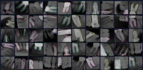
The data set collection phase is the most fundamental phase of the study. Dental X-ray images are used in the study. At this stage, data were collected from two different platforms in accordance with the data selection criteria as shown in Figure 1.
Figure 1. Sample images of the data set
The datasets used in the study were obtained from Roboflow and Kaggle platforms. Roboflow is a tool that facilitates the task of computer vision in deep learning. It enables developers to build computer vision applications regardless of their skill set or experience. It supports object detection and classification models [16]. Kaggle enables users to find and publish datasets, explore and build models in a web-based data science environment, work with other data scientists and machine learning engineers, and participate in competitions to solve data science challenges [17]. These platforms provide datasets containing dental X-ray images and different dental diseases. The data collected from two different platforms were merged and the most suitable ones were selected for the study. The merged version of the dataset is shown in Figure 1. When selecting a dataset, it is important to ensure that the dataset is up to date so that the results of the data analysis are more accurate. The completeness of the data set ensures that the data analysis results are more accurate. If the data set is suitable for the purpose of data analysis, the data analysis results will be more meaningful [18].
The researched data sets were brought together on the Roboflow platform and analyzed one by one. As a result of the examinations, the data suitable for the study were selected and merged. Unnecessary data were deleted. In this way, a smoother data set was obtained for model training. In this merged dataset, care was taken to ensure that the same images were not repeated, that there were no non-diseased tooth images, and that the images were free of noise, similarity of tissue and bone images, reflection and glare, low contrast and distortion. Preparation and labeling of the dataset is one of the most critical steps for model training. The data were examined one by one, and the preparation phase transitioned to the labeling phase. In the preparation phase, it was decided how to classify the data. These stages are visualized in Figure 2.
Figure 2. Preparation stages of the data set
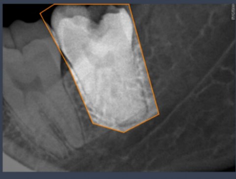
Figure 3. Labeled data image
In the labeling process, the diseases in each image are identified and labeled, as shown in Figure 3. This process aims to ensure the correct classification of the data to be used in the training of the model.
2.2 Eigen class activation map
Eigen Class Activation Map (Eigen-CAM) is a method for visualizing the decision processes of deep learning models. Eigen-CAM uses eigen-decomposition to extract the most meaningful and discriminative information from the model's feature maps. This method aims to explain the internal mechanisms of the model by shedding light on the important regions that the model considers in classification tasks [19]. During visualization, it provides valuable information to both researchers and practitioners by revealing from which areas the model draws information and how it shapes its decisions.
The basic working principle of Eigen-CAM is to analyze the feature maps prior to the final layers of the deep learning model. Using the eigenvalue decomposition method, the eigenvector corresponding to the largest eigenvalue is determined from these feature maps. This eigenvector allows highlighting the areas that are most influential in the model's decision mechanism. Unlike Grad-CAM and other similar class activation maps, this method has the advantage of reducing noise in visualizations and providing clearer results [20]. In particular, it makes it possible to obtain a more precise and reliable visualization on complex image data.
In this study, the Eigen-CAM method is investigated by applying it to various neural network architectures. In particular, the effects of model performance on visualization quality are investigated. The eigenvalue decomposition based structure of the method provides a remarkable level of explainability even for high accuracy architectures. It has been observed that Eigen-CAM improves both classification performance and visualization quality by using feature maps. This suggests that the method can find wide application in medical imaging, object detection and other computer vision applications.
An important advantage of Eigen-CAM is that it can be easily integrated into different deep learning architectures. For example, it is compatible with classical structures such as convolutional neural networks (CNNs), but it is also effective with newer architectures. This flexibility makes the method preferable in both research and application areas. Especially in areas where visualization quality is critical, the use of Eigen-CAM is expected to become increasingly widespread.
2.3 YOLOv8
YOLOv8 (You Only Look Once Version 8) is one of the latest versions of object detection algorithms and a major innovation in deep learning and computer vision. As the most advanced version of the YOLO family, YOLOv8 offers significant improvements over previous versions in terms of speed and accuracy in real-time object detection. This deep learning algorithm is based on convolutional neural networks (CNNs), enabling fast and accurate object detection. YOLOv8 shows an impressive performance by maintaining high accuracy rates, especially in real-time applications [21].
One of the most remarkable features of this algorithm is that it offers a series of model variations optimized for different use cases. The YOLOv8 series includes five different models, each with different capacity and processing speed: Nano (n), Small (s), Medium (m), Large (l) and Extra Large (x). The YOLOv8n model aims to achieve fast results with low hardware requirements and is therefore lighter and more compact. In contrast, the YOLOv8x model has an architecture that requires more parameters and processing time while achieving the highest accuracy. In between these two extremes, YOLOv8s, YOLOv8m and YOLOv8l models offer a balanced performance between speed and accuracy [22].
The performance of YOLOv8 is evaluated with various metrics to analyze the advantages and limitations of different variations. Metrics such as mean accuracy rate (mAP), latency and the number of parameters required by the model are the main indicators used in these evaluations. In particular, the performance metrics of the YOLOv8 models, shown in Table 2, provide an opportunity to compare the effectiveness of different models on various datasets. While YOLOv8x stands out with its superior accuracy on complex object detection tasks, YOLOv8n stands out in applications where speed is critical.
In this study, the overall performance of the YOLOv8 algorithm and its advantages in application domains are discussed. YOLOv8, which is used in different fields such as real-time object detection, medical imaging and autonomous systems, offers a wide range of applications thanks to its flexible and modular structure. The model's innovative architecture makes it possible to achieve high success in scenarios that require both speed and accuracy. Future work can focus on new optimizations and model enhancements to improve the performance of YOLOv8.
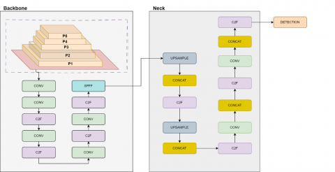
The metrics presented in Table 2 include input size (pixels), mean average precision (mAP), processing speeds (CPU ONNX and A100 TensorRT), number of parameters and number of floating point operations (FLOPs). There are notable differences in performance between the models, with larger models (e.g. YOLOv8x) requiring more computational resources while achieving high accuracy rates. In contrast, more compact models like YOLOv8n are suitable for applications needing speed and efficiency. Figure 4 shows the architecture of the YOLOv8 model.
Table 2. Variants of YOLOv8
|
Model |
Input Image Size (pixels) |
Mean Average Precision (mAP) (IoU 50-95) (%) |
Processing Time on CPU (milliseconds) |
Processing Time on NVIDIA A100 (TensorRT) (milliseconds) |
Number of Parameters (millions) |
Floating Point Operations (FLOPs) (billions) |
|
YOLOv8n |
640 |
37.3 |
80.4 |
0.99 |
3.2 |
8.7 |
|
YOLOv8s |
640 |
44.9 |
128.4 |
1.20 |
11.2 |
28.6 |
|
YOLOv8m |
640 |
50.2 |
234.7 |
1.83 |
25.9 |
78.9 |
|
YOLOv8l |
640 |
52.9 |
375.2 |
2.39 |
43.7 |
165.2 |
|
YOLOv8x |
640 |
53.9 |
479.1 |
3.53 |
68.2 |
257.8 |
Figure 4. YOLOv8 architecture [23]
2.4 Proposed method
In this study, a holistic approach is developed to improve data collection, model training and explainability of results in AI-assisted dental X-ray analysis. Using Roboflow and Kaggle platforms, images showing dental diseases and treatments are collected and labeled. With this process, it was aimed to maximize the training performance of the model and to create a quality data set.
Various variants of YOLOv8 (n, s, m, l and x) were used in the model training phase. Metrics such as accuracy, precision and F1 score were used during training. Data augmentation and preprocessing techniques were used to maximize the performance of the model. While the YOLOv8x model stands out with its accuracy rates, the YOLOv8n model stands out in terms of speed and efficiency. After the completion of the model training process, the Eigen-CAM method was used to visualize and explain the decision processes. This technique aims to help dentists and students better understand the results by visually showing from which regions the model receives information. Finally, the model is integrated into a web interface where users can upload X-ray images to get reliable and fast analysis results. Both in terms of technical accuracy and user experience, the proposed technique aims to make a significant contribution to dental practice and education.
In this study, the performance of YOLOv8 models (n, s, m, l, x) on dental X-ray images is analyzed in detail with various performance evaluation metrics. The main objective of the study is to demonstrate the effectiveness of different YOLOv8 architectures in dental X-ray analysis and to evaluate the advantages of these models compared to traditional methods. Table 3 summarizes the results obtained by the YOLOv8 models according to various metrics.
Each YOLOv8 model was trained for a specified number of epochs and the accuracy and loss values were monitored during training. Accuracy, precision, sensitivity and F1 scores for each trained model were calculated from the Confusion Matrices. The Confusion Matrices provided a better understanding of the performance by detailing the model's true positive, false positive, true negative and false negative classifications. In the study analysis, it was seen that the Yolov8n model provided the best performance.
Table 3. Trained model results
|
Model (%) |
Accuracy (%) |
Precision (%) |
Recall (%) |
F1-Score (%) |
|
YOLOv8n |
0.86 |
0.64 |
0.60 |
0.62 |
|
YOLOv8s |
0.84 |
0.60 |
0.62 |
0.60 |
|
YOLOv8m |
0.81 |
0.58 |
0.63 |
0.58 |
|
YOLOv8l |
0.79 |
0.56 |
0.60 |
0.58 |
|
YOLOv8x |
0.75 |
0.55 |
0.57 |
0.57 |
In the analysis, the YOLOv8n model is the prominent model of the study in terms of both speed and performance metrics as seen in Table 3. Especially with its low hardware requirement and speed advantage, it offers a preferable option in applications that require fewer resources compared to other models. In addition, although the YOLOv8x model requires more parameters and computation time, it offers a useful structure in multi-class analysis and extracting more features in complex tasks.
In addition, analysis of the Confusion Matrices obtained from YOLO models showed that fast and accurate predictions were obtained in dental disease detection. By analyzing the incorrect predictions in detail, it was determined on which images the model had difficulty and it was concluded that data diversity should be increased in these areas. The Confusion Matrix of the YOLOv8n model was obtained as shown in Figure 5.
The confusion matrix of the YOLOv8n model is presented in Figure 5. When the distribution of correct and incorrect predictions of the model is examined in detail, it is seen that 170 samples are correctly predicted in the “dentin cavity” class. In the “dental filling” class, 288 samples were correctly predicted, while 23 samples were misclassified as “dental crown.” The “dental implant” class had 330 correct predictions. The “root canal treatment” class was among the most successful with 444 correct predictions, while the “dental crown” class achieved the highest success rate with 510 correct predictions. These errors appear to be due to the structural similarities of some of the classes.
Figure 5. Confusion matrix of YOLOv8n model
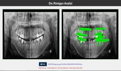
The trained model was made accessible through a user-friendly web interface. This is intended to significantly increase the speed and accessibility of the process. The web interface is designed in such a way that dentists and students can easily use it, and users can access the analysis results quickly and reliably by directly uploading dental X-ray images. As can be seen in Figure 6, the detected diseases or previously applied procedures can be instantly displayed on the uploaded images. In this way, faster transition to treatment processes is ensured and future complications can be prevented by early diagnosis in many diseases. The web interface has been designed in a user-friendly structure to help rapid decision-making in clinical settings and to increase the suitability of the model for practical use.
Figure 6. Web interface
As can be seen in Figure 6, it is possible to determine which disease is present in the dental X-ray whose image is uploaded or which procedures have been performed on this tooth structure before. Thus, it is aimed to provide a faster transition to the treatment process. It is thought that early diagnosis and treatment of many diseases can prevent future problems.
With the YOLOv8 model used in the study, dental X-rays were analyzed and dental procedures and problems were identified. The results obtained from the model were evaluated through performance metrics. Training was performed using the n, s, m, l, x models of YOLOv8. Accuracy and loss values were monitored during training. The confusion matrix of each model was analyzed and performance metrics were calculated for the models. Accuracy, precision, sensitivity and F1 scores were calculated for each model and class individually.
In order to evaluate the overall performance of the models, the performance metrics of each model were macro-averaged to obtain overall metric results. According to the results obtained, the different versions of YOLOv8 (n, s, m, l, x) provided very satisfactory performances. The trained model was made accessible through a web interface, which allowed dentists and dental students to upload X-ray images and detect dental procedures and diseases quickly and reliably.
Fast and accurate disease detection with the results obtained from the models is of great importance for early diagnosis in the treatment of dental diseases. In addition, thanks to its accuracy and efficiency in disease detection, a system that is thought to be useful for students in the learning process has been designed. The study aims to reduce the burden of dentists while providing a great benefit to students in terms of detection.
This work is supported by the Scientific Research Project Fund of FIRAT ÜNİVERSİTESİ (Grant No.: ADEP.23.09).
[1] Arıkan, A., Özkan, G., Pirinçci, S., Abacıgil, F., Keleş, S., Okyay, P. (2019). Hekim adaylarının ağız-diş sağlığı alışkanlıkları ve bilgi düzeylerinin değerlendirilmesi. Atatürk Üniversitesi Diş Hekimliği Fakültesi Dergisi, 29(2): 189-196. https://doi.org/10.17567/ataunidfd.496011
[2] Ünsal, Ü., Adem, K. (2022). Diş görüntüleri üzerinde görüntü işleme ve derin öğrenme yöntemleri kullanılarak çürük seviyesinin sınıflandırılması. Uluslararası Sivas Bilim ve Teknoloji Üniversitesi Dergisi, 2(2): 30-53.
[3] Güneç, H.G., Gökyay, S.S., Kaya, E., Aydın, K.C. (2022). Toplum yapay zeka ile dental tanı konmasına hazır mı? Selcuk Dental Journal, 9(1): 200-207. https://doi.org/10.15311/selcukdentj.915522
[4] Kayadibi, İ., Köse, U., Güraksın, G.E. (2024). Görüntü işleme teknikleri ve evrişimsel sinir ağı kullanılarak bilgisayar destekli diş segmentasyonu. Pamukkale Üniversitesi Mühendislik Bilimleri Dergisi, 30(7): 924-933. https://doi.org/10.5505/pajes.2024.22237
[5] Kahurke, S. (2023). Artificial intelligence algorithms and techniques for dentistry. In 2023 1st International Conference on Cognitive Computing and Engineering Education (ICCCEE), Pune, India, pp. 1-4. https://doi.org/10.1109/ICCCEE55951.2023.10424481
[6] Sur, J., Bose, S., Khan, F., Dewangan, D., Sawriya, E., Roul, A. (2020). Knowledge, attitudes, and perceptions regarding the future of artificial intelligence in oral radiology in India: A survey. Imaging Science in Dentistry, 50(3): 193. https://doi.org/10.5624/isd.2020.50.3.193
[7] Razaghi, M., Komleh, H.E., Dehghani, F., Shahidi, Z. (2024). Innovative diagnosis of dental diseases using YOLO V8 deep learning model. In 2024 13th Iranian/3rd International Machine Vision and Image Processing Conference (MVIP), Tehran, Iran, pp. 1-5. https://doi.org/10.1109/MVIP62238.2024.10491172
[8] Karakuş, R., Öziç, M.Ü., Tassoker, M. (2024). AI-assisted detection of interproximal, occlusal, and secondary caries on bite-wing radiographs: A single-shot deep learning approach. Journal of Imaging Informatics in Medicine, 37: 3146-3159. https://doi.org/10.1007/s10278-024-01113-x
[9] Brahmi, W., Jdey, I. (2024). Automatic tooth instance segmentation and identification from panoramic X-ray images using deep CNN. Multimedia Tools and Applications, 83(18): 55565-55585. https://doi.org/10.1007/s11042-023-17568-z
[10] Hasnain, M.A., Malik, H., Asad, M.M., Sherwani, F. (2024). Deep learning architectures in dental diagnostics: A systematic comparison of techniques for accurate prediction of dental disease through X-ray imaging. International Journal of Intelligent Computing and Cybernetics, 17(1): 161-180. https://doi.org/10.1108/IJICC-08-2023-0230
[11] George, J., Hemanth, T.S., Raju, J., Mattapallil, J.G., Naveen, N. (2023). Dental radiography analysis and diagnosis using YOLOv8. In 2023 9th International Conference on Smart Computing and Communications (ICSCC), Kochi, Kerala, India, pp. 102-107. https://doi.org/10.1109/ICSCC59169.2023.10335023
[12] Cha, J.Y., Yoon, H.I., Yeo, I.S., Huh, K.H., Han, J.S. (2021). Peri-implant bone loss measurement using a region-based convolutional neural network on dental periapical radiographs. Journal of Clinical Medicine, 10(5): 1009.
[13] Kurt Bayrakdar, S., Orhan, K., Bayrakdar, I.S., Bilgir, E., Ezhov, M., Gusarev, M., Shumilov, E. (2021). A deep learning approach for dental implant planning in cone-beam computed tomography images. BMC Medical Imaging, 21(1): 86. https://doi.org/10.1186/s12880-021-00618-z
[14] Chen, H., Zhang, K., Lyu, P., Li, H., Zhang, L., Wu, J., Lee, C.H. (2019). A deep learning approach to automatic teeth detection and numbering based on object detection in dental periapical films. Scientific Reports, 9(1): 3840. https://doi.org/10.1038/s41598-019-40414-y
[15] Lee, J.H., Kim, D.H., Jeong, S.N., Choi, S.H. (2018). Diagnosis and prediction of periodontally compromised teeth using a deep learning-based convolutional neural network algorithm. Journal of Periodontal & Implant Science, 48(2): 114-123. https://doi.org/10.5051/jpis.2018.48.2.114
[16] Roboflow: Computer vision tools for developers and enterprises. (t.y.). Geliş tarihi 16 Aralık 2024, gönderen https://roboflow.com/.
[17] Kaggle: Your Home for Data Science. (t.y.). Geliş tarihi 16 Aralık 2024, gönderen https://www.kaggle.com/
[18] Pipino, L.L., Lee, Y.W., Wang, R.Y. (2002). Data quality assessment. Communications of the ACM, 45(4): 211-218. https://doi.org/10.1145/505248.506010
[19] Selvaraju, R.R., Cogswell, M., Das, A., Vedantam, R., Parikh, D., Batra, D. (2017). Grad-cam: Visual explanations from deep networks via gradient-based localization. In 2017 IEEE International Conference on Computer Vision (ICCV), Venice, Italy, pp. 618-626. https://doi.org/10.1109/ICCV.2017.74
[20] Muhammad, M.B., Yeasin, M. (2020). Eigen-cam: Class activation map using principal components. In 2020 International Joint Conference on Neural Networks (IJCNN), Glasgow, UK, pp. 1-7. https://doi.org/10.1109/IJCNN48605.2020.9206626
[21] Terven, J., Córdova-Esparza, D.M., Romero-González, J.A. (2023). A comprehensive review of yolo architectures in computer vision: From yolov1 to yolov8 and yolo-nas. Machine Learning and Knowledge Extraction, 5(4): 1680-1716. https://doi.org/10.3390/make5040083
[22] Afdhal, A., Saddami, K., Sugiarto, S., Fuadi, Z., Nasaruddin, N. (2023). Real-time object detection performance of yolov8 models for self-driving cars in a mixed traffic environment. In 2023 2nd International Conference on Computer System, Information Technology, and Electrical Engineering (COSITE), Banda Aceh, Indonesia, pp. 260-265. https://doi.org/10.1109/COSITE60233.2023.10249521
[23] Karim, M.J., Nahiduzzaman, M., Ahsan, M., Haider, J. (2024). Development of an early detection and automatic targeting system for cotton weeds using an improved lightweight YOLOv8 architecture on an edge device. Knowledge-Based Systems, 300: 112204. https://doi.org/10.1016/j.knosys.2024.112204