Şeyma Çeçen*![]() | Songül Çeribaşı
| Songül Çeribaşı![]() | Merve Erkuş
| Merve Erkuş![]() | Ahmet Bedri Özer
| Ahmet Bedri Özer![]() | Taner Tuncer
| Taner Tuncer![]() | Ahmet Çınar
| Ahmet Çınar![]()
© 2024 The authors. This article is published by IIETA and is licensed under the CC BY 4.0 license (http://creativecommons.org/licenses/by/4.0/).
OPEN ACCESS
The present study aims to accurately classify estrus cycle by using images of the uterus from female rats. Convolutional neural network-based deep learning techniques were utilized for the classification process. While the human menstrual cycle spans 28 days, in rats, it completes within 4-5 days. Female rats are particularly preferred in studies related to the female reproductive system due to being a model organism. In the study, sections stained with Hematoxylin and Eosin from the uterine tissue of female rats were examined under a light microscope, and their images were digitized. The obtained images were used to histologically classify the estrus cycles in rats. Following the examination, an artificial intelligence-based model was proposed for the classification of estrus cycles in rats using images obtained from uterine sections. The study classifies estrus cycles into four stages: proestrus, estrus, metestrus, and diestrus. In the proposed model, the classification success of sub-models belonging to the YOLOv5 algorithm, such as YOLOv5n, YOLOv5s, YOLOv5m was compared with histological results. The YOLOv5m model achieved an accuracy of 98.3%, precision of 99%, recall of 98%, and an F1-score of 98% in classification. By using the YOLOv5m architecture, a 98% accuracy in classifying estrus cycles was achieved, providing a robust deep learning approach for tissue analysis. The obtained results indicate that the proposed model can offer a second opinion support to expert pathologists in analyzing microscopic images.
deep learning, histopathology, estrus cycle, estrus staging, pathological image, classification, uterus, YOLOv5
Owing to factors such as ease of care and feeding, shorter lifespans and gestation periods, and the ability to serve as models for other organisms, rats are commonly used in experimental studies. In a female reaching puberty, or the adolescent period, controlled by hormones, various changes occur in the ovarian and uterine tissues. Mammals exhibit estrus at different intervals depending on their species and ages. The period between the beginning of two consecutive estrus cycle is referred to as the estrus cycle or estrus period [1]. Due to the fact that in rats, the estrus cycle typically lasts 4-5 days compared to the 28-day menstrual cycle in humans, this animal species is often preferred as a model organism in studies related to the female reproductive system. In rats, the estrus cycle consists of four main stages: proestrus, estrus, metestrus, and diestrus [2, 3].
Artificial neural network-based deep learning studies have become widely used in various fields, including veterinary science and medicine, just as in many other areas nowadays [4, 5]. Within the scope of the study, recent research in this field present in the literature has been examined. The reviewed studies particularly focus on the field of pathology, which investigates the changes occurring in tissues, cells, and organs, as well as their causes. In the literature based on convolutional neural network techniques, there are numerous studies utilizing deep learning methods on various tissues. İnik et al. [6] proposed a new method based on ESA architecture for follicle counting from ovarian tissue images. In the study, a total of 1750 images with different magnification levels (x10, x20, x40) were classified into 5 groups. They mentioned that their proposed model in follicle classification through segmentation process was more successful compared to the Faster R-CNN model. Sun et al. [7] classified pathological diagnoses from images of endometrial (uterine lining) tissue into 4 groups. They stated achieving successful results in the classification field with their proposed model. Yan et al. [8], on the other hand, proposed a hybrid convolutional deep neural network model for classifying breast cancer tumor types using an increased number of images. They reported good results with 91.3% accuracy in a 4-class study conducted through image augmentation methods. Additionally, Huo et al. [9] employed the YOLOv5 model for the classification of parasite eggs in microscopic images. They highlighted the model's high average accuracy in an 8-class study containing 281 sample images. Drioua et al. [10] reported that effective results were achieved for the early detection of cancerous lesions in breast tissue images using the YOLOv5 model for classification. Huang et al. [11] proposed a two-stage method to enhance the rapid detection and screening effectiveness of abnormal prostate cancer cells. In their study, they applied the ResNet50 and YOLOv5 models to pathological cell images. They mentioned that in other models, due to the presence of numerous parameters and the complexity of model structures, overfitting occurred during training. However, they highlighted that the applied model, with its smaller number of parameters, reduced computational cost, and simpler structure, reduced the inference time. Guo et al. [12] conducted a study on the pathological detection of stomach cancer cells using the YOLOv5 model, aiming for deep learning-based classification. The study concluded that it could provide decision support for clinical judgment. On the other hand, Xu et al. [13] worked on deep learning-based cervical cell classification. They used a global average pooling layer to improve the recognition accuracy of cell images. They mentioned designing a superficial feature enhancement network to enhance the localization and recognition of weak cells. Additionally, data augmentation methods were implemented to enhance the model's detection capability. According to experimental results, the proposed model was stated to outperform CenterNet, YOLOv5, and Faster R-CNN algorithms in certain aspects such as time consumption and high recognition accuracy. Yu et al. [14] conducted research on pathological whole-slide images of uterine smooth muscle tumors. While the ResNet model was used as a classification network for cytologic atypia and necrosis, the YOLOv5 model was employed for mitotic count detection. The researchers mentioned achieving successful results with different parameter values. On the other hand, Çeçen and Özer [15] worked on the classification of pathological breast cancer images. They utilized the YOLOv5 model for a two-class grouped dataset consisting of 1693 image samples. According to experimental results, they expressed that the chosen model for classifying tumor types in pathological images was successful. When reviewing the studies in the literature, the focus was on parameters such as tissue type, image size, methodology used, number of classes, utilized model or algorithm, and the achieved accuracy value. The parameters used in the studies are presented in Table 1.
Table 1. An overview of deep learning studies with pathological images
|
Reference |
Tissue Type |
Image Size |
Method |
Number of Classes |
Model |
Accuracy |
|
İnik et al. [6] |
Ovary |
1024×1024 |
Segmentation, Classification |
6 |
Faster R-CNN |
95.35% |
|
Sun et al. [7] |
Endometrium |
224×224 |
Classification |
4 |
HIENet |
84.50% |
|
Yan et al. [8] |
Breast |
2048×1536 |
Classification |
4 |
Inception-V3 |
91.3% |
|
Huo et al. [9] |
Feces |
512×512 |
Object Detection |
7 |
YOLOv5 |
99.4% |
|
Drioua et al. [10] |
Breast |
512×512 |
Object Detection, Segmentation |
2 |
YOLOv5 |
89% |
|
Huang et al. [11] |
Prostate |
200×200 |
Classification, Object Detection |
5 |
ResNet50, YOLOv5 |
71.1% |
|
Guo et al. [12] |
Stomach |
640×640 |
Object Detection |
9 |
YOLOv5 |
61.1% |
|
Xu et al. [13] |
Cervical cell |
512×512 |
Object Detection |
8 |
GAN |
99.81% |
|
Yu et al. [14] |
Uterine smooth muscle |
224×224 |
Classification, Object Detection |
3 |
ResNet, YOLOv5 |
92% |
|
Çeçen and Özer [15] |
Breast |
224×224 |
Classification |
2 |
YOLOv5 |
95.3% |
1.1 Significance of the study
Numerous studies have been conducted on determining estrus cycles in animals, encompassing methods such as smear tests, cytological analyses, and histological examinations [16-18]. Furthermore, there are detailed publications defining the histological characteristics of estrus cycles specifically in rats [19, 20]. Emphasis has been placed on the effects of drugs or chemicals on the reproductive functions of female rats, highlighting that these effects often manifest as alterations in the normal morphology of the reproductive system or disruptions in the duration of specific stages within the estrus cycle. It has been stated that these changes can be accurately and consistently described only when a pathologist possesses sufficient knowledge and expertise regarding the continuously varying histological appearance of various components within the uterus throughout the estrus cycle. Despite the existence of comprehensive reports illustrating the normal appearance during the estrus cycle of rats for many years, it has been reported that they are often insufficient in defining different criteria to distinguish the end of one phase from the beginning of another [19]. Based on personal experience and literature references, aiming to address the shortcomings in determining the stages of the estrus cycle, a practical method was intended to be developed for histologists and pathologists involved in studies related to the female reproductive system. In this study, a convolutional neural network-based model is proposed for the classification of estrus cycles based on pathological images. Determining the estrus cycles in female rats provides fundamental information about the reproductive system. The analysis in the study was conducted using deep learning techniques on sections obtained from uterine tissue. Deep learning methods such as the YOLOv5 model were preferred for this analysis. The significance of the proposed model can be summarized as follows:
1.2 Contribution and novelty
When examining the studies in the literature, it is observed that convolutional neural network (CNN) based models and architectures have achieved success in the classification field with high accuracy rates and low loss rates. The utilization of CNN-based layered architecture enhances the inference of different features from images, thereby increasing the success and predictive power of the model.
The most significant benefit of digitized pathological images is their utilization as input data for artificial intelligence-supported systems [21].
To sum up, the contributions of this study to the literature are as follows:
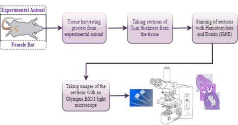
Figure 1. Obtaining process of images related to uterine tissue
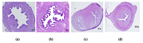
Figure 2. Samples related to the estrus cycle
The study utilized 78 Sprague Dawley rat uterine tissues, for which ethical approval was obtained from the Local Animal Experiments Ethics Committee of Gazi University with the ethical approval code G.U.ET-23.038 for the dataset used. Tissues were processed through routine histological procedures, stained using the Hematoxylin and Eosin (H&E) method, and photographed at 4x magnification using an Olympus BX 51 light microscope. The images of the captured uterine tissue were classified and grouped by an expert pathologist based on the estrus cycle stages of the rats (proestrus, estrus, metestrus, diestrus). The process of obtaining images related to the tissue is depicted in Figure 1. The obtained images are in different sizes such as 5184 × 3456, 3071 × 2126, and 2040 × 1536, and have been grouped according to classes with the guidance of an expert pathologist. The dataset comprises a total of 195 images, divided into proestrus (37), estrus (59), metestrus (44), and diestrus (55) phases. Within the dataset, the images were divided for the model's optimal performance into 70% for training, 10% for testing, and 20% for validation. For each phase of the estrus cycle, Figure 2 shows pathological tissue samples respectively classified as (a) Proestrus, (b) Estrus, (c) Metestrus, and (d) Diestrus.
2.1 Proposed method
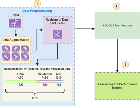
Deep learning constitutes a multi-layered structure that makes data meaningful and generates predictions about new data [22]. In this study, the detection of tissue classes is presented using the YOLOv5 algorithm based on Convolutional Neural Networks (CNNs), which is one of the deep learning methods. Compared to previous YOLO models, the YOLOv5 model is enhanced with the CSPDarknet53 backbone and PANet (Path Aggregation Network) structure for feature extraction. It enables better and faster processing of high-resolution images, exhibiting efficient and rapid operation supported by both CPU and GPU/TPU. In the study, pathological images of uterine tissue sections are used as input data for the model. A structure is proposed using the YOLOv5 model to accurately classify stages of the estrus cycle based on tissue images. The proposed method consists of three stages as depicted in Figure 3: data preprocessing, classification using the YOLOv5 architecture for feature extraction, and assessment of performance metrics.
Figure 3. Stages of the proposed method
2.1.1 Data preprocessing
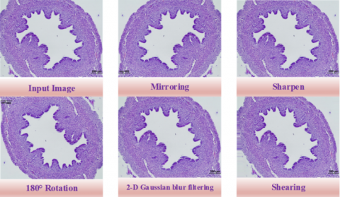
The data preprocessing stage aimed to prepare the dataset for use in the study. A sequential series of operations was applied to the created dataset, including increasing the number of images, resizing images, and partitioning data into training, testing, and validation sets. Augmenting the number of images was performed to enhance the volume of the dataset, thereby strengthening the convolutional neural network structure. In the study, data augmentation techniques such as mirroring, rotation, shearing, sharpening, and 2-D Gaussian blur filtering were applied, increasing the total number of available data from 195 to 1755 instances. Sample images illustrating the outputs of the data augmentation processes mentioned in Figure 4 are provided.
As the second step of the data preprocessing stage, the augmented data was uniformly resized. It is crucial for the effective functioning of the model that images have equal aspect ratios and balanced class distributions. Therefore, since images with a 4x magnification rate in the dataset were of various sizes, each image was resized to the optimal working size of the model, which is 224×224 pixels. As the final step of the data preprocessing stage, the dataset was divided into 70% for training, 10% for testing, and 20% for validation purposes. Accordingly, the dataset was distributed as 1227 instances for training, 178 instances for testing, and 350 instances for validation, thus completing the data preparation process.
Figure 4. Application of data augmentation methods
2.1.2 Classification with YOLOv5 architecture
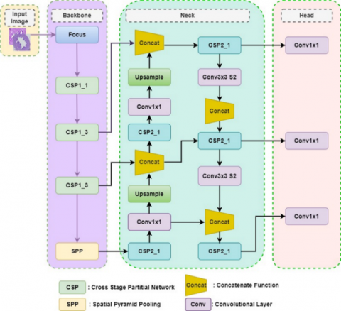
The YOLOv5 model architecture is the latest version of the YOLO (You Only Look Once) model family, designed for object detection and classification. YOLOv5 stands out as a fast and accurate object detection model developed with the PyTorch library. The architectural network of the YOLOv5 method consists of three main sections named backbone, neck, and head. The focus of the backbone section lies in extracting feature information from input images. The neck section combines the extracted feature information to generate a three-scale feature map. Meanwhile, the head section is responsible for classifying the objects within the created feature map. The backbone network is a convolutional neural network that generates feature maps of different sizes from the input image through multiple convolution and pooling steps. Within the YOLOv5 architecture, as depicted in Figure 5, there are layers of the feature map created within the backbone network. The neck network merges feature maps at different levels to obtain more correlated information from feature maps of different sizes and reduce information loss [23].
The core focus of the architecture is based on slicing each image and merging to better extract features during subsampling. The CBL (Convolution, Batch Normalization, Leaky ReLU) module consists of three main modules: convolution, normalization, and Leaky ReLU activation function. The CSP (Cross-Stage Partial) module, which comes in two variations within the backbone and neck networks, is employed in the architecture. The CSP network aims to reduce model size while maintaining accuracy and boosting inference speed. There exists a slight difference between the two types of CSP networks utilized. The CSP network in the backbone consists of one or more residual units, while in the neck, the CSP network replaces residual units with CBL modules. Here, dimension reduction operations are conducted in the pooling step to represent image features at a higher level of abstraction. Essentially, this process involves compressing the input feature map, reducing the complexity of computations within the network. On the other hand, feature compression facilitates the extraction of fundamental characteristics [23]. The Concat module aids in combining feature maps obtained from multiple convolutional layers to acquire deeper and more detailed features, contributing to a more accurate perception and classification of objects. Additionally, the Concat module enables the network to achieve higher accuracy with fewer parameters. Dropout layers are employed in deep learning models to mitigate overfitting. However, the YOLOv5 architecture, in its general structure, does not include dropout layers. Instead, the head module layers are utilized to enhance feature extraction and control overfitting.
Figure 5. YOLOv5 model network architecture
2.1.3 Assessment of performance metrics
In the study, evaluating the performance of the applied model in detecting estrus cycle phases requires assessment based on specific criteria. For this purpose, assessment metrics such as accuracy, precision, recall, and F1 score are utilized. Among the methods used to summarize the success of the classification algorithm is the confusion matrix [24]. The confusion matrix is created based on parameter values such as accuracy, precision, recall, and F1 score.
Accuracy parameter is one of the metrics that explains the accuracy of the algorithm in the classification process [25]. The accuracy value is expressed as the ratio of the number of matching samples to the total number of samples. This parameter is considered a criterion for assessing how well the model performs. It ranges between 0 and 1, and as the value approaches 1, the success rate increases [19]. The calculation formula is provided in Eq. (1).
Accuracy $=\frac{T P+T N}{T P+E N+E P+T N}$ (1)
The Precision parameter is used to measure the success of positive cases in a convolutional neural network [26]. It is defined as the ratio of true positive predictions to the total positive predictions made by the model. As this value increases, the success of the network also increases accordingly. It is calculated according to Eq. (2).
Precision $=\frac{T P}{T P+F P}$ (2)
The Recall parameter signifies how much of the positive given classes were predicted as positive or how much of the negative given classes were predicted as negative. It is calculated according to the formula given in Eq. (3).
Recall $=\frac{T P}{T P+E N}$ (3)
F1 Score parameter is expressed as the harmonic mean of precision and recall metrics. It is calculated according to the formula in Eq. (4).
F1 Score $=\frac{2 *(\text { Precision } * \text { Recall })}{(\text { Precision }+ \text { Recall })}$ (4)
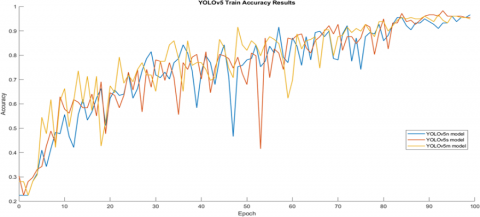
Training a model in deep learning is a complex and computation-intensive task. Utilizing GPU support in the working environment has made model training faster, leading to better results. Studies conducted using the YOLOv5 model indicate that as the number of parameters increases, the accuracy value rises [27]. In this study, aiming to classify stages of the estrus cycle, three sub-models of the YOLOv5 model, namely YOLOv5n, YOLOv5s, and YOLOv5m, were employed. The training outputs and results of these models were examined. The results of the training performed based on input images of size 224×224 for YOLOv5n, YOLOv5s, and YOLOv5m models are presented in Table 2. The study was conducted on a server with a Tesla T4 GPU and 16 GB of RAM within the Google Colab environment. The number of epochs for training was set to 100. The training time for the three applied models ranged from 8 to 10 minutes. The YOLOv5m model achieved the highest accuracy, reaching 98.3% based on the Accuracy parameter. According to the results, an increase in the number of layers was observed to correlate with an increase in the accuracy value. The training accuracy values obtained for 100 epochs using YOLOv5n, YOLOv5s, and YOLOv5m models are depicted in Figure 6. Additionally, the loss graphs for training and test data concerning YOLOv5n, YOLOv5s, and YOLOv5m models are provided in Figure 7.
Adjusting hyperparameters during model training in deep learning significantly affects the model's performance. The hyperparameters used in training the YOLOv5 model in the study are provided in Table 3. The learning rate is crucial in determining the amount of update to model weights. A high learning rate can provide speed but might result in variable and unstable weight updates. The number of epochs determines how many cycles the model will be trained for. It's crucial to properly set the number of epochs for a good model training. Depending on the number of epochs, the model might exhibit overfitting or underfitting. The number of epochs should be chosen carefully to balance these unwanted conditions. In the study, to prevent the mentioned conditions, the number of epochs was set to 100. The batch size represents the number of samples processed in each epoch and is adjusted based on the dataset's size. The images were resized to the pixel size where classification worked most efficiently according to the YOLOv5 model. The Adam optimization algorithm, a gradient-based optimization algorithm used to achieve rapid convergence and reduce overfitting issues, was employed.
Table 2. Training metrics of YOLOv5 sub-models on the uterus dataset
|
Model |
Size (Pixel) |
Accuracy (%) |
Training Duration (Hour) |
Number of Training Layers |
Number of Accuracy Layers |
|
YOLOv5n |
224 |
96.6 |
0.136 |
149 |
117 |
|
YOLOv5s |
224 |
96.6 |
0.145 |
149 |
117 |
|
YOLOv5m |
224 |
98.3 |
0.161 |
212 |
166 |
Figure 6. Training accuracy graph according to the YOLOv5 model
Figure 7. Training-test loss graphs according to the YOLOv5 model
Table 3. Training parameter values for YOLOv5 model
|
Parameters |
Value |
|
Rate of learning |
0.001 |
|
Number of Epochs |
100 |
|
Batch size |
64 |
|
Image size |
224 |
|
Optimization Algorithm |
Adam |
Table 4 presents the distributions and evaluation metrics of the results obtained from data trained on the YOLOv5n model. It's observed that the overall accuracy value according to the YOLOv5n model is 96.63%. Regarding the distribution by classes, the most successful class based on the recall metric is the 'diestrus' stage.
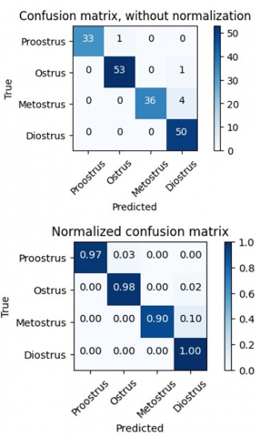
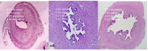
When examining the prediction rates of data trained on the YOLOv5n model by classes, it was observed that among 50 image data belonging to the 'diestrus' stage, 27 images had an accuracy rate of 90% and above. Among randomly selected images, the lowest accuracy rate was 60%, and the lowest recall rate according to the model was 94%, associated with the 'estrus' stage. In Figure 8, prediction rates for the 'diestrus' stage and randomly selected images with a 4x optical zoom ratio are provided. Additionally, non-normalized and normalized confusion matrices created for performance evaluation of the YOLOv5n model are shown in Figure 9.
Table 4. Test results of data trained with YOLOv5n model
|
Stage |
Number of Images |
Accuracy (%) |
Precision |
Recall |
F1 Score |
|
Whole |
178 |
96.63 |
0.96 |
0.96 |
0.96 |
|
Proestrus |
34 |
98.31 |
0.94 |
0.97 |
0.96 |
|
Estrus |
54 |
98.31 |
1.0 |
0.94 |
0.97 |
|
Metestrus |
40 |
98.88 |
1.0 |
0.95 |
0.97 |
|
Diestrus |
50 |
97.75 |
0.93 |
1.0 |
0.96 |
Figure 8. Sample images of the diestrus stages trained in the YOLOv5n model
Figure 9. Confusion matrices for the YOLOv5n model
The distribution and evaluation metrics of the results obtained from data trained on the YOLOv5s model are presented in Table 5. An accuracy of 96.63% was achieved with the YOLOv5s model. According to the confusion matrix, it's observed that all 50 images for the 'diestrus' stage were predicted correctly, supported by the sensitivity parameter.
Upon examining the prediction rates of data trained with the YOLOv5s model by classes, it was observed that among a randomly selected 50 image data belonging to the 'diestrus' stage, 33 images had a prediction rate of 90% and above. Among the randomly selected images, the lowest accuracy rate was 60%, and the lowest accuracy rate compared to the overall model was 90%, associated with the 'metestrus' stage. Figure 10 illustrates the prediction rates for the 'diestrus' stage and random image samples. Additionally, non-normalized and normalized confusion matrices created for the performance evaluation of the YOLOv5s model are shown in Figure 11. According to the confusion matrix, it's observed that all 50 images in the 'diestrus' stage with the highest prediction rate were predicted correctly. For the 'metestrus' stage, out of the allocated 40 images, 4 images were predicted as 'diestrus,' which affected the class's successful prediction rate.
The distributions and evaluation metrics of the results obtained from data trained on the YOLOv5m model are presented in Table 6. According to the YOLOv5m model, the classes predicted with the highest success rates based on the recall parameter are 'diestrus,' 'estrus,' and 'proestrus' stages.
Figure 10. Sample images of the diestrus stage trained in the YOLOv5s model
Figure 11. Confusion matrices for the YOLOv5s model
Table 5. Test results of data trained with YOLOv5s model
|
Stage |
Number of Images |
Accuracy (%) |
Precision |
Recall |
F1 Score |
|
Whole |
178 |
96.63 |
0.97 |
0.96 |
0.97 |
|
Proestrus |
34 |
99.44 |
1.0 |
0.97 |
0.99 |
|
Estrus |
54 |
98.88 |
0.98 |
0.98 |
0.98 |
|
Metestrus |
40 |
97.75 |
1.0 |
0.90 |
0.95 |
|
Diestrus |
50 |
97.19 |
0.91 |
1.0 |
0.95 |
Table 6. Test results of data trained with YOLOv5m model
|
Stages |
Number of Images |
Accuracy (%) |
Precision |
Recall |
F1 Score |
|
Whole |
178 |
98.31 |
0.99 |
0.98 |
0.98 |
|
Proestrus |
34 |
100 |
1.0 |
1.0 |
1.0 |
|
Estrus |
54 |
100 |
1.0 |
1.0 |
1.0 |
|
Metestrus |
40 |
98.31 |
1.0 |
0.93 |
0.96 |
|
Diestrus |
50 |
98.31 |
0.94 |
1.0 |
0.97 |
Figure 12. Sample images for classes trained in YOLOv5m model
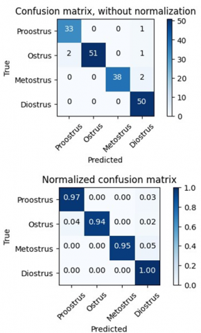
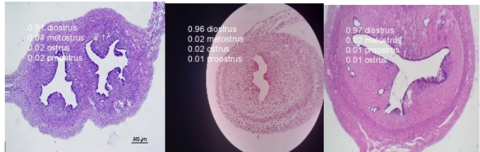
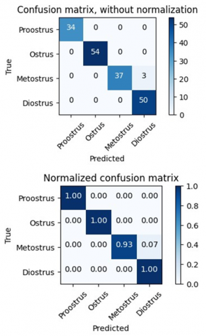
In the examination of data trained according to the YOLOv5m model, when analyzing the prediction rates for classes, a significantly high accuracy was achieved for image data belonging to the proestrus, estrus, and diestrus stages in our 4-class dataset. Comparing with YOLOv5n and YOLOv5s models, the increase in the total success rate along with the number of correctly predicted classes demonstrates that the YOLOv5m model is more successful than other models. This situation indicates that the success rate is influenced by the increase in parameters and layers of the applied model. In Figure 12, randomly selected sample images belonging to diestrus, estrus, and proestrus stages with high prediction accuracy are provided. The non-normalized and normalized confusion matrices created for evaluating the performance of the YOLOv5m model are given in Figure 13.
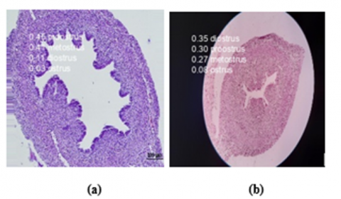
When test data is evaluated according to the YOLOv5 model, low accuracy in the classification process for certain image examples is presented in Figure 14. The estrous cycle occurs gradually in four stages: proestrus, estrus, metestrus, and diestrus, respectively. In Figure 14, for instance, image (a) is expected to be predicted as a 1st-stage image, but it is observed that the prediction closely aligns with the 3rd stage. Similarly, for image (b), although it is expected to be predicted as the 3rd stage, the prediction ratio indicates proximity to the 2nd and 4th stages. Considering the high similarity in some classes, it is apparent that image quality and resolution play a decisive role.
When comparing our study with similar studies in the literature, as shown in Table 7, for instance, Sun et al. [7] conducted a classification process for endometrial tissue using deep learning techniques in 2019. Similar to our study, a dataset consisting of 4 classes and 224x224-sized images was used. However, the success of the proposed HIENet model was found to be 84.5%. Our study, employing the YOLOv5 model, achieved a high accuracy rate of 98.3%, showcasing the success of the model compared to similar studies. Similarly, in the study by Yu et al. [14] in 2023, focusing on uterine smooth muscle tissue, the YOLOv5 model was preferred. The study, conducted on 3-class 224×224-sized images, achieved a success rate of 92%. According to the results, it's evident that the number of classes has an impact on model success. In the study conducted by Çeçen and Özer [15] in 2023, classification was performed for the tumor class in two groups based on pathological biopsy images of breast tissue. It was reported that a success rate of 95.3% was achieved using the YOLOv5 model in the preferred study. Our study's dataset and model seem to yield more successful results compared to similar studies. Important factors affecting model success include the size, resolution, and quantity of images used in the dataset.
Figure 13. Confusion matrices for YOLOv5m model
Figure 14. Low prediction confidence for estrus periods
Table 7. Comparison between the study conducted and other methods
|
References |
Tissue Type |
Image Size |
Method |
Number of Classes |
Model |
Accuracy |
|
Sun et al. [7] |
Endometrium |
224×224 |
Classification |
4 |
HIENet |
84.50% |
|
Yu et al. [14] |
Uterus smooth muscle |
224×224 |
Classification, Object Detection |
3 |
ResNet, YOLOv5 |
92% |
|
Çeçen & Özer [15] |
Breast |
224×224 |
Classification |
2 |
YOLOv5 |
95.3% |
|
The present study 2023 |
Uterus |
224×224 |
Classification |
4 |
YOLOv5 |
98.3% |
The pathological sections taken from the experimental animal in the study were examined under a microscope and digitized. This method is crucial for the detection of potential diseases or monitoring expected conditions in the reproductive system of the organism. In this study focusing on the classification of the relevant tissue using deep learning techniques, the aim was to provide expert pathologists with a second opinion. The YOLOv5 model was employed for the estimation of estrus cycle stages in tissue classification. A success rate of 98.3% was achieved by evaluating the results of the YOLOv5 model's performance based on its YOLOv5n, YOLOv5s, and YOLOv5m sub-models. Utilizing Google Colab as the working environment with GPU support expedited model training in the study.
The originality of the dataset created using sections from uterine tissue for classifying estrus stages within the scope of this study will contribute to the literature due to the scarcity of studies in this field. In the literature, it is observed that studies preferring the YOLOv5 architecture mainly focus on object detection. Particularly, the limited number of studies in the field of classification using the preferred architecture renders our study unique.
The results were evaluated in conjunction with expert opinions, and the proposed model provides the opportunity for tissue classification with minimum time cost, low error rate, and high accuracy value. In this respect, it enhances the potential preference for artificial intelligence-based decision support systems in pathological tissue classification.
The dataset used in this study has obtained ethical approval with the ethical committee approval number G.U.ET-23.038 from the Gazi University Local Ethics Committee for Animal Experiments.
This article was produced from the thesis number 810570 titled "Classification of Oestrus Periods in Rats with the YOLOv5 Model Using Deep Learning Techniques" and approved by Fırat University Institute of Science and Technology under the supervision of Prof Dr Ahmet Bedri ÖZER and Associate Professor Songül ÇERİBAŞI (Master's Thesis, Firat University 2023).
Access to the dataset created for this study is available at (URL: https://www.kaggle.com/datasets/seymacecen/classification-of-estrus-cycles-in-rats).
The authors declare no conflict of interest.
[1] Bozoğlu, H. (2013). Deneysel Hipertiroidi Oluşturulmuş Sıçanlarda Östrus Siklusunun Değişik Evrelerinde Dişi Genital Organlarda (Ovaryum Ve Uterus) Östrojen Ve Progesteron Reseptör Dağılımının İmmünohistokimyasal Olarak İncelenmesi. Edirne: Trakya Üniversitesi, Yüksek Lisans Tezi.
[2] Ekizceli, G., İnan, S., Öktem, G., Onur, E., Özbilgin, K. (2015). Sıçanlarda östrus döngüsü ile ilişkili ovaryum ve uterusların histolojik değerlendirmesi. Uludağ Üniversitesi Tıp Fakültesi Dergisi, 41(2): 65-72.
[3] Yiğit, A.A., Böyük, G., Kabakçi, R. (2019). Dişi ratlarda üreme fizyolojisi. Dicle Üniversitesi Veteriner Fakültesi Dergisi, 12(2): 163-167.
[4] Aytaç, Z., Iseri, İ., Dandil, B. (2021). Derin öğrenme kullanarak tiroid kanseri teşhisi. Avrupa Bilim ve Teknoloji Dergisi, (29): 292-298. https://doi.org/10.31590/ejosat.1011166
[5] Işık, A.H., Özmen, Ö., Eskicioğlu, Ö.C., Işık, N., Melenli, S. (2023). Classification and diagnosis of mammary tumors in dogs using deep learning techniques. Traitement du Signal, 40(4): 1747-1754. https://doi.org/10.18280/ts.400444
[6] İnik, Ö., Ceyhan, A., Balcıoğlu, E., Ülker, E. (2019). A new method for automatic counting of ovarian follicles on whole slide histological images based on convolutional neural network. Computers in Biology and Medicine, 112, 103350. https://doi.org/10.1016/j.compbiomed.2019.103350
[7] Sun, H., Zeng, X., Xu, T., Peng, G., Ma, Y. (2019). Computer-aided diagnosis in histopathological images of the endometrium using a convolutional neural network and attention mechanisms. IEEE Journal of Biomedical and Health Informatics, 24(6): 1664-1676. https://doi.org/10.1109/JBHI.2019.2944977
[8] Yan, R., Ren, F., Wang, Z., Wang, L., Zhang, T., Liu, Y., Rao, X.S., Zheng, C.H., Zhang, F. (2020). Breast cancer histopathological image classification using a hybrid deep neural network. Methods, 173: 52-60. https://doi.org/10.1016/j.ymeth.2019.06.014
[9] Huo, Y., Zhang, J., Du, X., Wang, X., Liu, J., Liu, L. (2021). Recognition of parasite eggs in microscopic medical images based on YOLOv5. In 2021 5th Asian Conference on Artificial Intelligence Technology (ACAIT), Haikou, China, pp. 123-127. https://doi.org/10.1109/ACAIT53529.2021.9731120
[10] Drioua, W. R., Benamrane, N., Sais, L. (2022). Breast cancer detection from histopathology images based on YOLOv5. In 2022 7th International Conference on Frontiers of Signal Processing (ICFSP), Paris, France, pp. 30-34. https://doi.org/10.1109/ICFSP55781.2022.9924866
[11] Huang, H., You, Z., Cai, H., Xu, J., Lin, D. (2022). Fast detection method for prostate cancer cells based on an integrated ResNet50 and YoloV5 framework. Computer Methods and Programs in Biomedicine, 226: 107184. https://doi.org/10.1016/j.cmpb.2022.107184
[12] Guo, Q., Yu, W., Song, S., Wang, W., Xie, Y., Huang, L., Wang, J., Jia, Y., Wang, S. (2023). Pathological detection of micro and fuzzy gastric cancer cells based on deep learning. Computational and Mathematical Methods in Medicine. https://doi.org/10.1155/2023/5147399
[13] Xu, L., Cai, F., Fu, Y., Liu, Q. (2023). Cervical cell classification with deep-learning algorithms. Medical & Biological Engineering & Computing, 61(3): 821-833. https://doi.org/10.1007/s11517-022-02745-3
[14] Yu, H., Luo, S., Ji, J., Wang, Z., Zhi, W., Mo, N., Zhong, P.P., He, C.Y., Wan, T., Jin, Y. (2022). A deep-learning-based artificial intelligence system for the pathology diagnosis of uterine smooth muscle tumor. Life, 13(1): 3. https://doi.org/10.3390/life13010003
[15] Çeçen, Ş., Özer, A. B. (2023). Patolojik meme kanseri görüntülerinin YOLOv5 algoritması ile sınıflandırılması. International Conference on Pioneer and Innovative Studies, 1: 13-18. https://doi.org/10.59287/icpis.798
[16] Dörtbudak, M.B., Çetin, N., Yildirim, S., Sağlam, Y.S. (2021). Köpek uteruslarında sitolojik ve histopatolojik incelemeler. Erciyes Üniversitesi Veteriner Fakültesi Dergisi, 18(3): 213-217. https://doi.org/10.32707/ercivet.1015843
[17] Hayiroğlu, A.E. (2013). Streptozotosin İle Deneysel Diyabet Oluşturulan Sıçanlarda Östrus Siklusunun Değişik Evrelerinde Ovaryum Ve Uterus Dokularında Mast Hücrelerinin Dağılımının Histokimyasal Ve İmmünohistokimyasal Olarak İncelenmesi. Trakya Üniversitesi Sağlık Bilimleri Enstitüsü, Yüksek Lisans Tezi.
[18] Ajayi, A.F., Akhigbe, R.E. (2020). Staging of the estrous cycle and induction of estrus in experimental rodents: An update. Fertility Research and Practice, 6(1): 1-15. https://doi.org/10.1186/s40738-020-00074-3
[19] Westwood, F.R. (2008). The female rat reproductive cycle: A practical histological guide to staging. Toxicologic Pathology, 36(3): 375-384. https://doi.org/10.1177/0192623308315665
[20] Dixon, D., Alison, R., Bach, U., Colman, K., Foley, G. L., Harleman, J.H., Yoshida, M. (2014). Nonproliferative and proliferative lesions of the rat and mouse female reproductive system. Journal of Toxicologic Pathology, 27(3-4 Suppl), 1S. https://doi.org/10.1293/tox.27.1S
[21] Aksoy, S., Gürcan, M.N. (2022). Patoloji ve yapay zekâ. https://sarkac.org/2022/02/patoloji-ve-yapay-zeka/, accessed on Apr. 20, 2023.
[22] Aydin, A., Salur, M.U., Aydin, İ. (2021). Fine-tuning convolutional neural network based railway damage detection. In IEEE EUROCON 2021-19th International Conference on Smart Technologies, Lviv, Ukraine, pp. 216-221. https://doi.org/10.1109/EUROCON52738.2021.9535585
[23] Zhu, L., Geng, X., Li, Z., Liu, C. (2021). Improving YOLOv5 with attention mechanism for detecting boulders from planetary images. Remote Sensing, 13(18): 3776. https://doi.org/10.3390/rs13183776
[24] Çinar, A., Erkuş, M., Tuncer, T. (2023). Türk işaret dilindeki harflerin tespiti için derin öğrenme tekniğinin kullanimi. In International Conference on Scientific and Innovative Studies, 1(1): 360-365. https://doi.org/10.59287/icsis.626
[25] Dang, A.T. (2021). Accuracy and Loss: Things to Know about The Top 1 and Top 5 Accuracy. https://towardsdatascience.com/accuracy-and-loss-things-to-know-about-the-top-1-and-top-5-accuracy-1d6beb8f6df3.
[26] İnik, Ö. (2019). Derin Öğrenme Kullanarak Ovaryum Foliküllerinin Sınıflandırılması. Konya: Konya Teknik Üniversitesi Doktora Tezi.
[27] YOLOv5 Classification. https://github.com/ultralytics/yolov5, accessed on Mar. 10, 2023.