Vejendla Lakshman Narayana*![]() | Vistamsetty Sujatha
| Vistamsetty Sujatha![]() | Kurra Santhi Sri
| Kurra Santhi Sri![]() | Vellalachervu Pavani
| Vellalachervu Pavani![]() | Tumati Venkata Naga Prasanna
| Tumati Venkata Naga Prasanna![]() | Katakam Ranganarayana
| Katakam Ranganarayana![]()
© 2023 IIETA. This article is published by IIETA and is licensed under the CC BY 4.0 license (http://creativecommons.org/licenses/by/4.0/).
OPEN ACCESS
The sudden appearance of the COVID-19 pandemic as a major health threat is a serious concern for global health professionals. The world's most pressing problem has now been revealed to be a deadly virus. Because of the limited supply of test kits and the need to screen and diagnose patients quickly, a self-operating detection strategy is required for the detection of COVID-19 infections and disorders. SARS-CoV-2 can be adequately screened to lessen the impact on healthcare systems. Models that incorporate a multitude of factors can predict the likelihood of infection. Deep convolutional neural networks (DCNN) use a full-resolution Convolutional network to partition the effected region for easier identification and classification. Use of an existing patient dataset with trained and tested samples for recognition, segmentation and classification is used to evaluate the proposed model. For clinicians worldwide, especially those in countries with little resources in the healthcare sector, new technologies are being developed. Computer Tomography (CT) testing results can be improved by using larger datasets from outside the field. There is a considerable possibility that CT scan interpretation could benefit from knowledge gained from out-of-the-field training. In order to accurately classify and predict COVID-19 from CT scans, an effective Interconnected Antecedence Clustering Model employing DCNN (IACM-DCNN) is proposed in this research. There are a number of datasets taken into account by the proposed model, including https://andrewmvd.kaggle.com/datasets and https://mosmed.ai/datasets and https://github.com/UCSDAI4H/COVID-CT/tree/master. When compared to current models, the proposed model's detection accuracy is better.
COVID-19, CT images, segmentation, feature extraction, clustering, classification, deep convolutional neural network
Residents of Wuhan in Hubei Province, China, have been struck down by COVID-19, an unusual coronavirus infection discovered by the World Health Organization (WHO). According to prior studies, the virus may have originated in a fish market in Wuhan, and bats may have served as the vehicle for its transmission from bats to humans [1]. The World Health Organization has classified this severe and fast-spreading virus from the Coronavirus family, an emergency because of the lack of health facilities and resources [2]. COVID-19, which originated from the SARS-CoV-2 virus, has posed a serious and immediate threat to human health. Since its first appearance in China's Hubei province in early December, the flu virus has quickly spread around the world [3]. With more than 170 nations reporting cases as of December, 20, 2021, it is probable that the true number of infected people is far higher. Nearly 3,457,000 people have been died because of COVID-19 [4].
There has been a significant shortage of medical supplies and an increase in the demand for hospital beds as a result of this pandemic. Therefore, it is essential to make rapid clinical decisions and make effective use of healthcare resources. COVID-19 RT-PCR [5], which has been in short availability in poorer countries for a long time, is the most extensively utilized diagnostic test [6]. Because of this, preventative actions are delayed, which raises the probability of infection. The effects on healthcare systems can be reduced if COVID-19 is diagnosed quickly and accurately by screening [7].
In the face of dwindling healthcare resources, medical professionals and organizations are relying on these models to help them prioritize their patients' care in a more informed manner [8]. CT scans, for example, are included in some of these models, as is the integration of these aspects. Because previous models relied on inaccurate data from inpatients [9], it is difficult to conduct SARS-CoV-2 screenings in the general public [10]. As may be seen in Figure 1, a high-resolution CT scan of a lung is represented.
Deep learning algorithms can tell the difference between a healthy individual and a COVID-19 patient based on a CT scan of the suspected patient. COVID-19 diagnostic systems are being built using deep learning algorithms. DenseNet121 [11], VGG16 and EfficientNet are just a few of the algorithms that use multiclass classification [12]. COVID-19-positive patients as well as ordinary patients and others are considered. CT scans indicate disease of the lungs, such as pneumonia and influenza, which should be considered a distinct disease. COVID-19's detection inaccuracy and lack of testing kits need the development of other testing procedures, including x-rays and chest CT scans [13]. Using these tools, it is feasible to identify the disease's radiographic characteristics [14]. Hospitals typically have x-ray machines; therefore, radiologists prioritize using them. When it comes to the chest, X-ray scans can't tell the difference between hard and soft tissues [15]. It is for this reason that chest CT scan images are used since they can detect soft tissues with more accuracy and produce results with greater speed Using deep learning techniques [16], researchers have been able to automatically analyze CT scan images and determine if a person is COVID positive or not [17].
Two algorithms that make extensive use of lung imaging in their diagnostic processes are the convolutional neural network (CNN) and the deep neural network (DNN). The CT scan image is the first step in corona virus detection [18]. A number of processes will be applied to the image to help identify the issue. There are three steps involved in feature-based classification: pre-processing, morphological building [19], and image classification. Instead of using sophisticated data collection strategies, features are manually gathered in an experiment and supplied into a classifier. The most common types of ensemble learning methods are banded, boosted, and layered [20].
Ensemble learning has the potential to significantly improve the generalization of a learning system. In order to develop classification model, the most common approach is to apply multiple learning algorithms to the same dataset. Distinct classifiers can be constructed using this method. Using the same supervised learning on different training data is also a possibility [21]. As a basic classifier, this approach produces a homogeneous classifier. One of the most important aspects of classification is the integration of learning techniques and averages into the overall strategy. Based on the target use of integrated learning, a variety of combination tactics are frequently employed. Individual learners' predictions are averaged or weighted when integrated learning is used to estimate regression [22]. Using integrated learning approaches, the classification findings must be voted on individually in order to arrive at a final classification. Absolute and relative majorities are the two sorts of majorities that can be voted on [23]. As a result of the overall majority voting technique, the final classification of integrated learning output is selected by more over half of the individual learners [24]. The relative majority technique, used to determine the largest number of unique learners who provide a specific classification result, is used to classify an integrated learning output. Figure 2 depicts the COVID prediction process utilizing the ensemble technique with hidden layers.
Figure 1. Lung CT images
Figure 2. COVID prediction process
COVID-19 disease, also known as COVID-19, is detected using chest CT images and a novel deep neural network method. Here, a convolutional neural network (CNN) was used to analyze CT scans of the lungs. This is accomplished with the help of ensemble learning and the DenseNet201 CNN architecture. The design's effectiveness in classifying CT images requires the use of multiple activation strategies [25]. To reduce diagnostic time, radiologists are increasingly turning to deep learning models trained on CT images. Radiologists were able to better distinguish COVID-19 abnormalities from chest CT pictures with the help of CNN designs, prompting the initial proposal of natural image analysis [26]. Regular CT scans and a huge dataset of illnesses not caused by the COVID virus or bacterium [27] were also examined by the researchers alongside the COVID-19 X-ray images. For precise COVID diagnosis, the suggested technique employs a deep learning model, which aids medical practitioners in administering timely, effective treatment.
When trained on a small sample of photos, a simple CNN design can outperform more complicated networks like Xception and DenseNet. Even though the classification accuracy of CNNs is high, therapists should hold off on drawing any conclusions about the input image until they can examine the region that the CNN identified as being relevant. In this research, an Interconnected Antecedence Clustering Model employing DCNN model is proposed that accurately extracts the features for prediction of COVID-19. The proposed model considers image dataset and performs segmentation on the images. The bagging ensemble model based prediction set is generated for the accurate prediction of COVID.
For the assessment and categorization of the COVID-19 disease, transcriptase polymerase could be a more useful, quick, and trustworthy technique. In order to speed up the discovery of the COVID-19 outbreak, a computerized system for identifying CT images of chests is currently needed. Castiglione et al. [1] offered an improved Convolutional neural network model (ADECO-CNN) to separate infected patients from uninfected ones, and they present their results briefly. Pretrained CNN-based VGG19, GoogleNet, and ResNet models are compared to the ADECO-CNN technique as well.
Ai et al. [2] analysed data from patients in Wuhan, China, who received a chest CT and an RT-PCR test between January 6 and February 6, 2020. The diagnostic accuracy of chest CT for the identification of COVID-19 was assessed using RT-PCR as a gold standard. For patients with a delay of four days or longer between testing, the dynamic conversion of data was assessed using a comparison to serial chest CT scans. The average time between the first and second negative RT-PCR findings was 5.1 1.5 days, whereas the average time between the first and second positive RT-PCR results was 6.9 2.3 days. Sixty percent to ninety percent of the 1014 people who had a positive CT scan for COVID-19 also had positive RT-PCR results.
Predictions about novel virus-based illnesses are still in their infancy, and there are no real-world data samples to support them. Therefore, the hardest part is picking a machine learning-based forecasting model that can make the best predictions with the data available to it during training. Ketu and Mishra [4] suggested an MTGP regression model that would improve predictions of coronavirus (COVID-19) outbreaks. The purpose of the MTGP regression model is to foretell the spread of COVID-19 over the world. Governments can alleviate some of the pressure brought on by an infectious disease's rapid spread by improving the planning of preventative measures.
A new convolutional neural network (CNN) model using end-to-end training was proposed by Ismael and Şengür [6]. 180 COVID-19 photos and 200 healthy chest X-ray images were used in the experiment. The success of the study was measured by how well it was able to categorize data. The experimental results show that deep learning can be useful for COVID-19 detection using chest X-ray images. The best results were achieved using an SVM classifier using a Linear kernel function and deep features obtained from the ResNet50 model, with an accuracy score of 94.7 percent. Complete CNN model training yielded a 91.6% success rate for the optimized ResNet50 model. When evaluating the efficiency of local texture descriptors versus alternative deep techniques for identifying COVID-19 from chest X-ray images, the results showed that the deep approaches were fairly effective.
Regular chest CT scans and deep learning can reliably detect COVID-19 pneumonia infections. To examine the Radiopeadia data, Khan et al. [10] create a convolutional neural network architecture with 15 layers. The max-layer-detail method is used to merge the deep features gathered from two layers, the global average pool and the entirely linked layers. Then, the features with the highest discriminatory power are chosen using entropy.
Pneumonia, caused by an infection in the lungs, is a common disease among children and adults. Preparing for a probable treatment technique to control and cure pneumonia requires an early diagnosis of the condition. Dey et al. [12] created a Deep-Learning System (DLS) to analyse chest X-ray images and detect abnormalities in the lungs. Conventional chest radiographs and threshold-filtered chest radiographs are used in the proposed work. Traditional DLS, such as AlexNet, VGG16, VGG19, and ResNet50 with a SoftMax classifier, was used for the initial experimental evaluation. When compared to other approaches, VGG19 had a classification accuracy of 86 percent. An ensemble feature scheme (EFS) that blends handmade features with the Deep-Features (DF) produced by Transfer-Learning (TL) practice is then presented for the VGG19 network.
In order to detect problems in the lungs, non - linearities in the lung can indeed be identified via feature extraction. Many feature extraction algorithms have been activated in order to better detect COVID-19. More often used methods for customized feature extraction include the Discrete Wavelet Transform (DWT), Gray Level Co-Occurrence Matrix (GLCM), and Haralick texture features. CNN-based feature models have also been taken into consideration in the extraction process.
By keeping only the most relevant data, feature selection reduces the amount of data that isn't strictly necessary. Several algorithms have been developed to prune irrelevant data from feature representations. The Stochastic Gradient Descent (SGD) model could be used with some fiddling with the learning rate, speed, and batch size. A 512×512 image was delivered to the model after the RGB channels were rearranged. We scaled the images to 512×512 for the training set, then randomly split them into 480×480 squares, and standardized the results. The images were then normalized after being flipped horizontally. Some of the features extracted from the images are id, diagnosis, radius_mean, texture_meanperimeter_mean, area_mean, smoothness_mean, compactness_mean, concavity_mean, concave points_mean, symmetry_mean, fractal_dimension_mean, radius_se, texture_se, perimeter_se, area_se, smoothness_se, compactness_se, concavity_se, concave points_se, symmetry_se, fractal_dimension_se, radius_worst, texture_worst, perimeter_worst, area_worst, smoothness_worst, compactness_worst, concavity_worstconcavepoints_worst, symmetry_worst, fractal_dimension_worst.
Using a majority voting-based ensemble learning technique, the final class predicted by either model is employed in this study. The inceptionV4 model is capable of factoring both convolutional and multiple-size filters at once. It performs a 1x1 convolution before each of these filters in order to limit the number of input channels. As a result of scaling the neurons in each layer consistently, the proposed model results in a more uniform scaling of the network's depth and width. The proposed model work flow is shown in Figure 3.
Figure 3. Proposed model workflow
In this analysis, Bagging is employed, shorthand for the bootstrap aggregating ensemble. This deep learning ensemble approach was created to improve classification accuracy and consistency. When compared to the Boosting ensemble method, bagging helps avoid overfitting problems because the Boosting algorithm only uses the misclassified samples from the previous phase as training data. Using the Bagging ensemble technique, we fine-tune the mode to predict the class probabilities of observations in the test set, and then we use these refined models to compute an average probability score. In this research, an efficient CT Image based Interconnected Antecedence Clustering Model using DCNN (IACM-DCNN) is proposed for accurate classification and prediction of COVID-19 from CT images. The process of COVID detection is clearly explained in the algorithm.
Algorithm IACM-DCNN
{
Input: CT Image Dataset{CTIDS}
Output: Prediction Set
Step-1: Load the image from the dataset and perform image segmentation to divide the image into partitions for accurate extraction of features from the image. The image segmentation process is performed as
$\begin{aligned} \text { contrast } & =\sum_{p, q=0}^N \operatorname{CTIDS}(p)_{p, q}(\operatorname{MinP}-\operatorname{Maxq}+\operatorname{greylevel}(\operatorname{img}(p))\end{aligned}$
$\begin{gathered}\text { Dissimilarity }=\sum_{\substack{p, q=0 \\}}^N \operatorname{img}|p-q|+\operatorname{MinP}(p, q) -\operatorname{sim}(q, p) \end{gathered}$
$\begin{array}{r}\text { Entropy }=\sum_{p, q=0}^N-\ln \left(\operatorname{Min} P_{p q}\right) * \operatorname{Min} Q_{p q}+\max (\operatorname{contrast}(q, p))\end{array}$
$\operatorname{SegSet}(\operatorname{CTIDS}(P, Q))=$
$\sum_{P, Q=0}^M \frac{\operatorname{Min} P(p, q)+\sum_{p, q=0}^{M-1}\, \,\,\,\operatorname{MaxQ}_\,\,{p \,\,q}\,\,\,(q-p)^2}{(q-p)^2+T} + \sum_{p, q} \frac{\mid \text { maxgreylevel }(\mathrm{q}, \mathrm{p}) \mid}{|\operatorname{sizeof}(C T I D S)|}\,\,\,\,+\max (entropy)$
Here $\operatorname{Min} P, \operatorname{Max} P$ are the minimum and maximum intensity levels of an image, p and q are the image pixel coordinates.
Step-2: The segments are considered for feature extraction and the features are extracted from the image that are used for training the ensemble bagging model. The feature extraction is performed as
$\begin{aligned} & \operatorname{Attr}(\operatorname{SegSet}(p))= \Sigma_{p=1} \frac{\text { CTIDS(img(i) ) }+ \text { minrange (Segset }(\mathrm{p}, \mathrm{p}+1)}{\text { maxintensity }(\operatorname{SegSet}(\mathrm{p}))}+ \max (\operatorname{contrast}(p, p+1)) \\ & \end{aligned}$
$\begin{aligned} & \text { FeatVec }[L] =\sum_{\substack{\text { img } \in \text { CTIDS }(p) }} \frac{\operatorname{sim}(\operatorname{Attr}(p, p+7)-\text { minintensity }(\operatorname{SegSet}(p, p+7)))}{\operatorname{sizeof}(\operatorname{Attr})}\,\,\,\,-\min (\text { entropy) }\end{aligned}$
Step-3: The clustering model is applied on the extracted features so that similar values are grouped together that are used for processing and disease detection. The clustering process is performed as
$\begin{aligned} & \text { IS }(\text { FeatVec }[L])= \\ & \frac{\sum_{p=1}^N(\operatorname{sim}(\operatorname{Attr}(p, p+1))+\operatorname{diff}(\text { FeatVec }(\min , \max ))-\min (\text { int ensity })}{\sqrt{\sum_{p=1}^N \max (\text { FeatVec }(\operatorname{SegSet}(p)))+\operatorname{sizeof}(\text { FeatVec })}} \\ & + \text { range }(\operatorname{sim}(\text { Attr }))\end{aligned}$
$\begin{aligned} \operatorname{Clset}(I S(L))= & \operatorname{diffrange}(\operatorname{IS}(p, q))+\sum_{p=1}^N \operatorname{diffAttr}(\operatorname{maxFeatVec}(p, p+1))+\operatorname{unique}(\operatorname{FeatVec}(p+1, p+2))\end{aligned}$
Step-4: The Bagging ensemble model is considered that works based on the voting model used to train the model by considering the 3×3 convolution hidden layers. The layers consideration is performed as
$\begin{array}{rl}H L(L, C)=\sum_{p=1}^N & C l \operatorname{set}(\operatorname{getAttr}(p, p+1)) -\operatorname{diffAttr}(p+1, p+2)+\text { FeatVec }(\operatorname{sim}(p, q))))+\frac{3 * \sqrt{\operatorname{sizeof}(C T I D S[L])^{q-p}}}{\operatorname{sizeof}(\text { Clset })}\end{array}$
Step-5: The feature class specification as COVID and non-COVID is performed by the last 1×1 convolution layer considered as output in the class labelling process. The class specification base model training and cross validation for function approximation is performed as
$\begin{aligned}\operatorname{Pset}(\operatorname{Clset}(L))=\sum_{p=0}^N \max (\operatorname{FeatVec}(q, p))+\frac{(\operatorname{Max}(\text { FeatVec }((\operatorname{sim}(q, p))))-\operatorname{Min}(\operatorname{FeatVec}(\operatorname{sim}(q, p)))}{\left(\frac{N+\lambda^* p}{3}\right)\left(\frac{3^* C-\lambda^* q}{3}\right)}+\,\,\operatorname{Th}\end{aligned}$
Here Th is the threshold value considered for feature class specification calculation process. λ is the max intensity value. The features that are dissimilar to the original values to predict the COVID is evaluated.
Step-6: The meta training model that reduces the cross functional approximations are trained with the base model training reducing the false predictions is performed as
$\begin{aligned} & \text { baseModel }(\text { Pset }(L))=\left(\frac{\lambda}{2^*} \text { Maxrange }(\text { FeatVec })\right)^{\frac{1}{\lambda}}+\left[\sum_{p=1}^N\left(\frac{1}{(\lambda-\max \operatorname{Attr}(\operatorname{Pset}(p)))}\right)^{\lambda / 3}\right]\end{aligned}$
MetaModel(baseModel(C))
$\begin{aligned} & \Rightarrow \frac{N}{\lambda^2}- \left(\sum_{p, q, i=1}^N \min \operatorname{Attr}(\text { baseModel }(L(p, p+1)))+\left(\exp \left(\frac{\left\|p_i-q_i\right\|^2}{\lambda^2}\right)\right)\right) \end{aligned}$
Step-7: The predictions based on the bagging ensemble model with the meta training is generated as
$\begin{aligned} & \text { Pr edSet } \\ & =\frac{\sum_{i=1}^N \operatorname{Min}\left(Clu_ \_\operatorname{Set}(i)\right)+\operatorname{sim}(\max (\text { weight }(i), \text { weight }(i+1))}{\max (\operatorname{Weight}(i))}\end{aligned}$
Step-8: Display the prediction set.
}
Chest CT scan pictures are fed into a deep learning algorithm for the purpose of diagnosing COVID-19. The dataset includes images of lung CT scans. CT scans utilise cutting-edge X-ray technology to provide precise diagnoses of the body's delicate internal organs. This study incorporates both COVID and non-COVID data. COVID patients fall into the COVID category, whereas healthy individuals fall into the non-COVID category. The collection consists of 1958 COVID CT scans and 1915 non-COVID CT scans. Due to a lack of data, this method's main weakness is that it cannot be utilized to illustrate the method's robustness and generalizability. The proposed model is implemented in python and executed in Google Colab. The datasets are considered from the links https://www.kaggle.com/datasets/andrewmvd/covid19-ct-scans, https://mosmed.ai/datasets/covid19_1110/ and https://github.com/UCSD-AI4H/COVID-CT/tree/master/Images-processed. The proposed CT Image based Interconnected Antecedence Clustering Model using DCNN (IACM-DCNN) is compared with the traditional Automatic Detection of the Novel Coronavirus Disease from CT Images Using an Optimized Convolutional Neural Network (ADECOCNN) [1] and optimized deep learning (DL) scheme (ODLS) [3].
The detection of COVID-19 infections using images from thoracic CT has been made easier by using an existing optimal CNN architecture that takes into account the aforementioned considerations. To begin with, there is image processing Convolution of Data Upon Entry Pooling Classification of Connected Output Features Datasets with COVID-19 CT Images that have been pre-processed Segmentation and precise edge detection are employed to distinguish between normal and diseased tissues. An available dataset containing CT images belonging to COVID-19 patients was used for the evaluation of the existing model (ADECO-CNN inside the complete manuscript, Automatic Detection of new Coronavirus illness from CT images using Optimized Convolutional Neural Network). The ADECO-CNN model uses the CNN architecture to identify preprocessed CT images based on their specificity and sensitivity.
CNN has seen extensive use in picture classification jobs and is widely considered as a potent technological tool. Several uses have brought attention to it, including image identification, analysis, detection of objects, and other computer vision tasks. CNN is effective at feature extraction because its hierarchical nature allows it to deal dynamically with pictures. There are three dimensions in which these layers can be categorized depth, width, and height. In this paradigm, connections between neurons in different layers are quite restricted. In the final output layer, scoring will be done using a single vector probability. Using characteristics extracted from CT scans prior to further processing, we are able to rectify misclassifications of COVID + ve and COVID ve cases [1].
The ADECO-CNN model has undergone rigorous testing for its performance on the CT images dataset. There is a 70-30 ratio between the training and testing sets. Five-fold cross-validation was used to prevent overfitting. Both the raw data and the normalized version of images have been used in analyses of the ADECO-CNN model and other transfer learning methods. After images have been normalized, coronavirus disease can be detected using preprocessing techniques. According to the formula "Sensitivity=TP/(TP + FN)," this is a measure of the index test's ability to correctly identify patients. "Specificity = TN/(FP+TN)" is the formula used to measure the efficacy of a disease-free inspection.
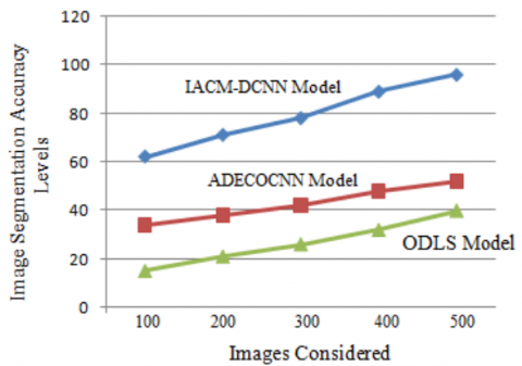
The image is divided into smaller subsets called Image segments for simpler examination and evaluation. Segmentation is the process of assigning labels to individual pixels. The percentage of an image's pixels that have the proper labels is another metric that may be used to assess the quality of a segmentation's semantics. Each category's pixels can be evaluated singly or as a group. The image segmentation accuracy levels of the existing and proposed models are shown in Figure 4 and epochs levels are represented in Table 1.
Figure 4. Image segmentation accuracy levels
Table 1. Segmentation epochs levels
|
|
IACM-DCNN Model |
ADECOCNN Model |
ODLS Model |
|
100 |
62 |
34 |
15 |
|
200 |
71 |
38 |
21 |
|
300 |
78 |
42 |
26 |
|
400 |
89 |
48 |
32 |
|
500 |
96 |
52 |
40 |
Feature extraction is the process of transforming raw data into numerical features that can be processed while maintaining all of the original information. When employing deep learning on raw data, it is more effective. Measured data is used to create relevant and non-redundant features that aid in later learning and generalization processes, as well as improving human observations in some cases. The Feature extraction accuracy levels of existing and proposed models are shown in Figure 5.
Figure 5. Feature extraction accuracy levels
Table 2. Fetaure extraction epochs
|
|
IACM-DCNN Model |
ADECOCNN Model |
ODLS Model |
|
100 |
60 |
40 |
15 |
|
200 |
70 |
45 |
22 |
|
300 |
80 |
50 |
28 |
|
400 |
85 |
55 |
35 |
|
500 |
97 |
60 |
42 |
Figure 6. Feature extraction time levels
Table 3. Bagging model epochs
|
|
IACM-DCNN Model |
ADECOCNN Model |
ODLS Model |
|
100 |
6 |
18 |
30 |
|
200 |
10 |
21 |
35 |
|
300 |
12 |
25 |
40 |
|
400 |
14 |
28 |
45 |
|
500 |
20 |
30 |
50 |
The prototype for COVID detection is trained using the extracted features. In the dimension reduction process, raw data is separated and reduced into smaller and more manageable groups by extracting features from images. The proposed model's feature extraction times are shorter than those of the proposed model. The Feature extraction time levels are shown in Figure 6 and feature extraction epochs levels are shown in Table 2 and Table 3 represents bagging model epochs.
Figure 7. Bagging model training accuracy levels
Table 4. Training accuracy levels
|
|
IACM-DCNN Model |
ADECOCNN Model |
ODLS Model |
|
100 |
62 |
22 |
7 |
|
200 |
75 |
34 |
11 |
|
300 |
84 |
42 |
16 |
|
400 |
92 |
53 |
20 |
|
500 |
97 |
62 |
25 |
In deep learning, bagging, also referred as Bootstrap aggregating, that is an ensemble learning strategy that helps to increase the accuracy and performance of the algorithm. This method is useful in dealing with trade-offs between bias and variance in a prediction model. The bagging model accuracy levels are illustrated in Figure 7. Table 4 represents training accuracy levels.
Predictions are divided by the proportion of positive predictions in order to calculate a model's classification accuracy statistic. For evaluating classifier models, it is the most often used statistic because it is easy to calculate and interpret. The COVID CT image features are classified for prediction of the disease. The classification accuracy levels are represented in Figure 8.
Based on the classifications done, the proposed ensemble model predicts the COVID disease from the training performed using the classes generated. The proposed model detection accuracy is high than the existing ones. The disease detection accuracy levels are shown in Figure 9. Table 5 shows the classification accuracy levels. Table 6 shows the disease detection accuracy levels.
Table 5. Classification accuracy levels
|
|
IACM-DCNN Model |
ADECOCNN Model |
ODLS Model |
|
100 |
45 |
22 |
3 |
|
200 |
66 |
26 |
7 |
|
300 |
76 |
32 |
13 |
|
400 |
86 |
45 |
18 |
|
500 |
96 |
55 |
22 |
Figure 8. Classification accuracy levels
Figure 9. Disease detection accuracy levels
Table 6. Disease detection accuracy levels
|
|
IACM-DCNN Model |
ADECOCNN Model |
ODLS Model |
|
100 |
62 |
25 |
15 |
|
200 |
75 |
36 |
22 |
|
300 |
82 |
56 |
35 |
|
400 |
92 |
60 |
42 |
|
500 |
98 |
65 |
50 |
During the current outbreak of COVID-19, automated image diagnosis approaches could play an important role in reducing the pressure on health care systems with a limited number of skilled clinicians. Recognition, segmentation, and classification are all part of the planned COVID-19 diagnostic system. The respiratory system is the focus of the proposed diagnostic method for locating COVID-19 hotspots. Coronavirus infection sites can be detected with CT scans of the lungs. To accurately predict and classify COVID-19 from CT scans, a DCNN-based interconnected antecedence clustering model is described here. Using trained algorithms for identification will aid in making treatment decisions for patients who have been diagnosed with COVID-19. Ensemble Learning is used here so that COVID-19 can be discovered rapidly and precisely. The only way to stop this pandemic in its tracks is to use Deep Learning technology for early detection and diagnosis of COVID-19 at the lowest possible cost and risk. According to the study's findings, diagnostic and treatment methods can be optimised using DL algorithms to improve healthcare outcomes. COVID-19 DL diagnostic models need to be trained on huge datasets that cover the whole data space to avoid overfitting and to guarantee that they are as generic and useful as feasible. The proposed model observes 98% accuracy in COVID detection. It is possible to lower the number of feature samples even further in the future to minimize the disease detection time complexity levels. Increasing the amount of images that can be used as training samples can help improve the accuracy of the system.
[1] Castiglione, A., Vijayakumar, P., Nappi, M., Sadiq, S., Umer, M. (2021). COVID-19: Automatic detection of the novel coronavirus disease from CT images using an optimized convolutional neural network. IEEE Transactions on Industrial Informatics, 17(9): 6480-6488. https://doi.org/10.1109/TII.2021.3057524
[2] Ai, T., Yang, Z., Hou, H., Zhan, C., Chen, C., Lv, W., Tao, Q., Sun, Z., Xia, L. (2020). Correlation of chest CT and RT-PCR testing for coronavirus disease 2019 (COVID-19) in China: A report of 1014 cases. Radiology, 296(2): E32-E40. https://doi.org/10.1148/radiol.2020200642
[3] Khan, M.A., Hussain, N., Majid, A., Alhaisoni, M., Bukhari, S.A.C., Kadry, S., Nam, Y., Zhang, Y.D. (2021). Classification of positive COVID-19 CT scans using deep learning. CMC-Computers Materials & Continua, 66(3): 2923-2938. http://dx.doi.org/10.32604/cmc.2021.013191
[4] Ketu, S., Mishra, P.K. (2021). Enhanced Gaussian process regression-based forecasting model for COVID-19 outbreak and significance of IoT for its detection. Applied Intelligence, 51: 1492-1512. https://doi.org/10.1007/s10489-020-01889-9
[5] Singh, V., Chandna, H., Kumar, A., Kumar, S., Upadhyay, N., Utkarsh, K. (2020). IoT-Q-Band: A low cost internet of things based wearable band to detect and track absconding COVID-19 quarantine subjects. EAI Endorsed Transactions on Internet of Things, 6(21). http://dx.doi.org/10.4108/eai.13-7-2018.163997
[6] Ismael, A.M., Şengür, A. (2021). Deep learning approaches for COVID-19 detection based on chest X-ray images. Expert Systems with Applications, 164: 114054. https://doi.org/10.1016/j.eswa.2020.114054
[7] Jain, R., Gupta, M., Taneja, S., Hemanth, D.J. (2021). Deep learning based detection and analysis of COVID-19 on chest X-ray images. Applied Intelligence, 51: 1690-1700. https://doi.org/10.1007/s10489-020-01902-1
[8] Kaur, M., Kumar, V., Yadav, V., Singh, D., Kumar, N., Das, N.N. (2021). Metaheuristic-based deep COVID-19 screening model from chest X-ray images. Journal of Healthcare Engineering, 2021: 8829829. https://doi.org/10.1155/2021/8829829
[9] Song, Y., Zheng, S., Li, L., Zhang, X., Zhang, X., Huang, Z., Chen, J., Wang, R., Zhao, H., Chong, Y., Shen, J., Zha, Y., Yang, Y. (2021). Deep learning enables accurate diagnosis of novel coronavirus (COVID-19) with CT images. IEEE/ACM Transactions on Computational Biology and Bioinformatics, 18(6): 2775-2780. https://doi.org/10.1109/TCBB.2021.3065361
[10] Khan, M.A., Kadry, S., Zhang, Y.D., Akram, T., Sharif, M., Rehman, A., Saba, T. (2021). Prediction of COVID-19-pneumonia based on selected deep features and one class kernel extreme learning machine. Computers & Electrical Engineering, 90: 106960. https://doi.org/10.1016/j.compeleceng.2020.106960
[11] Singh, D., Kumar, V., Kaur, M. (2021). Densely connected convolutional networks-based COVID-19 screening model. Applied Intelligence, 51: 3044-3051. https://doi.org/10.1007/s10489-020-02149-6
[12] Dey, N., Zhang, Y.D., Rajinikanth, V., Pugalenthi, R., Raja, N.S.M. (2021). Customized VGG19 architecture for pneumonia detection in chest X-rays. Pattern Recognition Letters, 143: 67-74. https://doi.org/10.1016/j.patrec.2020.12.010
[13] Jaiswal, A., Gianchandani, N., Singh, D., Kumar, V., Kaur, M. (2021). Classification of the COVID-19 infected patients using DenseNet201 based deep transfer learning. Journal of Biomolecular Structure and Dynamics, 39(15): 5682-5689. https://doi.org/10.1080/07391102.2020.1788642
[14] Ko, H., Chung, H., Kang, W.S., Kim, K.W., Shin, Y., Kang, S.J., Lee, J.H., Kim, Y.J., Kim, N.J., Jung, H., Lee, J. (2020). COVID-19 pneumonia diagnosis using a simple 2D deep learning framework with a single chest CT image: Model development and validation. Journal of Medical Internet Research, 22(6): e19569. https://doi.org/10.2196/19569
[15] Hu, S., Gao, Y., Niu, Z., Jiang, Y., Li, L., Xiao, X., Wang, M., Fang, E.F., Menpes-Smith, W., Xia, J., Ye, H., Yang, G. (2020). Weakly supervised deep learning for COVID-19 infection detection and classification from CT images. IEEE Access, 8: 118869-118883. https://doi.org/10.1109/ACCESS.2020.3005510
[16] Gao, K., Su, J.P., Jiang, Z.B., Zeng, L.L., Feng, Z.C., Shen, H., Rong, P.F., Xu, X., Qin, J., Yang, Y.X., Wang, W., Hu, D.W. (2021). Dual-branch combination network (DCN): Towards accurate diagnosis and lesion segmentation of COVID-19 using CT images. Medical Image Analysis, 67: 101836. https://doi.org/10.1016/j.media.2020.101836
[17] Ni, Q., Sun, Z.Y., Qi, L., Chen, W., Yang, Y., Wang, L., Zhang, X.Y., Yang, L., Fang, Y., Xing, Z.J., Zhou, Z., Yu, Y.Z., Lu, G.M., Zhang, L.J. (2020). A deep learning approach to characterize 2019 coronavirus disease (COVID-19) pneumonia in chest CT images. European Radiology, 30: 6517-6527. https://doi.org/10.1007/s00330-020-07044-9
[18] Horry, M.J., Chakraborty, S., Paul, M., Ulhaq, A., Pradhan, B., Saha, M., Shukla, N. (2020). COVID-19 detection through transfer learning using multimodal imaging data. IEEE Access, 8: 149808-149824. https://doi.org/10.1109/access.2020.3016780
[19] Ozturk, T., Talo, M., Yildirim, E.A., Baloglu, U.B., Yildirim, O., Acharya, U.R. (2020). Automated detection of COVID-19 cases using deep neural networks with X-ray images. Computers in Biology and Medicine, 121: 103792. https://doi.org/10.1016/j.compbiomed.2020.103792
[20] Islam, M.Z., Islam, M.M., Asraf, A. (2020). A combined deep CNN-LSTM network for the detection of novel coronavirus (COVID-19) using X-ray images. Informatics in Medicine Unlocked, 20: 100412.https://doi.org/10.1016/j.imu.2020.100412
[21] Voulodimos, A., Protopapadakis, E., Katsamenis, I., Doulamis, A., Doulamis, N. (2021). A few-shot U-net deep learning model for COVID-19 infected area segmentation in CT images. Sensors, 21(6): 2215. https://doi.org/10.3390/s21062215
[22] Ahsan, M., Based, M.A., Haider, J., Kowalski, M. (2021). COVID-19 detection from chest X-ray images using feature fusion and deep learning. Sensors, 21(4): 1480. https://doi.org/10.3390/s21041480
[23] Sharafeldeen, A., Elsharkawy, M., Alghamdi, N.S., Soliman, A., El-Baz, A. (2021). Precise segmentation of covid-19 infected lung from CT images based on adaptive first-order appearance model with morphological/anatomical constraints. Sensors, 21(16): 5482. https://doi.org/10.3390/s21165482
[24] Rehman, M.U., Shafique, A., Khalid, S., Driss, M., Rubaiee, S. (2021). Future forecasting of COVID-19: A supervised learning approach. Sensors, 21(10): 3322. https://doi.org/10.3390/s21103322
[25] Khan, M.A., Sharif, M.I., Raza, M., Anjum, A., Saba, T., Shad, S.A. (2022). Skin lesion segmentation and classification: A unified framework of deep neural network features fusion and selection. Expert Systems, 39(7): e12497. https://doi.org/10.1111/exsy.12497
[26] Rashid, M., Khan, M.A., Alhaisoni, M., Wang, S.H., Naqvi, S.R., Rehman, A., Saba, T. (2020). A sustainable deep learning framework for object recognition using multi-layers deep features fusion and selection. Sustainability, 12(12): 5037. https://doi.org/10.3390/su12125037
[27] Muzammil, S.R., Maqsood, S., Haider, S., Damaševičius, R. (2020). CSID: A novel multimodal image fusion algorithm for enhanced clinical diagnosis. Diagnostics, 10(11): 904. https://doi.org/10.3390/diagnostics10110904