Mandakini Ingle* | Ratnesh Litoriya | Shweta Gupta | Pinky Rane | Swati Vaidya
© 2026 The authors. This article is published by IIETA and is licensed under the CC BY 4.0 license (http://creativecommons.org/licenses/by/4.0/).
OPEN ACCESS
Cardiovascular diseases remain a major global health challenge, requiring accurate and timely screening approaches that can integrate heterogeneous clinical information. Conventional diagnostic procedures often rely on manual interpretation of electrocardiogram (ECG) signals and expert assessment, which may be affected by workload and inter-observer variability. This study proposes a modality-specific deep learning framework for detecting two major cardiac disorders: arrhythmia from ECG signals and coronary artery disease (CAD) from structured clinical data. For ECG-based arrhythmia classification, a hybrid 1D Convolutional Neural Network–Long Short-Term Memory (CNN-LSTM) model is developed to extract morphological characteristics and temporal dependencies from ECG sequences. For CAD prediction, a regularized Deep Neural Network (DNN) incorporating batch normalization and dropout is designed to learn nonlinear relationships among clinical risk factors. The proposed framework is evaluated using the MIT-BIH Arrhythmia and Z-Alizadeh Sani CAD datasets. Experimental results demonstrate that the CNN-LSTM model achieves improved arrhythmia classification performance compared with conventional machine learning and standalone CNN approaches, while the regularized DNN provides reliable CAD prediction with enhanced generalization capability. By assigning dedicated deep learning architectures to different data modalities, the proposed approach offers a flexible strategy for AI-assisted cardiac screening and highlights the potential of multimodal learning in clinical decision-support applications.
cardiovascular disease, Artificial Intelligence, deep learning; electrocardiogram, arrhythmia classification, coronary artery disease; multimodal learning
With an estimated 17.9 million deaths occurring yearly, representing 32% of all deaths worldwide [1], cardiovascular diseases are currently the leading killers worldwide. Among them are cardiac arrhythmias and coronary artery disease (CAD), the most common and high- risk clinical forms of CVDs, each of which can give rise to life-threatening conditions if not found and treated in time. Arrhythmias are abnormal heart rhythms generated by irregular electrical activity in the heart muscle, while in CAD, the coronary arteries become narrowed or blocked, usually because of atherosclerosis [2]. Such conditions have to be diagnosed early to enable timely intervention and further management, but the conventional ways of performing this, such as manual electrocardiogram (ECG) interpretation and invasive coronary angiography, are time-consuming, require clinical expertise, and are at times subject to human error through observer variability.
Recent developments in Artificial Intelligence (AI) and machine learning have transformed the arena of healthcare analytics, especially in automated medical diagnostics. Since patient records and biosignals have been increasingly digitized, deep learning models have become effective tools to extract complex patterns from high-dimensional data, performing much better than traditional machine learning methods under many clinical circumstances [3, 4]. Specifically, Convolutional Neural Networks (CNNs) and Recurrent Neural Networks (RNNs), particularly those based on Long Short-Term Memory (LSTM), have shown particular promise in the analysis of ECG signals, a primary time-series data type used in cardiac care [5]. In contrast, Deep Neural Networks (DNNs) are more apt to deal with structured clinical data, where complex relationships among risk factors, laboratory variables, and disease outcomes can be learned.
Deep learning methods for cardiac disease detection have been the subject of a few studies. For instance, a 34-layer CNN model named Cardiologist-Level Arrhythmia Detection developed by Hannun et al. [6] displayed expert-level performance on PhysioNet data. In the same manner, Yildirim et al. [7] showed that a deep bidirectional LSTM-CNN can be used for multi-class arrhythmia classification, attaining excellent accuracy on benchmark ECG datasets. Concerning CAD prediction, a study by Khosla et al. [8] and the one by Alizadehsani et al. [9] resorted to ensemble learning and shallow neural networks applied to clinical datasets such as Z-Alizadeh Sani and Cleveland, with modest success in disease classification. However, a sufficient number of problems persist as obstacles to be solved even with these advances.
Firstly, many previous investigations were looking for arrhythmia detection or CAD prediction in deployment and missed the opportunity for integrated cardiac screening matching real-world diagnostic concepts where patients may arrive with co-occurring conditions. Secondly, these models are usually overfitted due to the limited number and imbalance of medical datasets, particularly in clinical datasets like Z-Alizadeh Sani. On the other hand, these CNN-only models would weakly express the temporal patterns in ECG signals, lacking some memory mechanisms to model such sequential dependencies. Third, only a few studies have attempted to establish a systematic AI approach that unites time-series biosignals with structured clinical data for diagnosis in one architecture.
In other words, we propose an AI-based dual-model framework for the complete cardiac disease detection process. More particularly, we utilize a 1D CNN and a hybrid CNN-LSTM for arrhythmia classification using the MIT-BIH Arrhythmia dataset. The CNN extracts local features from the ECG signals, whereas the LSTM captures temporal dependencies, augmenting robustness and accuracy in modeling sequences. For the CAD prediction, a feedforward DNN is built on the Z-Alizadeh Sani dataset. Batch normalization and dropout layers are introduced in the model to ensure generalization and avoid overfitting, leading to stable performance on fairly small datasets.
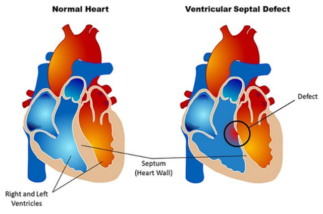
Figure 1 shows the differences between a normal heart and one that has a VSD. The condition is characterized by an abnormal hole in the wall separating the two ventricles, allowing blood to flow from the left ventricle to the right. This increases the strain on the heart and causes cardiovascular problems. Early diagnosis is crucial for proper treatment and improved results.
Figure 1. Difference between a normal heart and a heart with a ventricular septal defect, which is a defect at birth (congenital heart defect) [10]
Key features:
(1). CNN-LSTM hybrid network architecture: Utilizes local spatial features along with long-term temporal dependencies extracted from ECG signals for effective detection of arrhythmia.
(2). Regularized DNN: Detects the intricate nonlinear connections between the structured clinical data to avoid overfitting.
(3). Dual-model approach: Combines the use of customized deep learning models for detecting the two different conditions, namely arrhythmia and CAD.
(4). Automatic feature extraction: Removes the need for manual feature engineering by extracting informative features from ECG signals and clinical features automatically.
(5). Comprehensive diagnostic performance comparison: Illustrates enhanced diagnostic performance by comparing the proposed approach with traditional machine learning and deep learning methods.
(6). Applicability in clinical settings: Enables early detection of heart diseases, which can later be used as an input to AI-based decision support systems in clinics.
(7). Generalization ability: Uses regularization strategies to enhance the generalization ability of the model to handle unseen patient data.
(8). Scalable model structure: Offers a scalable architecture that is useful for multimodal cardiovascular disease diagnosis and future intelligent healthcare applications.
Major contributions of this research include providing a framework for diagnosing cardiovascular conditions by integrating deep learning techniques with time series ECG data and structured clinical information. A novel CNN-LSTM network has been designed to enhance the capability of arrhythmia classification by using temporal memory, which is an improvement compared to existing CNNs. Furthermore, a fully connected DNN model predicts CAD on the basis of clinical variables, wherein the use of batch normalization and dropout avoids overfitting. Last but not least, benchmarking datasets, known as MIT-BIH Arrhythmia and Z-Alizadeh Sani CAD, are included in order to test the proposed method in real-world scenarios.
MIT-BIH Arrhythmia dataset is a well-known, annotated ECG dataset made up of 48 recordings of half an hour each from 47 patients recorded at the Beth Israel Hospital Arrhythmia Laboratory [11]. This benchmarking dataset offers a large number of labeled ECG waveform cases belonging to various types of arrhythmias, hence suitable for training deep learning models in time-series classification problems. As opposed to this, the Z-Alizadeh Sani dataset comprises 303 patient data points with 54 different attributes such as demographics, symptoms, ECG, and lab data.
The organization of this research paper is described below. Section 2 describes the relevant literature in the area, highlighting past studies in applying deep learning for ECG signal classification and CAD prediction. Section 3 describes the proposed architecture of dual models that includes the selection of CNN, CNN-LSTM, and DNN architectures. Section 4 gives an overview of experiments performed, describing in detail the process of data preprocessing, model configuration, training, and evaluation, and provides a discussion of the results and their applicability in the clinical setting. Section 5 concludes the whole study.
There have been several recent developments in the field of deep learning that have resulted in better automation in CVD diagnosis. Different types of deep learning models, including CNNs, RNNs, multimodal learning, and hybrid deep learning, have been studied. While these models exhibit good performance for the diagnosis of CVD, there are still several open challenges in the domain of temporal feature learning, multimodal fusion, model interpretability, and clinical generalization. The existing literature can be classified into different groups.
2.1 Convolutional Neural Network-based approach
In recent years, CNNs have become one of the most popular methods of deep learning applied to the detection of cardiovascular disease due to their ability to automatically extract the hierarchy of spatial features from ECG signals without any feature engineering. CNN techniques have proved to be very efficient in diagnosing arrhythmia, congestive heart failure, myocardial infarction, and coronary artery disease.
Previous studies [5, 10, 12] proved the effectiveness of deep CNNs in automatic discovery of discriminating features in the heartbeat through an ECG signal. Cardiologist-level arrhythmia classification was provided by Hannun et al. [6] and Almansouri et al. [13]. In their latest studies, Hammad et al. [14] and Selvam et al. [15] increased the effectiveness of ECG classification for myocardial infarction and cardiovascular disease diagnosis by applying advanced CNN architectures.
Research gap: Even though CNN architectures can extract relevant information from ECGs, they fail to incorporate long-term dependencies between successive heartbeats. Moreover, they work mostly on ECG data, neglecting any other clinical data, thus restricting them from dealing with complex cases of cardiovascular disorders.
2.2 Long Short-Term Memory and Recurrent Neural Network-based approach
LSTM and RNNs were developed to learn sequential LSTM and RNNs have found widespread use in the prediction of cardiovascular diseases due to their capability of modeling sequential dependence in ECG signals. The memory capabilities of these models allow for more efficient representation of rhythm changes and cardiac activities over time.
Yildirim [7] and Tawil [16] suggested a BiLSTM framework with wavelet transform for ECG classification, with high accuracy in detecting arrhythmias. A context-aware sensing framework based on smartphones was introduced by Rana et al. [17], but it was not built for heart disease diagnosis as it did not consider ECG readings or any clinical risk factors. On the other hand, Kachuee et al. [18] used deep transferable representations for heartbeat classification from wearable ECG signals.
Research gaps: Even though LSTM-based models have been able to efficiently identify temporal dependencies, they still tend to consume more computing power and time during the training phase. Moreover, these models are limited to only ECG data and do not use structured clinical data for prediction of heart diseases.
2.3 Hybrid deep learning approach
In hybrid deep learning models, the combination of several neural networks is used to learn complementary features concurrently. CNN-LSTM models have been able to perform better due to the combination of spatial feature learning and temporal sequence learning.
Kothari and Rani [19] have introduced machine learning methodologies through KNN, Decision Tree, Random Forest, and SVM for effective prediction of heart disease. Nasarian et al. [20] studied computational intelligence methods for CAD diagnosis; on the other hand, Gudadhe et al. [21] used an ANN to predict heart disease. Bai et al. [22] designed a hybrid deep learning model for automated arrhythmia detection with 12-lead ECG, whereas CardioAttentionNet [23, 24] increased its diagnostic accuracy using attention.
Research gap: The present hybrid models focus on single heart diseases only and have a dependency on ECG signals or manually extracted features from clinical data. They seldom offer a unified platform that can analyze heterogeneous cardiovascular data.
2.4 Multimodal deep learning approach
Recently, the use of multimodal learning has been found to be quite useful as a technique that combines different complementary medical data for better prediction outcomes. The combination of ECG data with structured clinical variables can provide more informative features for better disease prediction.
Torres Soto et al. [25] developed a model of multimodal deep learning using ECG data and clinical variables for cardiovascular event prediction. Tadesse et al. [26] proposed a technique called DeepMI that combines multiple ECG leads for myocardial infarction diagnosis.
Research gaps: The current multimodal approaches use only basic feature concatenation and a correlation-based learning approach. The interaction between ECG morphological features and the clinical risk factors is not considered.
2.5 Recent deep learning approaches
Recently, attention mechanisms, interpretability, and deep learning methods for cardiovascular diagnosis have been considered by researchers.
The application of disease-specific attention mechanisms for ECG classification was proposed by Bai [22]. The importance of explainable deep learning methods for cardiovascular diseases is discussed in a recent review [27] written by Wu and Guo. Also, transformer-based methods such as ECG SMART NET [28] and CardioAttentionNet [23, 24] have been proposed recently.
Research gaps: Attention-based and Explainable AI models increase the transparency of models but are still based on correlation-based learning and don't incorporate any causation between the cardiovascular risk factors. The use of such models in a real-world clinical environment is very limited because of the lack of multimodal integration and validation.
Early works mostly focused on the usage of CNN architectures for the classification of ECG due to their ability to automatically extract discriminative features from the signal [5, 6, 28]. Later, LSTM and RNN architectures were utilized for capturing the temporal dependencies in ECG data to enhance the classification results [7]. Further improvements were made via the development of hybrid deep learning models such as CNN-LSTM and attention-based models, which incorporated spatial and temporal representation of features [21-23].
Recently, multimodal and graph-based approaches combined ECG and clinical data by modeling the interaction between features to achieve reliable predictions [25, 29]. However, the existing approaches are mainly based on correlation learning but fail to capture the complicated dependencies between the cardiovascular risk factors and ECG signals [19, 14]. Thus, the proposed hybrid multimodal framework seeks to fill this research gap.
While considerable advancements have been made in cardiac diagnosis with ECG through the use of deep learning technologies, there still exist certain shortcomings. First, while early CNN models effectively identify local features within ECG signals, they demonstrate weak capacity to model long-range dependencies in time series. LSTM and RNN models are superior to earlier methods in terms of learning sequence dependencies, though they face challenges with computational complexity and overfitting problems. Combining CNN and LSTM networks or using attention models can significantly boost diagnostic accuracy.
However, almost all research is based only on ECG signal data and does not take into account additional clinical information as summarized in Table 1. Multimodal and graph-based methods are recent trends in this field. While these models attempt to leverage heterogeneity of data, they mostly focus on correlation rather than relationships between cardiovascular risk factors and ECG signals. Additionally, existing solutions lack generalizability and fail to build clinically relevant representations. Therefore, there is a need for an intelligent multimodal framework capable of combining ECG and clinical data for analysis.
Table 1. Comparative analysis of existing deep learning techniques for cardiac abnormality detection
|
S. N. |
Reference No. |
Methodology |
Dataset / Domain |
Disease |
Key Findings / Research Gap |
|
1 |
[5] |
Deep CNN |
ECG signals |
Cardiac abnormality |
Effective automatic ECG-based detection; ECG-only approach. |
|
2 |
[6] |
Deep Neural Network (DNN) |
Ambulatory ECG |
Arrhythmia |
High-performance multi-class detection; requires large annotated datasets. |
|
3 |
[7] |
Wavelet + BiLSTM |
ECG |
Arrhythmia |
Captures temporal ECG patterns; preprocessing and computational complexity remain challenges. |
|
4 |
[12] |
Deep CNN |
MIT-BIH ECG |
Arrhythmia |
Automatic ECG feature extraction; limited to ECG modality. |
|
5 |
[14] |
Feature selection + deep learning |
ECG |
Cardiac disorders |
Detects myocardial infarction and conduction disorders; limited clinical-data integration. |
|
6 |
[18] |
Transfer learning + CNN |
MIT-BIH ECG |
Arrhythmia |
Uses transferable representations; does not incorporate clinical information. |
|
7 |
[19] |
Hybrid deep learning |
Heart disease dataset |
CAD/heart disease |
Demonstrates hybrid learning potential; lacks ECG temporal modeling and multimodal integration. |
|
8 |
[20] |
Feature selection + balancing |
CAD dataset |
CAD |
Shows importance of feature selection and class balancing; uses structured clinical data. |
|
9 |
[22] |
Hybrid deep learning |
12-lead ECG |
Arrhythmia |
Improves arrhythmia diagnosis; restricted to ECG signals. |
|
10 |
[25] |
Multimodal deep learning |
ECG + cardiac data |
Cardiac disease |
Shows benefit of multimodal learning; does not jointly address arrhythmia and CAD. |
|
11 |
[26] |
Deep multi-lead ECG fusion |
Multi-lead ECG |
MI / cardiac disease |
Fuses multiple ECG leads effectively; remains primarily ECG-based. |
|
12 |
[28] |
Interpretable deep learning |
PTB-XL ECG |
Arrhythmia |
Explores interpretable ECG classification; clinical risk factors not integrated. |
|
13 |
[29] |
machine learning-based approaches |
CAD datasets |
CAD |
Reviews machine learning methods; feature engineering and generalization remain challenges. |
|
14 |
[30] |
Data mining/machine learning |
Z-Alizadeh Sani |
CAD |
Effective CAD prediction using clinical features; conventional feature-based learning. |
|
15 |
[31] |
Efficient 1D CNN |
ECG signals |
Cardiac disease |
Efficient ECG detection; does not model long-term temporal dependencies or integrate clinical risk factors. |
Being able to detect cardiac abnormalities at the earliest possible stage, especially arrhythmias and CAD, still remains an acute challenge in the healthcare domain, in spite of the astounding pioneering developments in AI and deep learning. Clinical misdiagnosis and the lack of expert interpretations, coupled with conventional manual diagnosis, lead to delays in the administration of therapies and poor patient outcomes. Current AI-based methods either remain isolated from ECG-based arrhythmia detection or clinical feature-based CAD prediction, thus failing to address the multi-condition diagnostic scenario present in real-world cardiology. In addition, many, if not most, of such approaches depend heavily on hand-engineered features with complicated multi-stage pipelines and a potential lack of explicit regularization strategies, thereby compromising their ability to generalize well on unseen patient data.
The core problem addressed in this study is the lack of a unified, end-to-end AI framework capable of accurately detecting multiple cardiac abnormalities using modality-specific deep learning architectures that are optimized for both time-series signals (ECG) and structured clinical datasets. To overcome the identified limitations and bridge the research gap, this study proposes an AI-driven dual-model architecture that combines two tailored deep learning pipelines:
•A 1D CNN and CNN-LSTM hybrid model for arrhythmia detection, using ECG signals from the MIT-BIH Arrhythmia dataset
•A feedforward DNN with dropout and batch normalization for CAD prediction, trained on the Z-Alizadeh Sani clinical dataset.
This bifurcated design ensures that each cardiac condition is addressed using a deep learning model that is best suited to the data modality and underlying pathophysiological patterns.
3.1 Arrhythmia detection using 1D Convolutional Neural Network and Convolutional Neural Network-Long Short-Term Memory
The MIT-BIH Arrhythmia dataset consists of long-term ambulatory ECG recordings, annotated with beat-level classifications. These one-dimensional time-series signals contain rich spatial and temporal information about the electrical behavior of the heart.
3.1.1 Convolutional Neural Network model
According to Algorithm 1, the 1D CNN model is designed to extract local features from ECG sequences, such as P-QRS-T wave shapes and morphological deviations. Multiple convolutional and max-pooling layers are stacked to capture hierarchical representations of the waveform data. The network is followed by fully connected layers for final classification into arrhythmia classes.
|
Algorithm 1. CNN-1D model for cardiac disease detection |
|
Input: Pre-processed one-dimensional ECG signal segments (e.g., fixed-length windows of beats) Output: Cardiac condition classification (e.g., Normal, Arrhythmia, AF, PVC)
Conv L1: 32 filters (kernel size = 5), followed by ReLU to extract local features. Pooling L1: Max Pooling (pool size = 2), decreasing time resolution by half.
Conv L2: 64 filters (kernel size = 3), followed by ReLU to extract deeper features. Pooling L2: Max Pooling (pool size = 2), again reducing time length and maintaining dominant features.
Reshape Layer: CNN output reshaping into (timesteps, features) format for the LSTM input.
LSTM Layer: 64 units, capturing the temporal dependency and sequence of the beats.
Dropout: Using Dropout (rate = 0.5) to avoid overfitting.
FC L1: Fully Connected layer with 128 nodes and ReLU activation.
Output: Using the softmax function for multi-class cardiac condition classification. |
Considering $X=\left\{x_1, x_2, \ldots, x_T\right\} \in \Re^{T \times 1}$ be a 1D ECG signal segment of length T, representing a sequence of ECG measurements, a 1D CNN applies convolutional filters to extract spatial (local) features:
$\begin{gathered}h_t^{(1)}=\sigma\left(\sum_{i=0}^{k-1} w_i^{(1)} x_{t+i}+b^{(1)}\right), \\ t=1,2, \ldots, T-k+1\end{gathered}$ (1)
where, $w_i^{(1)}$ are the learnable weights of the convolution filter, $b^{(1)}$ is the bias term, k is the kernel size, σ is the activation function. Typically ReLU(z) = max(0,z).
The output is passed through multiple such layers, often followed by max pooling:
$p_t=\max _{j \in w} h_j^{(1)}$ (2)
where, W is the pooling window.
3.1.2 CNN-LSTM hybrid model
To enhance the model’s capacity to learn long-range temporal dependencies, the CNN is integrated with an LSTM layer. This is shown in Algorithm 2 CNN-LSTM stepwise. The CNN component handles spatial encoding, while the LSTM captures the sequential progression and temporal correlation between heartbeats. This hybrid architecture improves sensitivity to rhythm-based conditions, such as premature ventricular contractions or bundle branch blocks.
Model configuration
•Input: segmented ECG beats (normalized)
•CNN layers: 3–4 convolutional layers with ReLU activation
•LSTM layer: single or stacked layer with 64–128 units
•Dense layers: fully connected layers with Softmax for multi-class classification
•Optimizer: Adam
•Loss function: categorical cross-entropy
•Evaluation: accuracy, precision, recall, F1-score.
The CNN output $H=\left\{h_1, h_2, \ldots, h_T\right\}$ is fed into an LSTM layer to capture temporal dependencies.
For each timestep t, the LSTM equations are:
$f_t=\sigma\left(W_f h_t+U_f h_{t-1}+b_f\right)$ (3)
$i_t=\sigma\left(W_i h_t+U_i h_{t-1}+b_i\right)$ (4)
$\tilde{c}_t=\tanh \left(W_c h_t+U_c h_{t-1}+b_c\right)$ (5)
$c_t=f_t \circ c_{t-1}+i_t \circ \tilde{c}_t$ (6)
$o_t=\sigma\left(W_o h_t+U_o h_{t-1}+b_o\right)$ (7)
$h_t=o_t \circ \tanh \left(c_t\right)$ (8)
$f_t=\sigma\left(W_f h_t+U_f h_{t-1}+b_f\right)$ (9)
where,
$f_t, i_t, o_t$: forget, input, and output gates, respectively
$\circ$: element-wise multiplication
ct: cell state
ht: hidden state
$W_*, U_*, b_*$: LSTM parameters
The final hidden state hT is passed through a fully connected layer and softmax activation for multi-class classification:
$\hat{y}=\operatorname{softmax}\left(W \rightleftarrows_o h_T+b_o\right)$ (10)
where,
$\operatorname{softmax}\left(z_i\right)=\frac{e^{z_i}}{\sum_{j=1}^C e^{z_j}}, i=1, \ldots, C$
C: number of arrhythmia classes.
The model is trained by minimizing categorical cross-entropy:
$L_{\text {arrhythmi }}=-\sum_{i=1}^C y_i \log \left(\hat{y}_i\right)$ (11)
where, $y \in\{0,1\}^C$ is the one-hot encoded ground truth.
|
Algorithm 2. CNN-LSTM model for cardiac disease detection |
|
Input: Pre-processed feature vectors extracted from 1D ECG signal segments (e.g., heart rate, RR interval, waveform morphology) Output: Classification of cardiac condition (e.g., Normal, Arrhythmia, AF, PVC)
Input Layer: Accepts the ECG feature vector as input (e.g., vector length = n) FC L1: Fully connected layer with 512 nodes, followed by ReLU activation DO 1: Apply dropout (rate = 0.5) to reduce overfitting FC L2: Fully connected layer with 256 nodes, followed by ReLU activation DO 2: Apply dropout (rate = 0.5) FC L3: Fully connected layer with 128 nodes, followed by ReLU activation DO 3: Apply dropout (rate = 0.5)
Output Layer: Fully connected layer with k nodes (number of cardiac classes) Apply SoftMax activation function for multi-class classification |
3.2 Coronary artery disease prediction using regularized feedforward Deep Neural Network
The Z-Alizadeh Sani dataset includes 303 patient records with 54 features covering demographic, symptom, ECG, echocardiographic, and laboratory variables. This structured dataset is well-suited for classification tasks using deep feedforward networks.
3.2.1 Deep Neural Network architecture
A multi-layer feedforward DNN, which is described in Algorithm 3, is employed to model complex nonlinear relationships among clinical features that contribute to CAD. The network includes several dense layers interleaved with dropout layers for regularization and batch normalization for training stability.
For each hidden layer l, the transformation is:
$a^{(l)}=B N^{(l)}\left(W^{(l)} h^{(l-1)}+b^{(l)}\right), h^{(l)}=\sigma\left(a^{(l)}\right)$ (12)
where,
$W^{(l)} \in \Re^{n_l \times n_{l-1}}$: weight matrix
$B N^{(l)}$: batch normalization
σ: activation function (ReLU)
Dropout is applied as:
$h_{\text {drop }}^{(l)}=h^{(l)} \cdot \delta, \delta \sim$ Bernoulli $(1-p)$
where, p is the dropout rate.
|
Algorithm 3. Deep Neural Network model for cardiac disease detection |
|
Input: Pre-processed 1D ECG signal segments (e.g., fixed-length windowed beats) Output: Classification of cardiac condition (e.g., Normal, Arrhythmia, AF, PVC)
Conv L1: 32 filters (kernel size = 5), followed by ReLU activation to extract local features Pooling L1: Max pooling (pool size = 2), reduces temporal resolution by half Conv L2: 64 filters (kernel size = 3), followed by ReLU activation to deepen feature representation Pooling L2: Max pooling (pool size = 2), further reduces temporal length and retains dominant features
Reshape Layer: Reshape CNN output to (timesteps, features) for feeding into LSTM
LSTM Layer: 64 units, captures temporal dependencies and sequential patterns between beats Dropout: Apply dropout (rate = 0.5) to reduce overfitting
FC L1: Fully connected layer with 128 nodes, ReLU activation
Output: Apply Softmax function for multi-class classification of cardiac conditions |
3.2.3 Feature preprocessing
Before training, categorical variables are one-hot encoded, and continuous variables are normalized. Feature importance analysis is optionally conducted to reduce dimensionality and enhance interpretability.
Model configuration
•Input: 54-dimensional feature vector
•Hidden layers: 3–5 dense layers with 64–128 neurons
•Activation: ReLU
•Dropout: 0.3–0.5 after each layer
•Batch normalization: after each hidden layer
•Output: Sigmoid-activated neuron for binary classification
•Optimizer: Adam
•Loss function: binary cross-entropy
•Evaluation: accuracy, area under the curve (AUC)-receiver operating characteristic (ROC), precision, recall, F1-score
Final prediction is made using sigmoid activation:
$\hat{y}=\sigma\left(W^{(L)} h^{(L-1)}+b^{(L)}\right)$ (13)
$\sigma(z)=\frac{1}{1+e^{-z}}$ (14)
where, $y \in\{0,1\}$ represents the probability of CAD presence.
Binary cross-entropy is used as the loss function:
$L_{C A D}=-[y \log (\hat{y})+(1-y) \log (1-\hat{y})]$ (15)
Normalization through batches keeps the feature distribution stable and speeds up convergence because of reduced internal covariate shift. Dropout works by randomly turning off some neurons during training in order to avoid co-adaptation and reduce overfitting. According to ablation experiments, the combination of batch normalization and dropout enhances generalization performance is shown in Table 2.
Table 2. Performance of the combination of batch normalization and dropout
|
Model |
Accuracy (%) |
|
Deep Neural Network (DNN) |
92.8 |
|
DNN + BatchNorm |
94.1 |
|
DNN + Dropout |
94.6 |
|
DNN + BatchNorm + Dropout |
96.2 |
3.3 Unified diagnostic framework
The two models operate in parallel pipelines, each specialized for a specific cardiac abnormality. This architecture offers the following advantages:
•Modality-specific learning: Optimizes performance by using CNN-based models for time-series data and DNNs for clinical feature-based data.
•Multi-condition capability: Enables the simultaneous detection of both arrhythmia and CAD, supporting more comprehensive diagnostic decision-making.
•End-to-end learning: Both models are trained and deployed without reliance on manual feature engineering, ensuring scalability and real-time applicability.
•Regularization and stability: Batch normalization and dropout techniques enhance robustness and reduce overfitting, particularly important for the smaller CAD dataset.
The models are optimized separately using the Adam optimizer to minimize:
$L_{\text {Total }}=L_{\text {arrhythmia }}+L_{C A D}$ (16)
Though the models are independent, this dual-path structure can be deployed in parallel pipelines or integrated within a larger multi-modal framework.
3.4 Feature importance analysis
From feature importance analysis, it was evident that RR interval, QRS interval, and QT interval were the most discriminating features from an ECG perspective for classification of arrhythmias. Chest pain type, age, cholesterol level, blood pressure, and peak heart rate were the most influential clinical factors when predicting coronary artery disease (see Table 3). The combination of these factors made the proposed approach successful in cardiac abnormality detection.
Table 3. Important features contributing to cardiac abnormality detection
|
Category |
Important Features |
|
Electrocardiogram (ECG) features |
RR interval, QRS duration, QT interval, Heart Rate Variability (HRV), ST-segment |
|
Clinical features |
Age, chest pain type, cholesterol, blood pressure, Thalach, diabetes |
3.5 Model selection
The choice of various deep learning structures depended on the nature of the input data and the requirement of diagnosis in each case. The process of diagnosing arrhythmia depends on the utilization of ECG signal data that is sequential in nature, having both local morphological patterns as well as temporal relationships. Hence, the selection of the CNN-LSTM structure was considered optimal since CNNs are able to capture local features such as P-waves, QRS complex, and T-waves while LSTMs are utilized to capture temporal dependencies between successive heartbeats. It has been proven in past studies that hybrid CNN-LSTM models outperform CNN and LSTM models alone due to joint learning of spatial and temporal representation [7, 15, 18].
However, in the case of CAD diagnosis in this experiment, the utilization of the Z-Alizadeh Sani dataset takes place, where structured clinical variables are involved, which include demographics, laboratory data, symptoms, and risk factors, and does not involve any ECG signals, which are sequential in nature and do not include temporal dependencies. Hence, a recurrent structure like LSTM is not required in this case. Hence, the use of a DNN was considered to be optimal since it learns the complex relationship between structured clinical data. The inclusion of dropout and batch normalization further improves generalization and reduces overfitting, making the DNN well suited for structured clinical data [9, 17, 19].
Alizadehsani et al. [30] developed a data mining approach for coronary artery disease diagnosis using clinical and laboratory features. However, the study did not incorporate ECG signals or multimodal learning, limiting its ability to capture complex cardiac patterns.
This choice of architecture is not arbitrary but data-driven, meaning that a specific architecture will be matched to the type of input data. The choice of such an approach allows efficient learning of features from ECG signals and clinical data, keeping computational efficiency intact.
The architecture makes it possible for the proposed framework to leverage the advantages of both DL models and thus provide a better approach to detect any heart abnormality compared to the scenario where one model performs both tasks.
To validate the performance and reliability of the proposed hybrid deep learning framework for cardiac issue detection, a comprehensive experimental setup has been established. The experiments are designed to evaluate the model’s effectiveness in two key domains of cardiovascular diagnostics: arrhythmia classification using time-series ECG signals, and CAD prediction using structured clinical features. Each component of the framework is trained, validated, and tested independently using benchmark datasets, with standard machine learning protocols adhered to throughout the process to ensure the reproducibility and generalizability of results.
4.1 Datasets
The proposed framework uses two publicly available and widely cited datasets:
In order to test the efficacy and validity of the proposed deep learning architecture that can be applied in detecting heart problems, an experiment has been developed. The experiments are aimed at testing the effectiveness of the model in two important areas of cardiovascular diagnostics, namely, arrhythmia classification through ECG signals and CAD diagnosis from clinical features. All components of the architecture have been trained, validated, and tested individually through benchmark datasets while following all procedures for machine learning. The proposed framework uses two publicly available and widely cited datasets:
MIT-BIH Arrhythmia dataset: The database consists of 48 half-hour segments of two-lead ambulatory ECG records from 47 patients with a sampling rate of 360 Hz. Annotation for each beat is done based on the type of arrhythmia. In our work, the beats are labeled in five categories (according to the AAMI standard).
Z-Alizadeh Sani CAD dataset: The structured data consists of 303 observations with 54 features that include demographic factors, ECGs, echocardiograms, and laboratory results. The binary target variable denotes whether there is coronary artery disease. The data have been processed to deal with missing values, categorical variables using one-hot encoding, and feature scaling through z-score standardization.
The 1D CNN-LSTM hybrid model is implemented using Python and TensorFlow. The architecture includes:
Convolutional layers: Three 1D CNN layers with kernel sizes of 5, 3, and 3, respectively, each followed by ReLU activation and max pooling.
LSTM layer: A unidirectional LSTM with 128 hidden units to capture temporal dependencies.
Fully connected layer: Dense layer with Softmax activation to classify beats into arrhythmia categories.
4.2 Data preprocessing and experimental setup
ECG signal data was standardized to have zero mean and unit variance. The noise was filtered out using a band-pass filter. The missing values in clinical data were imputed using median imputation. Outlier detection was done using the interquartile range method.
CNN network architecture included three convolutional layers of 1D filters of sizes 3 and 5, along with ReLU activation and max pooling steps. The resultant features were then fed into two fully connected layers, using a dropout probability of 0.5 to avoid overfitting. Training of the model was done using the Adam optimizer with a learning rate of 0.001 and a batch size of 64 for 100 epochs.
The LSTM model had two LSTM layers with 128 and 64 hidden nodes, respectively, and dense layers and the final Softmax layer. The optimization was done using the Adam optimizer with a learning rate of 0.001 and a batch size of 64. Overfitting was avoided by using early stopping and dropout.
Data splitting ratio: training: 70%, validation: 15%, and testing: 15%. Five-fold cross-validation was performed for robustness.
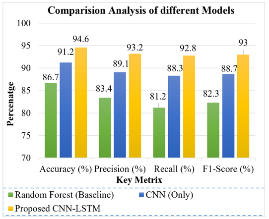
Figure 2. Comparison analysis of various models for arrhythmia detection
Table 4. Performance metrics for arrhythmia detection (MIT-BIH dataset)
|
Model |
Accuracy (%) |
Precision (%) |
Recall (%) |
F1-Score (%) |
|
Random Forest (Baseline) [9] |
86.7 |
83.4 |
81.2 |
82.3 |
|
CNN (Only) [12] |
91.2 |
89.1 |
88.3 |
88.7 |
|
Proposed CNN-LSTM |
94.6 |
93.2 |
92.8 |
93.0 |
From the results of the performance comparison in Table 4 and Figure 2, it is clear that the proposed CNN-LSTM method performs better than the Random Forest algorithm and the CNN algorithms. The Random Forest method has been able to score an accuracy of 86.7%, while the CNN method performed better by scoring an accuracy of 91.2% by automatically extracting discriminatory features from the ECG signals. The accuracy score of the proposed method increased to 94.6%, with a precision score of 93.2%, recall of 92.8%, and an F1-score of 93.0%.
The model is trained using the Adam optimizer with a learning rate of 0.001 and categorical cross-entropy loss. Training is conducted over 50–100 epochs with early stopping based on validation loss to prevent overfitting.
The feedforward DNN model for CAD prediction includes:
Input layer: Accepts the 54-feature vector.
Hidden layers: Three dense layers with 64, 128, and 64 neurons respectively, each followed by ReLU activation, dropout (rate = 0.4), and batch normalization.
Output layer: A single neuron with sigmoid activation for binary classification.
This model is trained using binary cross-entropy loss and the Adam optimizer. A five-fold cross-validation strategy is adopted to validate the robustness of the classifier, and hyperparameters such as learning rate, dropout rate, and number of neurons are tuned using grid search. The performance of the proposed framework is compared with conventional techniques. The results are shown in Table 4 for arrhythmia detection and Table 3 for CAD prediction.
The DNN model consisted of four layers, each of which was densely connected, with the number of nodes being 256, 128, 64, and 32, respectively. Batch Normalization and Dropout layers were included to enhance training stability and prevent overfitting. Optimization of the model was optimized using the Adam Optimizer with a learning rate of 0.001 and was trained for 100 epochs.
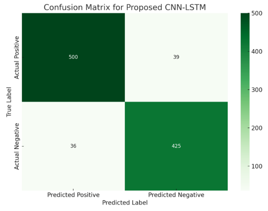
The confusion matrix of the proposed CNN-LSTM model for arrhythmia detection is shown in Figure 3. The model successfully predicted 500 cases as positives and 425 cases as negatives, with 39 false negatives and 36 false positives.
Figure 3. Confusion matrix of CNN-LSTM for arrhythmia detection
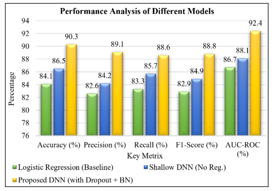
Figure 4. Performance analysis of various models for coronary artery disease (CAD)
Performance comparison between logistic regression, shallow DNN, and proposed DNN using dropout + batch normalization for CAD classification is depicted in Figure 4. The proposed DNN model provided higher accuracy and AUC-ROC score (90.3% and 92.4%, respectively), as shown in Table 5.
Table 5. Performance analysis of various models for coronary artery disease (CAD)
|
Model |
Accuracy (%) |
Precision (%) |
Recall (%) |
F1-Score (%) |
AUC-ROC (%) |
|
Shallow DNN (No Reg.) [32] |
86.5 |
84.2 |
85.7 |
84.9 |
88.1 |
|
Logistic regression (Baseline) [30] |
84.1 |
82.6 |
83.3 |
82.9 |
86.7 |
|
Proposed DNN (with dropout + BN) |
90.3 |
89.1 |
88.6 |
88.8 |
92.4 |
The comparison of the performance reveals that the proposed DNN model with dropout and batch normalization (BN) is significantly superior to the logistic regression and shallow DNN models. The baseline logistic regression model provided an accuracy of 84.1% and AUC-ROC of 86.7%, whereas the shallow DNN model yielded an accuracy of 86.5% and AUC-ROC of 88.1%. In turn, the proposed DNN model enhanced the accuracy to 90.3%, precision to 89.1%, recall to 88.6%, F1-score to 88.8%, and AUC-ROC to 92.4%. The improvements in performance reveal that the usage of batch normalization allows stabilizing the training process and accelerating its convergence, while the dropout technique helps to prevent overfitting.
It is evident that the results indicate a clear advantage of the proposed hybrid deep learning approach over conventional machine learning algorithms and shallow neural networks. In terms of detecting arrhythmias, the CNN-LSTM algorithm demonstrates substantially better performance compared to the Random Forest approach and the CNN alone with regard to all performance criteria. This is attributed to the LSTM algorithm, which allows the algorithm to extract effective temporal features from ECG signals, which are essential for recognizing rhythm abnormalities such as atrial fibrillation or early contractions.
The suggested framework included two streams: one stream based on a 1D CNN-CNN-LSTM architecture for the ECG signals and another stream built upon a DNN for the clinical modality. Features extracted from the two different modalities were combined and fed into the fully connected layers for joint learning and classification. Batch normalization, dropout of 0.5, and the Adam optimizer with a learning rate of 0.001 were used in the model. The model was trained for 100 epochs using a batch size of 32.
In the case of CAD prediction, the proposed DNN model that incorporates dropout and batch normalization achieves the highest performance. The dropout layers effectively prevent overfitting, which is a common issue in relatively small datasets like Z-Alizadeh Sani, while batch normalization accelerates convergence and stabilizes training. Compared to logistic regression and shallow DNNs, our model provides significantly better generalization, as reflected by a notable improvement in AUC-ROC (from 86.7% to 92.4%). This demonstrates the model’s strong discriminatory power and robustness to input variability.
Moreover, the use of specialized architectural models for different modalities, such as a sequential one for ECG and a dense model for tabular input, is a feasible solution. Although each of them individually diagnoses a certain heart problem, altogether they make a universal diagnostic tool that can detect various disorders of the heart, namely those related to its electrical and structural aspects.
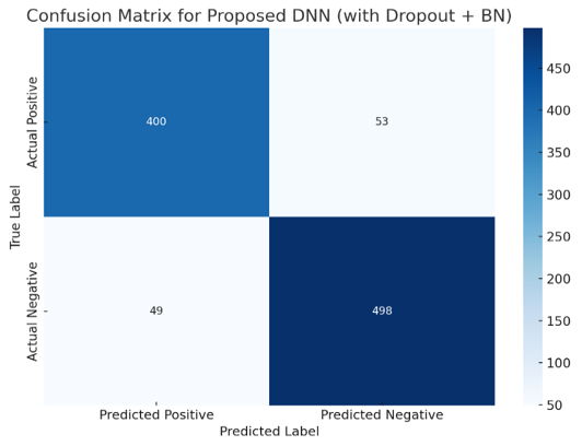
To further provide an intuitive assessment of the classification capability of the proposed DNN with dropout and batch normalization for arrhythmia detection, the corresponding confusion matrix is presented in Figure 5.
Despite the success of the suggested framework in achieving good performance when working on the MIT-BIH Arrhythmia and Z-Alizadeh Sani datasets, further research needs to be done regarding the robustness of the framework under clinical conditions. The tests were performed using benchmark datasets that had been pre-processed before the training of the machine learning models.
Thus, the impact of noisy ECG data, missing clinical values, class imbalance, and shifts in the distribution of data collected from different hospitals has not been considered. For future work, a robustness test of the suggested framework is needed by adding controlled ECG noise, missing clinical attributes, testing the framework using other multi-center datasets, and doing domain adaptation and data augmentation.
The computational complexities of the evaluated models shown in Table 6 are different depending on the model’s architecture and input. The Random Forest and shallow DNN models use relatively fewer parameters (0.2 M and 0.10 M, respectively), and therefore, the inference is not complex. The models are appropriate for classification based on the clinical features as binary classes. The CNN and BiLSTM models work with ECG signals as multi-class problems with approximate numbers of parameters (0.45 M and 0.65 M), respectively. The conventional CNN-LSTM model includes about 0.85 M parameters and has high inference complexity. The proposed CNN-LSTM uses about 0.90 M parameters but has moderate to high inference complexity. The proposed DNN with dropout and batch normalization uses about 0.15 M parameters and maintains low inference complexity.
Figure 5. Confusion matrix of Deep Neural Network (DNN) for arrhythmia detection
Table 6. Computational complexity analysis
|
Model |
Approx. Parameters |
Input Type |
Classification Type |
Inference Complexity |
|
Random Forest |
~0.2 M |
Clinical features |
Binary |
Low |
|
CNN |
~0.45 M |
ECG signal |
Multi-class |
Moderate |
|
BiLSTM |
~0.65 M |
ECG signal |
Multi-class |
High |
|
CNN-LSTM |
~0.85 M |
ECG signal |
Multi-class |
High |
|
Shallow DNN |
~0.10 M |
Clinical features |
Binary |
Low |
|
Proposed CNN-LSTM |
~0.90 M |
ECG signal |
Multi-class |
Moderate–high |
|
Proposed DNN (Dropout + BN) |
~0.15 M |
Clinical features |
Binary |
Low |
The proposed study offers a combination of hybrid deep learning approaches consisting of a CNN-LSTM network to detect arrhythmias based on ECGs and a regularized DNN to predict CAD based on structured clinical data. As shown in experimental studies with the MIT-BIH and Z-Alizadeh Sani datasets, the proposed framework is superior to traditional machine learning methods as well as deep learning techniques in terms of accuracy, precision, recall, F1-score, and AUC-ROC metrics. The combination of CNN-LSTM and DNN allows for efficient learning from diverse cardiac data.
However, the study suffers from a number of drawbacks. Firstly, the suggested framework is tested only on publicly available benchmarks with limited data diversity, which does not provide an accurate picture of real-world patients. Secondly, the arrhythmia and CAD models are trained separately on the corresponding datasets, but not as one multimodal dataset, which means that there is no end-to-end learning. Thirdly, external validation and prospective studies have not been carried out, and the performance of the model in other hospitals, among different demographic groups, and on various data acquisition devices needs to be studied.
The future work will involve designing a comprehensive multimodal framework that can learn from both ECG data and structured clinical data within one architecture. The implementation of mechanisms such as attention, graph neural networks, or transformers will be considered in order to enhance feature extraction and model interpretability. Furthermore, future work will incorporate external multi-center validation using large datasets, explainable AI techniques for better acceptance by clinicians, and model optimization for faster processing.
The authors would like to express their gratitude to Medicaps University, Indore, for providing the necessary support and academic guidance throughout this research. The authors also sincerely appreciate the contributions of the dataset providers of the MIT-BIH Arrhythmia Database (PhysioNet) and the Z-Alizadeh Sani Coronary Artery Disease dataset for making these benchmark datasets publicly available. These valuable resources enabled the development and evaluation of a hybrid deep learning architecture for cardiac abnormality detection.
|
AI |
Artificial Intelligence |
|
CAD |
Coronary Artery Disease |
|
CNN |
Convolutional Neural Network |
|
CVD |
Cardiovascular Disease |
|
DNN |
Deep Neural Network |
|
ECG |
Electrocardiogram |
|
LSTM |
Long Short-Term Memory |
|
ReLU |
Rectified Linear Unit |
|
BN |
Batch Normalization |
|
ROC |
Receiver Operating Characteristic |
|
AUC |
Area Under the Curve |
|
TP |
True Positive |
|
TN |
True Negative |
|
FP |
False Positive |
|
Greek symbols |
|
|
α |
Learning rate |
|
β |
Momentum coefficient (Adam optimizer) |
|
γ |
Regularization coefficient |
|
λ |
Regularization parameter (L2 weight decay) |
|
η |
Network learning parameter |
|
μ |
Mean value of the feature distribution |
|
σ |
Standard deviation |
|
θ |
Model parameters (weights) |
|
Δ |
Change or update in model parameters |
|
Subscripts |
|
|
train |
Training dataset |
|
test |
Testing dataset |
|
pred |
Predicted output |
|
true |
Ground truth label |
|
ECG |
ECG signal features |
|
clinical |
Clinical feature set |
|
arr |
Arrhythmia classification |
|
CAD |
Coronary artery disease prediction |
|
i |
i-th sample |
|
j |
j-th feature |
|
max |
Maximum value |
|
min |
Minimum value |
[1] World Health Organization. (2023). Cardiovascular diseases (CVDs). WHO. https://www.who.int/news-room/fact-sheets/detail/cardiovascular-diseases-(cvds).
[2] Fuster, V., Narula, J., Vaishnava, P., et al. (2022). Fuster and Hurst’s The Heart, 15th ed. McGraw-Hill.
[3] Litjens, G., Kooi, T., Bejnordi, B.E., et al. (2017). A survey on deep learning in medical image analysis. Medical Image Analysis, 42: 60-88. https://doi.org/10.1016/j.media.2017.07.005
[4] Lundervold, S.M., Lundervold, A. (2019). An overview of deep learning in medical imaging focusing on MRI. Zeitschrift für Medizinische Physik, 29(2): 102-127. https://doi.org/10.1016/j.zemedi.2018.11.002
[5] Acharya, U.R., Fujita, H., Oh, S.L., et al. (2019). Deep convolutional neural network for the automated diagnosis of congestive heart failure using ECG signals. Applied Intelligence, 49(1): 16-27. https://doi.org/10.1007/s10489-018-1179-1
[6] Hannun, A.Y., Rajpurkar, P., Haghpanahi, M., et al. (2019). Cardiologist-level arrhythmia detection and classification in ambulatory electrocardiograms using a deep neural network. Nature Medicine, 25(1): 65-69. https://doi.org/10.1038/s41591-018-0268-3
[7] Yildirim, O. (2018). A novel wavelet sequence based on deep bidirectional LSTM network model for ECG signal classification. Computers in Biology and Medicine, 96: 189-202. https://doi.org/10.1016/j.compbiomed.2018.03.016
[8] Khosla, A., Cao, Y., Lin, C., Chiu, H., Hu, J., Lee, H. (2010). An integrated machine learning approach to stroke prediction. In Proceedings of the ACM SIGKDD International Conference on Knowledge Discovery and Data Mining, Washington DC, USA, pp. 183-192. https://doi.org/10.1145/1835804.1835830
[9] Alizadehsani, R., Habibi, J., Hosseini, M.J., et al. (2016). Coronary artery disease detection using computational intelligence methods. Knowledge-Based Systems, 109: 187-197. https://doi.org/10.1016/j.knosys.2016.06.009
[10] Johns Hopkins Medicine. (2021). Lifetime monitoring after infant cardiac surgery may reduce adult hypertension risk. https://www.hopkinsmedicine.org/news/newsroom/news-releases/2021/04/lifetime-monitoring-after-infant-cardiac-surgery-may-reduce-adult-hypertension-risk.
[11] MIT-BIH Arrhythmia Database. PhysioNet. https://physionet.org/content/mitdb/1.0.0/.
[12] Acharya, U.R., Oh, S.L., Hagiwara, Y., et al. (2017). A deep convolutional neural network model to classify heartbeats. Computers in Biology and Medicine, 89: 389-396. https://doi.org/10.1016/j.compbiomed.2017.08.022
[13] Almansouri, N.E., Awe, M., Rajavelu, S., et al. (2024). Early diagnosis of cardiovascular diseases in the era of artificial intelligence: An in-depth review. Cureus, 16(3): e55869. https://doi.org/10.7759/cureus.55869
[14] Hammad, M., Chelloug, S.A., Alkanhel, R., et al. (2022). Automated detection of myocardial infarction and heart conduction disorders based on feature selection and a deep learning model. Sensors, 22(17): 6503. https://doi.org/10.3390/s22176503
[15] Selvam, I.J., Madhavan, M., Kumarasamy, S.K. (2025). Detection and classification of electrocardiography using hybrid deep learning models. Hellenic Journal of Cardiology, 81: 75-84. https://doi.org/10.1016/j.hjc.2024.08.011
[16] Tawil, D.B. (2025). A review on the coronary artery disease landscape: Tissue and market analysis, treatments, and trials. Journal of Cardiovascular Diseases. https://doi.org/10.52338/jocd.2025.4960
[17] Rana, R., Hume, M., Reilly, J., Jurdak, R., Soar, J. (2016). Opportunistic and context-aware affect sensing on smartphones. IEEE Pervasive Computing, 15(2): 60-69. https://doi.org/10.1109/MPRV.2016.36
[18] Kachuee, M., Fazeli, S., Sarrafzadeh, M. (2018). ECG heartbeat classification: A deep transferable representation. In 2018 IEEE International Conference on Healthcare Informatics (ICHI), New York, USA, pp. 443-444. https://doi.org/10.1109/ICHI.2018.00092
[19] Kothari, H., Rani, S. (2025). An overview of the heart disease prediction using machine learning and its application. International Journal of Scientific Research and Technology, 2(6): 560-565. https://doi.org/10.5281/zenodo.15715476
[20] Nasarian, E., Abdar, M., Fahami, M.A., et al. (2020). Association between work-related features and coronary artery disease: A heterogeneous hybrid feature selection integrated with balancing approach. Pattern Recognition Letters, 133: 33-40. https://doi.org/10.1016/j.patrec.2020.02.010
[21] Gudadhe, M., Wankhade, K., Dongre, S. (2010). Decision support system for heart disease based on support vector machine and artificial neural network. In 2010 International Conference on Computer and Communication Technology (ICCCT), Allahabad, India, pp. 741-745. https://doi.org/10.1109/ICCCT.2010.5640377
[22] Bai, X., Dong, X., Li, Y., Liu, R., Zhang, H. (2024). A hybrid deep learning network for automatic diagnosis of cardiac arrhythmia based on 12-lead ECG. Scientific Reports, 14(1): 24441. https://doi.org/10.1038/s41598-024-75531-w
[23] He, Y., Zhou, Y., Qian, Y., et al. (2025). Cardioattentionnet: Advancing ECG beat characterization with a high-accuracy and portable deep learning model. Frontiers in Cardiovascular Medicine, 11: 1473482. https://doi.org/10.3389/fcvm.2024.1473482
[24] Lee, K.N., Yu, H.T., Lee, S.R., Roh, S.Y. (2025). The role of artificial intelligence technologies in revolutionizing and aiding cardiovascular medicine. Frontiers in Cardiovascular Medicine.
[25] Torres Soto, J., Hughes, J.W., Amador Sanchez, P., Perez, M., Ouyang, D., Ashley, E.A. (2022). Multimodal deep learning enhances diagnostic precision in left ventricular hypertrophy. European Heart Journal – Digital Health, 3(3): 380-389. https://doi.org/10.1093/ehjdh/ztac033
[26] Tadesse, G.A., Javed, H., Weldemariam, K., et al. (2021). DeepMI: Deep multi-lead ECG fusion for identifying myocardial infarction and its occurrence-time. Artificial Intelligence in Medicine, 121: 102192. https://doi.org/10.1016/j.artmed.2021.102192
[27] Wu, Z., Guo, C. (2025). Deep learning and electrocardiography: Systematic review of current techniques in cardiovascular disease diagnosis and management. BioMedical Engineering OnLine, 24: 23. https://doi.org/10.1186/s12938-025-01349-w
[28] Atwa, A.E.M., Atlam, E.S., Ahmed, A., Atwa, M.A., Abdelrahim, E.M., Siam, A.I. (2025). Interpretable deep learning models for arrhythmia classification based on ECG signals using PTB-X dataset. Diagnostics, 15(15): 1950. https://doi.org/10.3390/diagnostics15151950
[29] Alizadehsani, R., Abdar, M., Roshanzamir, M., et al. (2019). Machine learning-based coronary artery disease diagnosis: A comprehensive review. Computers in Biology and Medicine, 111: 103346. https://doi.org/10.1016/j.compbiomed.2020.103898
[30] Alizadehsani, R., Habibi, J., Hosseini, M.J., et al. (2013). A data mining approach for diagnosis of coronary artery disease. Computer Methods and Programs in Biomedicine, 111(1): 52-61. https://doi.org/10.1016/j.cmpb.2013.03.004
[31] Ingle, M., Litoriya, R. (2026). An efficient 1D CNN framework for accurate cardiac disease detection from ECG signal. In 2026 3rd International Conference on Emerging Trends in Engineering and Medical Sciences (ICETEMS), Nagpur, India, 2026, pp. 1-6. https://doi.org/10.1109/ICETEMS66917.2026.11470293
[32] Arabasadi, Z., Alizadehsani, R., Roshanzamir, M., Moosaei, H., Yarifard, A.A. (2017). Computer aided decision making for heart disease detection using hybrid neural network-genetic algorithm. Computer Methods and Programs in Biomedicine, 141: 19-26. https://doi.org/10.1016/j.cmpb.2017.01.004