Syafri Arlis* | Musli Yanto | Vina Tri Septiana
© 2026 The authors. This article is published by IIETA and is licensed under the CC BY 4.0 license (http://creativecommons.org/licenses/by/4.0/).
OPEN ACCESS
Automated analysis of chest X-ray images has become an important approach for supporting the detection and classification of pulmonary diseases. However, existing deep learning (DL) models often face challenges in accurately capturing complex anatomical structures and preserving detailed boundaries during image segmentation. This study proposes FCNU-Net, a hybrid DL framework that integrates the encoder characteristics of Fully Convolutional Networks (FCNs) with the decoder structure of U-Net for lung disease segmentation and classification from chest X-ray images. The proposed model was evaluated using 876 chest X-ray images collected from Siti Rahmah Islamic Hospital, including normal, pneumonia, and tuberculosis cases. The experimental framework involved image preprocessing, semantic segmentation, and classification based on the modified convolutional architecture. The results demonstrate that FCNU-Net achieved an overall classification accuracy of 96.60%, with a precision of 90.7% and sensitivity of 85.4%. Furthermore, the model obtained an average Intersection over Union (IoU) of 0.8904 and Dice coefficient of 0.9417, indicating effective pixel-level segmentation performance. The proposed framework improves feature representation and boundary localization by combining the strengths of FCN-based dense prediction and U-Net-based spatial reconstruction. These findings suggest that FCNU-Net provides a reliable DL approach for automated lung disease analysis using chest X-ray images and may serve as a potential tool for computer-assisted medical diagnosis.
lung disease, chest X-ray imaging, FCNU-Net, deep learning, semantic segmentation, medical image analysis, Convolutional Neural Network
Lung disease is one of the most serious health conditions that threatens human life, particularly in developing countries [1]. The severity of this disease is attributed to its consequences, which can lead to fatal outcomes if early treatment and proper monitoring are not provided [2]. Lung disease is commonly caused by bacterial infections, one of which is Mycobacterium tuberculosis [3]. This bacterium can rapidly spread to other parts of the body, including bones, lymph nodes, the central nervous system, and kidneys [4].
Recent advances in information technology have enabled the development of various early intervention approaches through the application of Artificial Intelligence (AI), particularly by developing automated detection systems [5]. The implementation of AI in the detection process has demonstrated a considerable contribution to supporting diagnostic procedures [6]. The resulting outputs exhibit adequate accuracy, thereby assisting medical professionals in clinical decision-making [7]. AI is introduced in the diagnosis of lung diseases through learning-based approaches that enable systematic problem-solving [8]. Furthermore, the role of AI in detection processes has yielded significant results through the application of digital image processing techniques [9].
Image processing is a branch of AI that focuses on the processing and analysis of images for detection and object identification purposes [10]. This concept has contributed to advancements and progress in medical technology by addressing various diagnostic challenges that were previously difficult to resolve [11]. Previous studies have reported that image processing techniques have had a positive and significant impact on disease detection processes [12]. Several image processing techniques can be adopted for detection purposes, one of which is image segmentation [13]. Image segmentation is a technique used to separate specific regions or objects within an image [14]. This technique enables the division of objects into multiple segments, thereby facilitating more accurate and efficient analysis and detection processes [15].
The concept of segmentation can be integrated with deep learning (DL) approaches to perform detection and classification tasks [16]. Other studies have indicated that DL can be effectively adopted for detection processes, yielding results with a high level of accuracy [17]. DL–based segmentation methods, such as Convolutional Neural Networks (CNNs), have demonstrated effectiveness in capturing complex features and enabling early detection for timely intervention in lung disease patients [18]. DL models are capable of producing structured outputs across various tasks, including identification and classification processes [19]. Furthermore, DL has shown significant performance in detecting disease-related objects, thereby supporting medical professionals in clinical decision-making [20]. DL can also be utilized to recognize specific patterns associated with various types of lung diseases [21]. Through model-based analysis, DL enables object detection with improved accuracy [22] and facilitates early detection in lung disease identification, contributing to disease control and prognosis management [23].
Previous studies have demonstrated that the application of DL provides effective and efficient models for lung disease detection processes [24]. Comparisons of CNN architectures, such as ResNet and Inception, have been conducted to modify DL models and evaluate their performance in detection tasks [25]. In addition, the development of multichannel DL models has been shown to produce significant results in detecting lung disease objects [26], with reported accuracy reaching 93.75% based on the proposed DL architecture [27]. The development of DL models within hybrid frameworks has also yielded optimal performance outcomes [28]. Furthermore, studies applying DL to detection tasks have reported accuracy levels ranging from 94% to 99.5% through modifications of unsupervised learning approaches [29]. Modifications of CNN architectures for lung disease classification have also been shown to be effective based on the redesigned models [30]. Moreover, the implementation of correlation-based feature selection methods to optimize detection performance has resulted in the highest accuracy of 95.56% using the Support Vector Machine (SVM) method, followed by CNN at 92.11% and K-Nearest Neighbor (K-NN) at 88.40% [31].
Based on a review of previous research findings, this study aims to address similar challenges in the detection and classification of lung diseases. The proposed work focuses on developing a DL-based model using a U-Net architecture integrated with a Fully Convolutional Neural Network (FCNU-Net). The development of the FCNU-Net architecture is intended to introduce novelty in DL-based detection and classification models. This architectural innovation is expected to contribute to improved DL performance in lung disease detection and classification processes based on predefined evaluation parameters.
The detection and classification processes are conducted based on the development of a DL model using the FCNU-Net architecture through several experimental stages to achieve optimal results. The dataset used in this study for the detection and classification of lung diseases was obtained from chest X-ray examinations of lung patients at Siti Rahmah Islamic Hospital, Padang City. A total of 876 X-ray images were collected, representing various lung conditions, including normal, pneumonia, and tuberculosis.
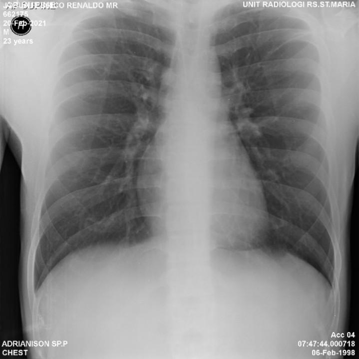
The dataset used in this study was categorized into three diagnostic classes representing different lung conditions. The Normal class consisted of 617 chest X-ray images, representing healthy individuals without radiological signs of pulmonary abnormalities. The Pneumonia class included 197 images showing radiographic manifestations of pneumonia, while the Tuberculosis class comprised 62 images containing characteristic findings associated with pulmonary tuberculosis. Consequently, the dataset exhibited an imbalanced class distribution, with the Normal class representing the largest proportion of samples and the Tuberculosis class containing the fewest images. This imbalance poses additional challenges for the DL model, as it may bias the learning process toward the majority class. Samples of chest X-ray images from patients diagnosed with lung diseases are presented in Figure 1.
Figure 1. Illustration of the image annotation and training process
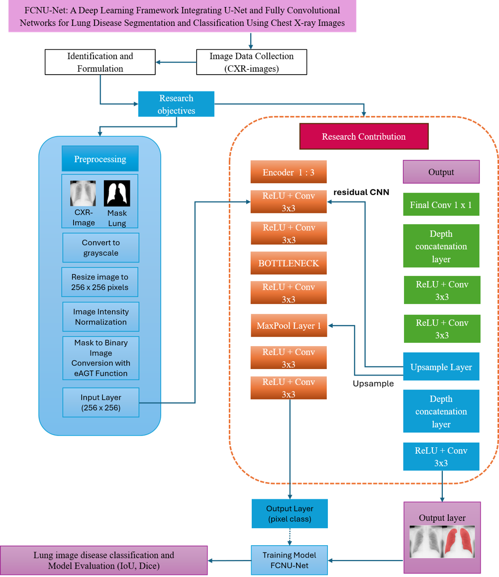
Figure 1 illustrates the chest X-ray images used as the research dataset in the lung disease detection and classification process. Before being utilized for DL training in detection and classification tasks, the images undergo a preprocessing stage to remove noise and irrelevant artifacts that may still be present. Subsequently, the dataset is divided into training and testing sets, comprising 676 images for training and 200 images for testing. These experiments are organized into multiple phases, including preprocessing, segmentation, and DL learning. The preprocessing stage is applied to enhance the quality of the input images. Subsequently, the preprocessed images undergo a segmentation process to determine the regions of interest for object detection. The resulting segmented images are then used as input for the DL learning process to perform detection and classification tasks. The overall research framework is illustrated in Figure 2.
Figure 2. Research framework
Figure 2 presents the research framework for the detection and classification process. This framework highlights the novelty of the proposed DL model, which is embodied in the FCNU-Net architecture employed in this study. The development of the CNN architecture in this research focuses on the design of a Nested FCNU-Net network, which modifies the classical CNN structure into a Fully Convolutional Network (FCN) comprising four main components: (1) a three-level encoder–decoder architecture with a bottleneck consisting of 512 filters, (2) nested convolution blocks involving two successive 3 × 3 convolution operations at each level to enhance feature representation, (3) multi-level skip connections between the encoder and decoder to preserve spatial details, and (4) the use of transposed convolution as a learnable upsampling mechanism. The entire architecture is constructed modularly using a layer graph approach, allowing flexible and conflict-free inter-block connections. This architectural development results in a segmentation model capable of accurately predicting lung masks at full image resolution. Furthermore, the development of the DL model based on the FCNU-Net architecture for detection and classification purposes is illustrated in Figure 3.
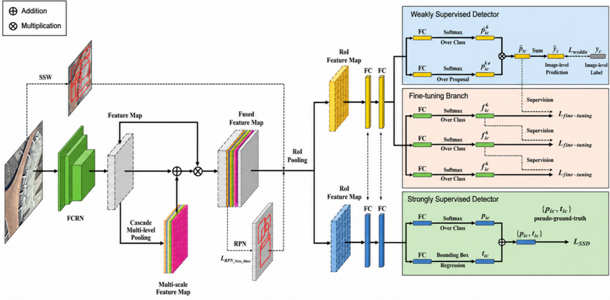
Figure 3 illustrates the FCNU-Net architecture in the development of a DL model for lung disease detection and classification. The FCNU-Net architecture is modified by integrating the encoder component of an FCN with a U-Net–based decoder. This architectural design results in a robust model for image segmentation and classification, offering the ability to handle images with objects of varying sizes, improved computational efficiency, and enhanced precision in producing accurate segmentation and classification outputs.
Figure 3. Development of the Fully Convolutional Neural Network (FCNU-Net) deep learning (DL) architecture for lung disease detection and classification
The proposed architecture provides a robust and effective framework for image segmentation and classification by integrating advanced DL techniques capable of accurately identifying and classifying objects with varying scales and complex visual characteristics. One of the main advantages of this architecture is its ability to handle objects of different sizes more effectively while maintaining high computational efficiency. Furthermore, the model is designed to produce more precise and sharper segmentation boundaries, thereby improving the quality of object delineation in the resulting segmentation maps. The novelty of this study lies in the development and implementation of a DL-based segmentation model that enhances feature extraction and object representation, leading to improved segmentation performance compared to conventional approaches.
During the training phase, the DL model was configured using a learning rate of 0.003, which determines the magnitude of weight updates during the optimization process. This learning rate was selected to achieve a balance between convergence speed and training stability, enabling the model to minimize the loss function efficiently while reducing the risk of unstable parameter updates. In addition, a batch size of 4 was employed, meaning that four training samples were processed simultaneously in each iteration. Although relatively small, this batch size allows more frequent parameter updates, which can improve the model's generalization capability, particularly when working with datasets of limited size or high-resolution images that require substantial GPU memory.
The training process was carried out over 2,000 epochs, allowing the model to repeatedly learn from the training data through multiple forward and backward propagation cycles. Such an extensive training duration was intended to ensure that the network sufficiently captured both low-level and high-level features present in the dataset while gradually optimizing the model parameters to achieve convergence. Repeated exposure to the training data enables the model to better understand complex patterns and relationships among image features, thereby improving segmentation and classification accuracy.
All experiments were conducted on a computer equipped with an AMD Ryzen 7 5800H processor with Radeon Graphics, which provided the computational resources necessary to execute the DL framework. The hardware supported the complete training pipeline, including forward propagation, loss computation, backpropagation, and optimization of network parameters throughout the training process. Although the experiments were performed without a dedicated high-end GPU, the available processing capability was sufficient to conduct extensive training and evaluate the proposed architecture under consistent experimental conditions. Overall, the selected hyperparameter configuration, together with the computational environment, was expected to facilitate stable model convergence, improve learning effectiveness, and achieve optimal predictive performance in both image segmentation and classification tasks.
The improvement of DL performance in the detection and classification of lung diseases using X-ray images is based on modifications to the CNN architectural model by integrating the roles of U-Net and FCNU-Net. The process begins with a preprocessing stage aimed at generating noise-free images. The preprocessing stage involves several techniques, including image transformation, image resizing, image masking, and image intensity conversion using the eAGT function. The output results of the preprocessing stage are presented in Figure 4.
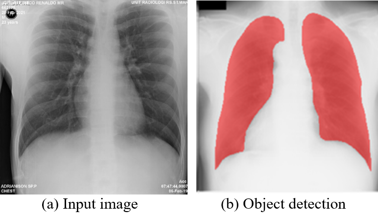
Figure 4 presents a preprocessed image, which aims to remove irrelevant objects and noise from the original image. The results of preprocessing play a crucial role in supporting the detection and classification processes to achieve optimal performance. The effectiveness of the detection and classification processes is closely related to the initial concept of semantic segmentation utilizing the role of CNNs, which were later modified. The results of the detection and classification processes are presented in Figure 5.
Figure 4. Preprocessing result
Figure 5. Detection and classification results
Figure 5 presents the testing results of the detection and classification process based on the modified CNN architecture. These results demonstrate accurate object localization through semantic-based segmentation. The findings further confirm that performance improvements in CNN-based detection and classification are achieved through architectural modifications designed to optimize the detection and classification processes.
This modification was made to develop a better CNN architecture model for detection and classification purposes. The development of this model is to maximize the DL performance of the CNN architecture in detection and classification. The modified CNN architecture proposed in this study is presented in Algorithm 1.
|
Algorithm 1. Modified CNN Architecture |
|
Input: Chest X-ray image I (256×256×1) Output: M lung mask prediction (256×256, binary) start ---------- PREPROCESSING ---------- If I has 3 channels (RGB) then: Convert I to grayscale If size of I is not 256×256 then: Resize I to 256×256 Normalize intensity of I to range [0,1] ---------- ENCODER ---------- // Encoder 1 F11 ← ReLU(Conv3x3(I, 64)) F12 ← ReLU(Conv3x3(F11, 64)) Save F12 as skip level 1 feature P1 ← MaxPool2x2(F12) // Encoder 2 F21 ← ReLU(Conv3x3(P1, 128)) F22 ← ReLU(Conv3x3(F21, 128)) Save F22 as skip level 2 feature P2 ← MaxPool2x2(F22) // Encoder 3 F31 ← ReLU(Conv3x3(P2, 256)) F32 ← ReLU(Conv3x3(F31, 256)) Save F32 as skip level 3 feature P3 ← MaxPool2x2(F32) // ---------- BOTTLENECK ---------- B1 ← ReLU(Conv3x3(P3, 512)) B2 ← ReLU(Conv3x3(B1, 512)) // ---------- DECODER ---------- // Decoder 3 U3 ← TransposedConv2x2(B2, 256, stride=2) C3 ← Concatenate(U3, F32) // skip level 3 D31 ← ReLU(Conv3x3(C3, 256)) D32 ← ReLU(Conv3x3(D31, 256)) // Decoder 2 U2 ← TransposedConv2x2(D32, 128, stride=2) C2 ← Concatenate(U2, F22) // skip level 2 D21 ← ReLU(Conv3x3(C2, 128)) D22 ← ReLU(Conv3x3(D21, 128)) // Decoder 1 U1 ← TransposedConv2x2(D22, 64, stride=2) C1 ← Concatenate(U1, F12) // skip level 1 D11 ← ReLU(Conv3x3(C1, 64)) D12 ← ReLU(Conv3x3(D11, 64)) // ---------- OUTPUT ---------- O ← Conv1x1(D12, numClasses=2) P ← Softmax(O) // probability of each pixel class For every pixel (x,y): M(x,y) ← argmax_k P_k(x,y) // select the class with highest probability Output M as a lung segmentation mask end |
Algorithm 1 presents the modified CNN architecture developed for semantic-based segmentation in the context of lung disease detection and classification. The proposed development demonstrates that the CNN model incorporates architectural modifications aimed at achieving optimal object segmentation. The modified CNN learning process is capable of producing satisfactory performance, as evidenced by the output results illustrated in Figure 6.
Figure 6 presents the validation results of the performance of the modified CNN model in the learning process for lung disease detection and classification. The experimental results presented in Figure 6 demonstrate that the proposed model achieved an overall classification accuracy of 96.60%, indicating that the model was able to correctly classify the majority of the input samples with a high level of reliability. This high accuracy reflects the effectiveness of the proposed architecture in learning discriminative features from the dataset and performing accurate predictions across different image classes. The obtained result also suggests that the integration of the proposed segmentation and classification framework contributes positively to the overall predictive performance of the model.
Figure 6. Results of the modified Convolutional Neural Network (CNN) learning process
In addition to overall accuracy, several complementary evaluation metrics were employed to provide a more comprehensive assessment of the model's performance. The model achieved a precision of 90.7%, indicating that most of the samples predicted as belonging to the target class were indeed correctly classified. This high precision demonstrates the model's ability to minimize false positive predictions, which is particularly important in applications where incorrect positive classifications may lead to undesirable consequences.
Furthermore, the model obtained a sensitivity (recall) value of 85.4%, reflecting its capability to correctly identify the majority of actual positive samples within the dataset. The recall score indicates that the proposed model successfully detected a substantial proportion of the target objects, although a small number of positive instances were still misclassified as negatives. The combined results of accuracy, precision, and recall indicate that the proposed model not only produces highly accurate predictions but also maintains a balanced performance in minimizing both false positive and false negative errors. Overall, these evaluation metrics confirm the robustness and effectiveness of the proposed DL architecture for image segmentation and classification tasks. These findings demonstrate that the modified CNN model delivers performance that surpasses previous CNN-based models in the detection and classification process. Validation of the proposed approach is further supported by Intersection over Union (IoU) and Dice coefficient evaluations, as presented in Table 1.
Table 1 presents a sample evaluation of the IoU and Dice coefficient obtained from the modified CNN architecture using the FCNU-Nested model. The experimental results indicate that the proposed model achieved an average IoU value of 0.8904 and a Dice coefficient of 0.9417. These results demonstrate that the modified CNN architecture provides a significant performance improvement, thereby enhancing the effectiveness of the conventional CNN model. Additional experiments were conducted to further validate these findings by comparing them with results reported in previous studies on lung disease detection and classification. The comparative evaluation of related research findings is presented in Table 2, confirming the consistency and superiority of the proposed FCNU-Nested model in improving detection and classification performance.
Table 1. Evaluation of Intersection over Union (IoU) and Dice in modified Convolutional Neural Network (CNN) with FCNU-nested
|
Image |
IoU |
Dice |
Image |
IoU |
Dice |
Image |
IoU |
Dice |
|
1 |
0.87742 |
0.93471 |
11 |
0.90471 |
0.94997 |
21 |
0.89053 |
0.9421 |
|
2 |
0.89854 |
0.94656 |
12 |
0.90649 |
0.95095 |
22 |
0.84059 |
0.91339 |
|
3 |
0.8798 |
0.93606 |
13 |
0.93001 |
0.96373 |
23 |
0.80103 |
0.88953 |
|
4 |
0.91731 |
0.95687 |
14 |
0.89275 |
0.94333 |
24 |
0.88662 |
0.9399 |
|
5 |
0.85509 |
0.92188 |
15 |
0.89632 |
0.94533 |
25 |
0.85927 |
0.92431 |
|
6 |
0.90106 |
0.94795 |
16 |
0.88367 |
0.93824 |
26 |
0.92474 |
0.9609 |
|
7 |
0.872 |
0.93163 |
17 |
0.84985 |
0.91883 |
27 |
0.92846 |
0.96291 |
|
8 |
0.89689 |
0.94564 |
18 |
0.91083 |
0.95334 |
28 |
0.88575 |
0.93941 |
|
9 |
0.90435 |
0.94977 |
19 |
0.86544 |
0.92787 |
29 |
0.86094 |
0.92528 |
|
10 |
0.89794 |
0.94623 |
20 |
0.92894 |
0.96316 |
30 |
0.91979 |
0.95822 |
Table 2 presents a comparative analysis of several previous studies related to lung disease detection and classification using DL approaches. Based on the reported results, it can be observed that the proposed approach for improving CNN performance in this study produces competitive and superior outcomes. These findings confirm that enhancing CNN performance through the FCNU-Nested architecture is effective and capable of accurately performing lung disease detection and classification.
Table 2. The comparative evaluation of related research findings
|
No. |
Previous Research Results |
Results of Work Performed |
|
1. |
Research in lung disease detection presents an accuracy of 93.75% based on the developed deep learning (DL) architecture model [27]. |
The resulting performance accuracy has been confirmed at 96.60% based on CNN modification in lung disease detection and classification. |
|
2. |
The application of DL in detection also presents an accuracy level of 94%–99.5% by modifying unsupervised learning [29]. |
|
|
3. |
The implementation of the correlation selection method was also carried out to maximize the detection process based on DL performance, which has provided the best accuracy output from the Support Vector Machine (SVM) method of 95.56%, then the Convolutional Neural Network (CNN) 92.11%, and the K-Nearest Neighbor (K-NN) 88.40% [31]. |
The proposed architecture provides a robust and effective framework for image segmentation and classification by integrating advanced DL techniques capable of accurately identifying and classifying objects with varying scales and complex visual characteristics. One of the main advantages of this architecture is its ability to handle objects of different sizes more effectively while maintaining high computational efficiency. Furthermore, the model is designed to produce more precise and sharper segmentation boundaries, thereby improving the quality of object delineation in the resulting segmentation maps. The novelty of this study lies in the development and implementation of a DL-based segmentation model that enhances feature extraction and object representation, leading to improved segmentation performance compared to conventional approaches. During the training phase, the DL model was configured using a learning rate of 0.003, which determines the magnitude of weight updates during the optimization process. This learning rate was selected to achieve a balance between convergence speed and training stability, enabling the model to minimize the loss function efficiently while reducing the risk of unstable parameter updates. In addition, a batch size of 4 was employed, meaning that four training samples were processed simultaneously in each iteration. Although relatively small, this batch size allows more frequent parameter updates, which can improve the model's generalization capability, particularly when working with datasets of limited size or high-resolution images that require substantial GPU memory.
The training process was carried out over 2,000 epochs, allowing the model to repeatedly learn from the training data through multiple forward and backward propagation cycles. Such an extensive training duration was intended to ensure that the network sufficiently captured both low-level and high-level features present in the dataset while gradually optimizing the model parameters to achieve convergence. Repeated exposure to the training data enables the model to better understand complex patterns and relationships among image features, thereby improving segmentation and classification accuracy.
All experiments were conducted on a computer equipped with an AMD Ryzen 7 5800H processor with Radeon Graphics, which provided the computational resources necessary to execute the DL framework. The hardware supported the complete training pipeline, including forward propagation, loss computation, backpropagation, and optimization of network parameters throughout the training process. Although the experiments were performed without a dedicated high-end GPU, the available processing capability was sufficient to conduct extensive training and evaluate the proposed architecture under consistent experimental conditions. Overall, the selected hyperparameter configuration, together with the computational environment, was expected to facilitate stable model convergence, improve learning effectiveness, and achieve optimal predictive performance in both image segmentation and classification tasks.
[1] Javed, R., Abbas, T., Khan, A.H., Daud, A., Bukhari, A., Alharbey, R. (2024). Deep learning for lungs cancer detection: A review. Artificial Intelligence Review, 57(8): 1-39. https://doi.org/10.1007/s10462-024-10807-1
[2] Bhuiyan, M.S., Chowdhury, I.K., Haider, M., Jisan, A.H., Jewel, R.M., Shahid, R., Ferdus, M.Z., Siddiqua, C.U. (2024). Advancements in early detection of lung cancer in public health: A comprehensive study utilizing machine learning algorithms and predictive models. Journal of Computer Science and Technology Studies, 6(1): 113-121. https://doi.org/10.32996/jcsts.2024.6.1.12
[3] Orgeur, M., Sous, C., Madacki, J., Brosch, R. (2024). Evolution and emergence of Mycobacterium tuberculosis. FEMS Microbiology Reviews, 48(2): fuae006. https://doi.org/10.1093/femsre/fuae006
[4] Hwang, H., Lee, J., Heo, E.Y., Kim, D.K., Lee, H.W. (2023). The factors associated with mortality and progressive disease of nontuberculous mycobacterial lung disease: A systematic review and meta-analysis. Scientific Reports, 13(1): 1-16. https://doi.org/10.1038/s41598-023-34576-z
[5] Liu, J.A., Yang, I.Y., Tsai, E.B. (2022). Artificial intelligence (AI) for lung nodules, from the AJR special series on AI applications. American Journal of Roentgenology, 219(5): 703-712. https://doi.org/10.2214/ajr.22.27487
[6] Boddu, R.S.K., Karmakar, P., Bhaumik, A., Nassa, V.K., Bhattacharya, S. (2021). Analyzing the impact of machine learning and artificial intelligence and its effect on management of lung cancer detection in COVID-19 pandemic. Materials Today: Proceedings, 56: 2213-2216. https://doi.org/10.1016/j.matpr.2021.11.549
[7] Ramadevi, P., Das, R. (2024). An extensive analysis of machine learning techniques with hyper-parameter tuning by Bayesian optimized SVM kernel for the detection of human lung disease. IEEE Access, 12: 97752-97770. https://doi.org/10.1109/access.2024.3422449
[8] Chen, H., Sung, J.J.Y. (2020). Potentials of AI in medical image analysis in Gastroenterology and Hepatology. Journal of Gastroenterology and Hepatology, 36(1): 31-38. https://doi.org/10.1111/jgh.15327
[9] Wang, H.S., Liang, W.Y. (2024). Combining artificial intelligence and simplified image processing for the automatic detection of Mycobacterium tuberculosis in acid-fast stain: A cross-institute training and validation study. The American Journal of Surgical Pathology, 48(7): 866-873. https://doi.org/10.1097/PAS.0000000000002223
[10] Varela-Santos, S., Melin, P. (2021). A new modular neural network approach with fuzzy response integration for lung disease classification based on multiple objective feature optimization in chest X-ray images. Expert Systems with Applications, 168: 114361. https://doi.org/10.1016/j.eswa.2020.114361
[11] Muneeswaran, V., Nagaraj, P., Ijaz, M.F. (2022). An articulated learning method based on optimization approach for gallbladder segmentation from MRCP images and an effective IoT based recommendation framework. In Connected e-Health: Integrated IoT and Cloud Computing, pp. 165-179. https://doi.org/10.1007/978-3-030-97929-4_8
[12] Kora, P., Ooi, C.P., Faust, O., Raghavendra, U., Gudigar, A., Chan, W.Y., Meenakshi, K., Swaraja, K., Plawiak, P., Acharya, U.R. (2022). Transfer learning techniques for medical image analysis: A review. Biocybernetics and Biomedical Engineering, 42(1): 79-107. https://doi.org/10.1016/j.bbe.2021.11.004
[13] Riza, B.S., Na’am, J., Sumijan, S. (2022). Convolutional neural network as an image processing technique for classification of Bacilli tuberculosis extra pulmonary (TBEP) disease. TEM Journal, 11(3): 1331-1340. https://doi.org/10.18421/tem113-43
[14] Bhagat, N., Kaur, G. (2022). MRI brain tumor image classification with support vector machine. Materials Today: Proceedings, 51: 2233-2244. https://doi.org/10.1016/j.matpr.2021.11.368
[15] Austin, E.K., James, C., Tessier, J. (2021). Early detection methods for silicosis in Australia and internationally: A review of the literature. International Journal of Environmental Research and Public Health, 18(15): 8123. https://doi.org/10.3390/ijerph18158123
[16] Murugesan, M., Kaliannan, K., Balraj, S., Singaram, K., Kaliannan, T., Albert, J.R. (2022). A hybrid deep learning model for effective segmentation and classification of lung nodules from CT images. Journal of Intelligent & Fuzzy Systems: Applications in Engineering and Technology, 42(3): 2667-2679. https://doi.org/10.3233/jifs-212189
[17] Rana, M., Bhushan, M. (2022). Machine learning and deep learning approach for medical image analysis: Diagnosis to detection. Multimedia Tools and Applications, 82(17): 26731-26769. https://doi.org/10.1007/s11042-022-14305-w
[18] Kido, S., Hirano, Y., Mabu, S. (2020). Deep learning for pulmonary image analysis: Classification, detection, and segmentation. In Deep Learning in Medical Image Analysis, pp. 47-58. https://doi.org/10.1007/978-3-030-33128-3_3
[19] Kotwal, J., Kashyap, D., Pathan, D. (2023). Agricultural plant diseases identification: From traditional approach to deep learning. Materials Today: Proceedings, 80: 344-356. https://doi.org/10.1016/j.matpr.2023.02.370
[20] Armghan, A., Logeshwaran, J., Sutharshan, S., Aliqab, K., Alsharari, M., Patel, S.K. (2023). Design of biosensor for synchronized identification of diabetes using deep learning. Results in Engineering, 20: 101382. https://doi.org/10.1016/j.rineng.2023.101382
[21] Hasanah, S.A., Pravitasari, A.A., Abdullah, A.S., Yulita, I.N., Asnawi, M.H. (2023). A deep learning review of ResNet architecture for lung disease identification in CXR image. Applied Sciences, 13(24): 13111. https://doi.org/10.3390/app132413111
[22] Thanoon, M.A., Zulkifley, M.A., Zainuri, M.A.A.M., Abdani, S.R. (2023). A review of deep learning techniques for lung cancer screening and diagnosis based on CT images. Diagnostics, 13(16): 2617. https://doi.org/10.3390/diagnostics13162617
[23] Al-Salman, H., Hsu, C., Jawad, Z.N., Mahmoud, Z.H., Mohammed, F., Saud, A., Al-Mashhadani, Z.I., Abu Hadal, L.S., Kianfar, E. (2023). Graphene oxide-based biosensors for detection of lung cancer: A review. Results in Chemistry, 7: 101300. https://doi.org/10.1016/j.rechem.2023.101300
[24] Goyal, S., Singh, R. (2021). Detection and classification of lung diseases for pneumonia and Covid-19 using machine and deep learning techniques. Journal of Ambient Intelligence and Humanized Computing, 14(4): 3239-3259. https://doi.org/10.1007/s12652-021-03464-7
[25] Naik, R., Wani, T., Bajaj, S., Ahir, S., Joshi, A. (2020). Detection of lung diseases using deep learning. In Proceedings of the 3rd International Conference on Advances in Science & Technology (ICAST).
[26] Ravi, V., Acharya, V., Alazab, M. (2022). A multichannel EfficientNet deep learning-based stacking ensemble approach for lung disease detection using chest X-ray images. Cluster Computing, 26(2): 1181-1203. https://doi.org/10.1007/s10586-022-03664-6
[27] Alshmrani, G.M.M., Ni, Q., Jiang, R., Pervaiz, H., Elshennawy, N.M. (2022). A deep learning architecture for multi-class lung diseases classification using chest X-ray (CXR) images. Alexandria Engineering Journal, 64: 923-935. https://doi.org/10.1016/j.aej.2022.10.053
[28] Rao, G.E., Rajitha, B., Srinivasu, P.N., Ijaz, M.F., Woźniak, M. (2023). Hybrid framework for respiratory lung diseases detection based on classical CNN and quantum classifiers from chest X-rays. Biomedical Signal Processing and Control, 88: 105567. https://doi.org/10.1016/j.bspc.2023.105567
[29] Yadav, P., Menon, N., Ravi, V., Vishvanathan, S. (2021). Lung-GANs: Unsupervised representation learning for lung disease classification using chest CT and X-ray images. IEEE Transactions on Engineering Management, 70(8): 2774-2786. https://doi.org/10.1109/tem.2021.3103334
[30] Bhosale, Y.H., Patnaik, K.S. (2022). PulDi-COVID: Chronic obstructive pulmonary (lung) diseases with COVID-19 classification using ensemble deep convolutional neural network from chest X-ray images to minimize severity and mortality rates. Biomedical Signal Processing and Control, 81: 104445. https://doi.org/10.1016/j.bspc.2022.104445
[31] Abdullah, D.M., Abdulazeez, A.M., Sallow, A.B. (2021). Lung cancer prediction and classification based on correlation selection method using machine learning techniques. Qubahan Academic Journal, 1(2): 141-149. https://doi.org/10.48161/qaj.v1n2a58