B V Subbayamma* | K. Ramanjaneyulu | Haroon Rasheed | V. Sharmila | Anjaneyulu Naik R | Venkateswara Rao Cheekati | M. Babu Reddy | N. Jaya
© 2026 The authors. This article is published by IIETA and is licensed under the CC BY 4.0 license (http://creativecommons.org/licenses/by/4.0/).
OPEN ACCESS
Skin lesion classification plays a critical role in the early diagnosis of skin cancer; however, accurate discrimination between multiple lesion categories remains challenging due to high intra-class similarity and inter-class variability. Traditional machine learning approaches rely heavily on handcrafted features and often fail to generalize well across diverse dermoscopic imaging conditions. To address these limitations, this study proposes a transfer learning-based deep convolutional framework using the VGG16 architecture for multi-class skin lesion classification. The proposed method leverages pre-trained VGG16 weights learned from ImageNet and adapts them to dermoscopic image analysis through fine-tuning of higher-level convolutional layers. The HAM10000 dataset, comprising 10,015 dermoscopic images across seven lesion categories, is used for training and evaluation. A standardized preprocessing pipeline is applied, including image resizing to 100 × 100 resolution, normalization, and data augmentation techniques such as rotation, flipping, and zooming to improve model generalization. The dataset is split into 80% training and 20% testing subsets. The model is trained using the Adam optimizer with a learning rate of 0.0001 and categorical cross-entropy loss. Experimental results demonstrate that the proposed VGG16-based model achieves an overall classification accuracy of 94.2% on the HAM10000 dataset, outperforming several baseline Convolutional Neural Network (CNN) architectures. The model shows strong performance in distinguishing major lesion categories, although some misclassification occurs among visually similar classes such as melanoma and benign keratosis. The results indicate that transfer learning with VGG16 provides an effective and computationally efficient solution for automated skin lesion classification, with potential applications in computer-aided dermatological diagnosis systems.
skin lesion classification, transfer learning, VGG16, deep convolutional neural network, dermoscopic image analysis, HAM10000 dataset, medical image classification
Skin cancers are malignancies that show up on the skin, and honestly, they can be attractive or revolting. They happen when abnormal cells start growing, and they can invade nearby tissue or even travel, sort of like they can spread through the whole body [1]. The three most common kinds of skin cancer are melanoma, squamous-cell carcinoma, and basal-cell carcinoma, often shortened to basal-cell cancer (BCC) in some contexts. Brenner et al. [2] emphasized that nonmelanoma skin cancer (NMSC) includes the first two types of skin cancer as well as a few less prevalent types.
Although it is unlikely to spread or be fatal, basal-cell carcinoma can damage nearby tissue and grow slowly. It typically appears as a shiny, raised, ulcerated skin lesion with fine visible blood vessels traversing its surface and is often painless.
Skin cancer with squamous cells is more likely to spread, yes. A hard lump with a scaly top is the most common sign, though it can develop into an ulcer. Melanomas are the most aggressive types of cancer [3]. The mole's size, form, colour, uneven edges, presence of many colours, itching, or bleeding are warning indicators. Nearly 90% of cases come from being exposed to Ultraviolet (UV) light from the sun. The three main types of skin cancer are made more likely through this kind of exposure [4]. One of the most common kinds of cancer worldwide is skin cancer, and honestly, it shows up a lot, like more than people think, and its occurrence is rising quickly as a result of environmental causes, lifestyle modifications, and extended UV exposure. Because of its aggressive nature and high fatality rate if undiscovered, melanoma is one of the most dangerous skin tumors.
Accurate diagnosis is also necessary for earlier skin lesions, such as basal cell carcinoma, squamous cell carcinoma, and benign lesions like nevi, even when they seem “less serious” at first, to ensure the appropriate treatment plan. The timely uncovering of skin lesions through precise methods leads to both lower healthcare costs and better patient survival results. The process of manual diagnosis requires skin examination through dermoscopic analysis and visual inspection, which depends on dermatologists' expertise but results in subjective outcomes that take time to complete and lead to differences in assessment between observers.
Recent years have shown that artificial intelligence, together with deep learning algorithms. Image classification and segmentation with detection tasks present remarkable performance capabilities through Convolutional Neural Networks (CNNs). The CNN-based models eliminate the need to use manually created features, which computer vision systems need because they learn hierarchical features through unsupervised processing of image data. Deep learning models have shown positive results in dermatology because they can classify skin lesion images with accuracy that matches or exceeds the skills of experienced medical practitioners. Deep CNNs require extensive annotated medical imaging datasets and substantial computational resources, which most medical imaging applications do not possess.
Transfer learning has established itself as an effective solution to both insufficient data availability and high computational needs in medical image classification tasks. Transfer learning enables smaller dataset usage by transferring knowledge from extensive databases such as ImageNet to complete a specific task. Transfer learning improves generalization ability while accelerating training speed and decreasing overfitting risk through the application of existing weight knowledge. The method provides an efficient solution for skin lesion identification because high-quality dermoscopy image collection and expert-based image annotation processes require significant resources. Medical image analysis research frequently employs pre-trained CNN architectures because their VGG, ResNet, Inception, DenseNet, and EfficientNet systems demonstrate strong feature extraction abilities.
Within this architectural context, the deep learning model VGG16 continues to be one of the most significant and popular models for image classification tasks.
The deep yet straightforward architecture of Simonyan and Zisserman's VGG16, which consists of 16 weight layers with tiny (3 × 3) convolutional filters, is its defining feature. VGG16 uses a uniform architectural design that enables it to detect minute spatial details required for skin lesion analysis because even small variations in colour and texture and border irregularities can indicate cancer presence. VGG16 works well for medical applications that use transfer learning because its organized structure combines high representational power with extensive parameter capacity.
The classification of skin lesion images needs dependable and efficient learning systems to solve its existing problems. First, there is frequently a class disparity in skin lesion datasets, which contain far more benign cases than malignant ones. This disparity can impair classification performance and skew the learning process. Second, different imaging environments create noise and heterogeneity through their various lighting conditions, resolution settings, skin color patterns, and imaging equipment. Third, visual similarities between different lesion types can lead to misclassification, especially in early-stage melanoma detection. Therefore, an effective transfer learning system needs to be able to learn discriminative characteristics while remaining resilient to such changes.
The paper presents an effective VGG16-based transfer learning architecture that successfully addresses the challenges of skin lesion image classification. The system uses a pre-trained VGG16 model as its backbone for feature extraction, which is followed by customized fully connected layers that have been designed to optimize lesion classification. The model can modify high-level features to match the special characteristics of dermoscopic images through the fine-tuning process of specific VGG16 layers because it has already learned general visual patterns from extensive dataset training. Rotation, flipping, scaling, and colour normalization are among the data augmentation techniques used in the study to produce a variety of datasets that improve the model's ability to generalize.
The suggested framework shows its effectiveness through two essential factors, which are its ability to process computations and its capacity to classify objects. The framework is ideal for real-world clinical deployment since the training process is made faster and less resource-intensive by freezing early convolutional layers and fine-tuning only later layers. The automated feature extraction and categorization process becomes possible through the model's end-to-end learning capacity, which reduces the need for expert intervention and human analysis. The VGG16 transfer learning method demonstrates its effectiveness in separating different skin lesion types through performance evaluation, which uses Common evaluation criteria, including area under the ROC curve, F1-score, recall, accuracy, and precision.
The combination of VGG16 architecture and transfer learning creates an efficient skin lesion image classification system. The proposed solution uses pre-trained knowledge to address three major challenges medical imaging faces, which include limited data access, class imbalance, and complex visual elements. The system helps dermatologists enhance their clinical decision-making process while also supporting early detection methods, which result in improved patient outcomes.
Due to the ozone layer's depletion, exposure has increased. Tanning beds are another common source of UV radiation. Childhood exposure is, kind of, riskier for melanomas and basal cell cancers. For squamous cell skin cancers, total exposure seems more important than the moment when it first starts; 20 to 30 percent of melanomas are linked with moles. People with lighter skin are more susceptible, and so are those with a weakened immune system because of HIV or certain drugs.
Over 40% of all cancer cases worldwide are skin cancer, so it’s the most common kind. Nonmelanoma is also the leading skin cancer type, affecting at least two to three million people every single year, and it tends to be the “most usual” one overall [5]. This is merely an approximation because exact numbers are not available. Basal-cell carcinomas make up roughly 80% of NMSC, while squamous cell tumors account for about the remaining 20 [6]. Rarely do squamous- cell and basal-cell skin cancers lead to death, or even mortality, for most people.
A spot of skin that differs from the rest of your skin is called a skin lesion. Skin lesions are frequently caused by trauma or other skin injuries, like sunburn [7]. In certain situations, they may be a sign of underlying issues such as infections or autoimmune illnesses. Skin lesions may indicate more serious conditions, even though most of them are benign and noncancerous. Predicting malignant and non-cancerous skin lesions can be aided by classifying skin lesions according to their nature [8]. Our method uses the VGG16 CNN model to quickly and effectively classify skin lesion photos [9].
Due to the mounting pervasiveness of skin cancer and the increasing need for early and precise diagnosis, computer-aided diagnostic (CAD) systems have become ever more crucial in dermatology in recent years. Advancements in medical imaging and computational intelligence have developed automated systems that enable physicians to conduct more precise and dependable analyses of dermoscopic images. The visual complexity of skin lesions, together with their small differences between benign and malignant cases, requires reliable automated classification methods to support clinical decision-making while decreasing diagnostic errors.
The standard methods used for skin lesion analysis depend on human experts to establish their testing parameters through named features, which include colour distribution, texture patterns, border irregularities, and the various shapes of lesions. The techniques display certain effectiveness, but their practical application depends on feature extraction methods and the knowledge of professional users. The specialized features that researchers develop to study complex visual characteristics fail to identify high-level visual details because existing tumors possess similar appearances across all medical diagnosis groups. The limitations of the current system have led to an increase in deep learning methods that automatically generate distinguishing characteristics from original image material.
CNNs operate as advanced medical imaging tools because they can learn feature hierarchies through their training process. The CNN system uses stacked convolutional and pooling layers to extract basic edge and texture information, which enables the system to build up to higher-level semantic understanding of lesion anatomy. Because of this feature, CNNs are especially well-suited for dermoscopy image processing, where even small changes in colour, texture, and border structure can have a substantial diagnostic impact. However, large-scale annotated datasets and significant computational resources—which are frequently unavailable in medical domains—are necessary for training deep CNNs from scratch.
As a result, transfer learning has gained popularity as a way to overcome computational limitations and data shortages in medical image categorization. Transfer learning uses comparatively smaller datasets to adapt models pre-trained on big benchmark datasets, like ImageNet, to domain-specific tasks. This method lowers the chance of overfitting, speeds up training, and improves generalization performance. Transfer learning makes it possible to refine higher-level representations unique to dermoscopy patterns while reusing learnt visual features in the context of skin lesion classification.
Because of its straightforward and consistent architecture, VGG16 is still one of the most often used pre-trained CNN architectures for medical image analysis. The VGG16 architecture maintains detailed spatial details throughout its structure by implementing 3 cross 3 convolutional filters as its fundamental building blocks. This capability becomes especially useful in skin lesion analysis, which relies on detecting minute differences in lesion boundaries and pigmentation and texture variations to identify malignant and benign cases. VGG16 achieves effective transfer learning performance through its upper layer fine-tuning method because its first layer lock-in establishes better model performance in most scenarios despite its extensive parameter count. Data augmentation methods, which include rotation and flipping, scaling, and intensity normalization, help to enhance model robustness and generalization during the training process. The dataset diversity extends through these methods, which create real-world imaging condition variations that include changes in lighting and orientation and acquisition device usage. The proposed system, based on VGG16, effectively solves class imbalance and visual similarity challenges between different lesion types by using class-balancing methods and optimized fully linked layers.
The amalgamation of CNN-based deep learning and transfer learning creates a trustworthy skin lesion identification system that operates automatically and produces accurate results. The proposed method achieves better diagnostic results through VGG16 architectural adaptation because it maintains its computational efficiency during dermatological application testing. These intelligence tools could help doctors detect skin cancer early, lessen subjectivity in diagnosis, and eventually improve patient outcomes.
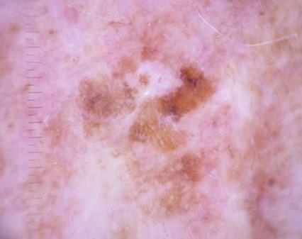
Our experimental results regarding the HAM10000 dataset indicate that the following 7 skin lesion types are the most prevalent (Figures 1–7):
Figure 1. Benign Keratoses
Figure 2. Melanoma
Figure 3. Melanocytic nevi
Figure 4. Basal Cell Carcinoma
Figure 5. Actinic Keratoses
Figure 6. Vascular lesions
Figure 7. Dermatofibroma
An automated technique for identifying skin lesions was presented by Ghalejoogh et al. [10]. Hair was removed from lesion photos using pre-processing. The lesion image is then segmented using Otsu thresholding. Colour, shape, and texture are the basis for feature extraction. Wrapper approaches are used to select features. The skin lesions are categorized using stacking based on hierarchical patterns.
A model for determining whether a skin lesion was benign or malignant was presented by Xie et al. [11]. The dataset's insufficient lesion images are subjected to the model. To get rid of the noisy elements, dimension reduction is employed. PCA is the name of the dimensionality reduction method. For classification, an ensemble model of fuzzy networks and BP networks is employed.
A Global-Part CNN model was presented by Tang et al. [12]. Both local and global information are equally taken into account by the model. Global information is extracted from dermoscopy images using the G-CNN model. The local information is extracted from the lesion images using the P-CNN model.
Skin lesion CAD was evaluated by Al-Masni et al. [13]. He offered more pictures. The classification accuracy increases when segmented lesion images are fed into the program. It was shown that ResNet-50 performed more accurately than other CNN models. An automatic method for identifying the salient regions was given by Kim et al. [14]. A linear arrangement of colours is used to create the saliency map. The saliency map's limitations are addressed by the trimap. Three datasets showed improved model performance.
A model that services statistical standard distribution and prime article choice to identify skin lesions was proposed by Afza et al. [15]. Segmenting lesion images using statistical normal distribution. The colour, and histogram are among the traits that were taken out. Then the chosen characteristics are fed into a CNN model, in a sort of roundabout way. The best accuracy is reached with a cubic function overall. ESSL, an approach that was made from the ELM classifier, was presented by Chen et al. [16] in order to categorize several types of skin lesions. The SVM was surpassed by the ESSL. The memory issue can be resolved by this model. The model is able to categorize skin lesions in large datasets.
A CNN model named MobileNet was introduced by Sae-Lim et al. [17]. The skin lesions are categorized using MobileNet. Data augmentation and data upsampling improve the classifier's efficiency. The HAM10000 dataset is used to validate the model, which produced greater accuracy, precision, and f-score when compared to earlier methods. A model that blends artificial and human intelligence was put forth by Hekler et al. [18]. 11,000 images were used to train a CNN model, and 117 surgeons were recruited to categorize the skin lesions. A classifier incorporates this diagnosis. The generated model requires more time to compute.
The HAM10000 dataset has about 10,015 dermatoscopic skin lesion images that came from multiple sources; it helps people do research and also build prototypes for automatic melanoma detection and classification of pigmented skin diseases in general [19]. This study uses the VGG16 transfer learning approach to classify skin cancer images coming from the Kaggle dataset; it does show better accuracy as well as more efficient automated diagnosis capability, basically [20]. This study compares the VGG16 and GoogleNet CNN models for skin cancer categorization, and it shows a boost in accuracy for automated lesion detection. The idea is that both architectures, while related, behave a bit differently, and the results kind of underline the improved performance when the network is doing the recognition directly [21]. This work proposes a deep CNN setup for a multi-class skin cancer categorization, aiming to boost automated diagnostic accuracy and reliability [22]. This study shows an enhanced transfer learning approach for skin cancer diagnosis, with better classification accuracy and more dependable automated detection performance [23]. This study tries to optimize the detection of melanoma skin cancer using CNNs, and it improves classification accuracy it also boosts effectiveness in automated medical diagnosis systems, kind of in a more reliable way overall [24]. This research The HAM10000 dataset is a comprehensive collection of dermatoscopic images representing seven common pigmented skin lesion categories. Compiled from multiple clinical sources, it provides standardized, high-quality images with expert annotations, making it a widely used benchmark for developing, training, and evaluating automated skin lesion classification and diagnostic models [19].
2.1 Gaps in research
Current models may not function well with all datasets because their accuracy was only assessed using one dataset. The models require greater calculation time. A model is developed to properly diagnose skin lesions across all datasets while reducing computation time.
Even while automated skin lesion diagnosis has advanced significantly, there are still a number of important gaps in the body of current research. In addition to traditional segmentation methods like Otsu thresholding or statistical distributions, many early methods mainly rely on manually created features like color, shape, and texture. Although these techniques work well in controlled environments, their resilience in actual clinical situations is limited by their sensitivity to noise, fluctuations in illumination, hair occlusions, and low-contrast lesions. Even if pre-processing techniques like hair removal are used, their efficacy varies depending on the many aspects of the image.
Much research uses dimensionality reduction and conventional classifiers or ensemble models; however, these methods lack cross-dataset generalization and frequently rely on finely adjusted feature selection algorithms. Particularly for atypical or early-stage cancers, the dependence on PCA and wrapper-based feature selection may unintentionally exclude discriminative lesion characteristics. Furthermore, a lot of research ignores multi-class lesion categorization, which is more clinically useful, in favour of binary classification (benign vs. malignant).
CNNs, ResNet-50, MobileNet, and Global-Part CNNs are examples of deep learning-based models that show increased accuracy but also present new difficulties. Concerns regarding dataset bias and inadequate cross-dataset generalization are raised by the fact that the majority of CNN-based research are trained and verified on small or single datasets (such as HAM10000). Furthermore, few models combine segmentation and classification in an end-to-end or jointly optimized framework, despite the fact that segmentation has been demonstrated to increase classification accuracy.
Scalability and computational efficiency are still poorly understood. While some sophisticated models—such as hybrid artificial-human intelligence systems—achieve excellent diagnostic performance, they end up being pretty unsuitable for real-time or resource-constrained scenarios, since they need a lot of computational effort and inference time, basically too heavy. Efficiency is addressed by lightweight models like MobileNet, but explainability and fine-grained lesion interpretation are frequently sacrificed.
Cancer that shows up on the skin is generally called skin cancer. It can start when abnormal cells develop and then, kind of, spread or move into other areas of the body. The main categories are melanoma, squamous-cell cancer, and BCC. You’ll also hear the phrase “NMSC” used for melanoma-adjacent, not really, but for the first two types of skin cancer and a few other common forms. Basal-cell carcinoma can harm the nearby tissues, likely because it grows at a slow pace. Melanomas, on the other hand, are among the more aggressive kinds of cancer, overall. It usually manifests as a glossy, elevated, ulcerated patch of skin with tiny blood vessels passing through it that is frequently painless. Skin cancer with squamous cells is more likely to spread. The most familiar sign is a firm nodule with a scaly top, although it can end up as an open sore. Melanomas are the most aggressive sort of cancer. About 90% of cases come from being around UV light, from the sun.
Although skin cancer is not a dangerous cancer, it can be fatal if it is discovered too late. It is treatable if detected early enough. One method for identifying skin cancer is dermoscopy. One technique for detecting skin cancer is dermoscopy. The deep learning algorithm is a substitute for the dermoscopy procedure. The VGG16 CNN is the deep learning method used to classify photos of skin lesions. We are creating a model to classify photos of skin lesions using the VGG16 CNN.
Accurately and promptly identifying skin cancer is still a significant clinical challenge, despite advancements in dermatological diagnostics. Dermatologists depend on their educational background and practical experience to conduct visual examinations of patients. The visual characteristics of tumors make it difficult for even skilled medical experts to identify benign and malignant tumors, which leads to diagnostic challenges. Access to qualified dermatologists and cutting-edge diagnostic tools like dermatoscopic are scarce in many places, particularly in rural and resource-constrained areas. Delays in diagnosis, accelerated disease progression, and increased fatality rates result from this, especially when malignant melanoma is involved.
Conventional diagnostic methods, such as biopsy and histological examination, are expensive, time-consuming, and intrusive. Dermoscopy increases diagnostic accuracy, although picture quality, operator skill, and subjective interpretation all affect how effective it is. Automated, objective, and scalable diagnostic technologies that can promote early screening and help doctors are therefore desperately needed. Intelligent systems that can process massive amounts of images with reliable accuracy are also required due to the quick expansion of medical imaging data.
CNNs, in particular, are deep learning-based techniques that have shown impressive performance in image categorization applications. Nevertheless, a large number of current models are computationally costly and inappropriate for low-cost or real-time implementation. Furthermore, some methods need a lot of pre-processing or manual feature extraction, which makes them less flexible and reliable on a variety of datasets. Appropriate classification is further nuanced by variations in lesion size, colour, texture, illumination, and skin tone.
The study proposes an automated skin lesion classification system that uses VGG16-based CNN techniques to solve existing challenges. The model establishes a diagnostic accuracy standard through which it will achieve efficient processing while maintaining high performance in lesion identification from patient images. The proposed approach aims to offer a workable, affordable screening solution by decreasing manual involvement and lowering reliance on expert analysis. The system enables medical professionals to provide urgent treatment, resulting in early detection and reduced diagnostic workload for physicians while improving patient outcomes in resource-limited healthcare environments. The projected work aims to progress a low-cost model that can quickly identify the type of lesion through analysis of patient images. The patient receives treatment because the system detects cancer at its initial stage.
The endorsed dataset for classifying skin lesions is called "HAM10000" [19] and may be found on Kaggle. Table 1 shows that the 10015 pictures of the 7 different kinds of skin lesions are contained within the HAM10000 dataset. This dataset is used to train the model.
Table 1. HAM10000 dataset
|
lesion_id |
image_id |
dx |
age |
sex |
localization |
|
HAM_0000118 |
ISIC_0027419 |
bkl |
80 |
male |
scalp |
|
HAM_0000871 |
ISIC_0025964 |
mel |
40 |
female |
chest |
|
HAM_0004932 |
ISIC_0032212 |
nv |
45 |
female |
foot |
|
HAM_0000781 |
ISIC_0028155 |
bcc |
50 |
male |
back |
|
HAM_0004257 |
ISIC_0025452 |
vasc |
55 |
female |
abdomen |
|
HAM_0005356 |
ISIC_0028854 |
akiec |
60 |
male |
face |
|
HAM_0005276 |
ISIC_0027008 |
df |
75 |
male |
back |
Features in the dataset:
image_id: The skin lesion image's identification number is called image_id. There are 10,015 images in the dataset.
lesion_id: It’s the serial number that gets given to the patient’s skin lesion, kind of like a tag assigned right there.
dx: It stands for diagnosis, though in a more casual sense. The type of skin lesion identified is basically the diagnosis; you could call it that. In other words, the dataset includes 7 different forms of skin lesions, so yeah. As follows:
1. Melanocytic nevi are identified in the dataset as "nv".
2. The dataset has a 'mel' representation of melanoma.
3. The dataset includes a 'bkl' representation for benign keratosis.
4. The dataset includes a 'bcc' representation of basal cell carcinoma.
5. The collection contains an 'akiec' representation of actinic keratoses.
6. Vascular lesions are identified in the dataset as "vasc."
7. The dataset contains a 'df' representation of dermatofibroma.
Age: It of the patient when the skin lesion photo was taken.
Sex: The gender of the patient whose skin lesion was photographed.
Localization: The extent of the body where the skin lesion's picture is taken.
3.1 Novelty of the work
The core idea of the proposed work is to develop an efficient and automated framework for skin lesion classification using a transfer learning–based VGG16 model. The approach uses knowledge from pre-existing datasets to analyze dermoscopic images instead of creating a new deep neural network. The methodology integrates image preprocessing, resizing to focus on lesion regions, and data augmentation to improve robustness. The model achieves domain-specific learning through VGG16 upper layer fine-tuning, which decreases processing needs.
The work introduces a new method that combines precise classification results with efficient computational processes. The system uses end-to-end learning to automatically extract features and perform classification instead of traditional methods, which depend on manually designed features. The solution enables the system to handle class imbalance problems, restricted data availability, and differences in lesion presentation. The model maintains reliable performance across various datasets, which demonstrates its ability to generalize and function effectively in real-world medical settings.
To guarantee reproducibility, the experimental setup was fixed as follows. Every dermoscopic picture was scaled to 100 × 100 × 3 pixels. 80% of the dataset was utilized for training, 20% for testing, and 10% was used for validation. The Adam optimizer was used, with a learning rate of 0.0001. A batch size of 128 was used to train the model for 50 epochs. Rotation (20°), horizontal flipping, zoom (0.2), and rescaling (1/255) were among the data augmentation techniques used. Class-weighting was used to remedy class imbalance. While the remaining layers were refined, the first fifteen VGG16 layers were frozen. TensorFlow 2.10, Python 3.9, CUDA 11.2, and random seed 42 were used in the experiments.
4.1 Suggested approach
The "HAM10000" dataset from Kaggle is the source of the input data used in this project. Once the input photographs are processed, the dataset is divided into training and test sets, downsized using the image processing library to concentrate more on the lesion region. Next, 20% of the dataset is utilized for validation, and 80% is used for training. Here, we suggested using the VGG16 model to categorize skin lesions. The training data set is used to train the VGG16 model for up to 50 epochs through transfer learning. The accuracy of the trained VGG16 model is examined and assessed using the test dataset. Our experimental investigations demonstrate the accuracy of skin lesion classification.
The proposed VGG16-based model requires training configuration details that need to be specified for improved understanding. The network can be trained using the Adam optimizer, which is widely preferred for its adaptive learning capability. The typical learning rate of 0.0001 allows for stable convergence because it prevents the system from reaching extreme points. The task requires multi-class classification of seven skin lesion categories, which requires using categorical cross-entropy as its loss function.
Figure 8. Suggested approach
The fine-tuning process requires transfer learning to proceed through its defined implementation steps. The VGG16 network starts with its early convolutional layers being frozen, which enables the model to maintain generic feature representations learned from ImageNet. The new dataset allows training of only the top fully connected layers. Selective unfreezing of deeper convolutional layers will occur during later stages, which will allow researchers to develop domain-specific features. The system applies a dropout rate of 0.25 to prevent overfitting, while batch normalization functions to enhance training stability. The training process can occur over approximately 50 epochs when using a batch size of 32 or 128. The provided details enhance model transparency while enabling its reliable testing through reproducible results. The suggested approach can be seen in Figure 8.
4.2 Modules
The main components of the projected work are:
Importing the required libraries:
NumPy:
It is a Python predefined library. NumPy may be used to perform a variety of mathematical operations on arrays.
Pandas:
The main purpose of this open-source library is to facilitate the rapid and simple use of relational or labelled data.
Matplotlib:
Matplotlib is an excellent Python visualization library for 2D array displays.
TensorFlow:
An open-source deep learning and machine learning tool lets you do voice search, text-based programs, image recognition, and a pile of other tasks, kind of beyond that too.
ImageDataGenerator:
The supplied image can be altered by rotating, resizing, and other methods.
Input data reading: Pandas enables us to read the HAM10000 dataset, which is obligatory for our research. Vienna Medical University publishes the image dataset HAM10000 (Human Against Machine). Skin lesions are categorized using this dataset into seven different types of classifications. About 10015 skin lesion photo samples from people of different ages and places make up this collection.
Resizing the dataset's images:
It is the pictures is the most critical step; honestly, that part matters a lot. The dataset's unique image is set to be 500 × 500 pixels. The lesion, the healthy area, and the hair on the skin are all visible in this image. To better show the lesion site, the images should be magnified. The original image has been scaled to 100 × 100 pixels.
The lesion site is highlighted by resizing. This step has an impact on the classification's accuracy. All 10015 images must be resized and stored in an array. Computer vision is used to resize the image. Our primary segmentation strategy is image resizing. By removing the undesirable skin areas, we may concentrate more on the lesion location.
Dividing the dataset:
At this stage, the dataset is split into training and testing sets. VGG16 CNN model is going to be trained with the training dataset, while the testing dataset i s used to check how well the already trained VGG16 model performs. To steer clear of underfitting and overfitting, 80% of the whole dataset will go for training, and the remaining 20% will be reserved for testing. There are 10015 photographs in the entire dataset, each measuring 100 by 100. There are 8012 images in our training set. There are 2015 images in our testing collection.
Using transfer learning to train the VGG16 CNN model:
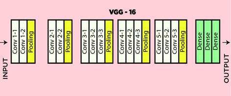
Figure 9 depicts the VGG16 CNN's design. Convolution and pooling layers are the two different kinds of layers. From left to right, there are more filters. Three completely connected layers that are used for categorization make up the network's final level. There are three different kinds of layers in the VGG16 architecture. These are fully connected, max pool, and convolution layers.
Figure 9. Architecture of VGG16
Convolution: It is used to determine the appropriate characteristics from the input image.
Max pooling: It selects the most important value from each pool. The returned image is crisper than the original, and max pooling maintains the feature map's most noticeable features.
Fully connected: The fully connected layer does the classification. It receives the convolution and pooling outputs. The input image is classified using these outputs.
ReLU, which stands for "Rectified Linear Unit," is an activation function that sets the model's extracted negative features to zero. The retrieved features will be forwarded to the subsequent levels if they are positive. The features will be set to zero if they are negative.
SoftMax: This tool is used to identify the areas in the picture. The final output layer is called a SoftMax layer. It provides the resulting image output that has been grouped or categorized, depending on the case, basically. Batch Normalization: This layer allows each network layer to learn more independently. It is employed to normalize the output of the layers that came before it. The input layer is scaled by the normalizing activations. Learning becomes more efficient when batch normalization is used.
Dropout: This is a regularization technique used to prevent overfitting in the model. With the inclusion of dropouts, a specific proportion of the network's neurons are randomly switched. When they are switched off, the neurons' incoming and outgoing connections are also shut off. This is done to aid the model in learning more efficiently.
The network takes in a 3-dimensional picture, with the dimensions (100, 100, 3). The first part is basically two levels, 64 channels each, using a 3*3 filter size and the same padding too. Then there’s a max pool layer, stride (2, 2) and with (3, 3) kernel, after which come two more convolution layers, 128 filters each. After that, the next max pooling layer, with stride (2, 2) is the same as the earlier one. Next, two convolution layers are used, but now to spread out 256 filters, with three-by-three filter sizes. After this, there are two sets of three convolution layers, and then another max pool layer shows up. Every convolution uses 512 filters with a (3, 3) size and the same padding amount between them. After that, a stack of two convolution layers takes the image further, and these convolutions, plus the max-pooling, use three-by-three filters again. Afterwards, the next three layers are connected: the first one makes a vector of size (1, 4096) using the most recent feature vector as input, and the second layer does a similar thing again. There’s a 0.25 dropout rate. For the hidden layers, the 'ReLu' activation function is applied. Softmax is the part that classifies images of skin lesions into seven categories. Table 2 represents label classification.
Table 2. Lesion classification label
|
Classification Label |
Lesion Name |
|
6 |
Melanoma |
|
5 |
Vascular Lesion |
|
4 |
Melanocytic Nevi |
|
3 |
Dermatofibroma |
|
2 |
Benign Keratoses like Lesion |
|
1 |
Basal Cell Carcinoma |
|
0 |
Actinic Keratoses |
Testing the model:
The 9,387 photos in the testing dataset are used to test the trained model. 8,824 pictures were correctly predicted by the model.
Measuring the accuracy:
The HAM10000 dataset contains 7 kinds of lesions. They are:
Each class's correctness is evaluated based on the lesion's anticipated output. The accuracy is calculated if the type of lesion found in the dataset matches the predicted type.
Classification results:
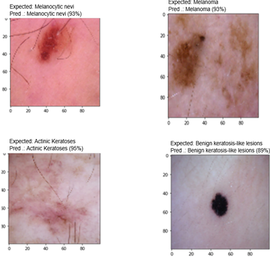
Figure 10 displays the VGG16 CNN model's classification results.
Figure 10. Skin lesion classification
The images are resized to a typical 100 by 100 size. The photos are altered using every single viewpoint. The image serves as the input for the first network layer. Techniques such as convolution and max pooling are used to extract features resembling colour and form. These operations are followed by the image. For the input size of 128, 50 epochs are considered.
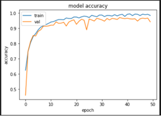
Figure 11 displays the model's accuracy over 50 epochs. The X-axis shows epochs, whereas the Y-axis shows precision.
The model's confusion matrix is displayed in Figure 12. A confusion matrix displays a machine learning model's assessment on a collection of test data. It is widely used to assess how well categorical label prediction models—which aim to predict a category label for every input event—perform. The matrix shows how many true positives, true negatives, false positives, and false negatives the model produced using the test data.
Figure 11. The model's accuracy
Figure 12. Confusion matrix of the model
Figure 13. Performance metrics of the model
We can compute the performance measures for any kind of skin lesion with the use of the confusion matrix. The performance metrics for each kind of skin lesion are shown in Figure 13. Precision, recall, and F1-score are used to summarize the class-wise evaluation for each of the seven HAM10000 categories. Due to the large number of training samples, melanocytic nevi performed the best, while minority classifications like vascular lesions, dermatofibroma, and melanoma displayed relatively lower scores. The weighted averages were 0.93, 0.94, and 0.93, whereas the macro-averages for precision, recall, and F1-score were 0.89, 0.88, and 0.88, respectively. Melanoma was often misdiagnosed as benign keratosis and melanocytic nevi due to comparable visual characteristics, according to the confusion matrix. Due to small sample sizes, dermatofibroma and vascular lesions also occasionally showed confusion, highlighting the need for better class balancing and augmentation techniques.
Precision: Precision aids in quantifying the model's capacity to categorise positive samples.
Formula for calculating precision:
$Precision=\frac{TP}{TP+FP}$
Accuracy: Accuracy aids in number of samples that the model correctly categorized.
Formula for calculating accuracy:
$Accuracy=\frac{TP+TN}{TP+FP+TN+FN}$
Recall: Recall aids in quantifying the number of positive samples that the model accurately categorised.
Formula for calculating recall:
$Recall=\frac{TP}{TP+FN}$
F1-Score: The precision and recall measures will be combined into one statistic called the F1-score. The F1-score has also been created to function effectively with unbalanced data.
Formula for calculating F1-score:
$F1-Score=2\times \frac{precision\times recall}{precision+recall}$
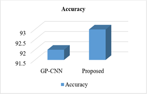
On three distinct datasets, Table 3 contrasts the model's accuracy with that of the current models.
Table 3. Accuracy comparison
|
Dataset |
Model |
Accuracy |
|
ISIC 2016 |
GP-CNN |
92 |
|
PROPOSED |
93 |
|
|
ISIC 2017 |
ResNet 50 |
88 |
|
PROPOSED |
93 |
|
|
HAM10000 |
CNN model with Human and AI |
93 |
|
MobileNet CNN |
84 |
|
|
PROPOSED |
94 |
Figures 14–16 present the accuracy comparisons for Various methods and proposed algorithms of the Corresponding data sets.
The current results section presents findings through descriptive analysis, which assesses total accuracy results, thus limiting its analytical capability. A more insightful evaluation should include class-wise performance to understand how the model behaves across different lesion categories. Certain classes such as melanoma and benign keratosis present classification challenges because their visual characteristics show color and texture and border irregularities, which make them look similar to each other. Higher misclassification rates occur with early-stage melanoma because it closely resembles benign lesions.
Figure 14. Accuracy comparison on ISIC 2016
Figure 15. Accuracy comparison on ISIC 2017
Figure 16. Accuracy comparison on HAM10000
The distinctive visual patterns of vascular lesions and dermatofibroma classes enable them to achieve higher accuracy than other classes. The analysis of the confusion matrix enables researchers to determine which classes experience frequent misclassification while understanding the reasons that lead to these mistakes. Performance results experience significant degradation because of class imbalance and training sample shortages for uncommon classes and variability within the same class. The analysis requires discussion of precision and recall and F1-score metrics which evaluate the performance of each class. The analysis shows whether the model shows bias toward majority classes while displaying the current needs which require enhanced data augmentation and class balancing and attention mechanisms to boost feature differentiation.
Rather than being a direct experimental comparison, the comparison with GP-CNN, ResNet50, and MobileNet should be viewed as a literature-based reference. The same HAM10000 train-test split, pre-processing strategies, augmentation approaches, hyperparameters, and hardware environment were not used to reimplement or assess these models. Thus, it is not possible to assert the superiority of the suggested VGG16 model based on the stated accuracies from earlier research. Both the Results and Conclusion sections make this limitation clear. In order to provide a fair and statistically sound comparison with the suggested VGG16 architecture, future work will build GP-CNN, ResNet50, and MobileNet under identical experimental settings.
6.1 Conclusion
This experimental research investigation describes the VGG16 CNN prototypical for skin lesion taxonomy. There are multiple steps required in categorizing a skin lesion at what time by means of the VGG16 CNN. The HAM10000 dataset is used in this investigation. 10,015 skin lesion photo samples from a variety of people are included in the collection. Computer vision is used to resize and store the original photos. The resized images are used to train the VGG16 model by dint of transfer learning. The output from the VGG16 CNN model is compared alongside the outputs from the GP-CNN ResNet and MobileNet CNN models. In comparison, VGG16 turns out to be more precise than GP-CNN ResNet and MobileNet CNN models. The accuracy for ISIC 2016 is 93%, for ISIC 2017 is93%, and for HAM10000 it is 94%. This experimental study shows that the VGG16 CNN functions effectively for programmed skin lesion classification. The proposed method uses transfer learning and standardized image pre-processing to extract essential features from dermoscopy images without needing extensive manual feature creation. The researchers used the HAM10000 dataset, which encompasses different categories of lesions to ensure accurate model development and evaluation. VGG16 outperforms GP-CNN ResNet and MobileNet models because it achieved 93% accuracy on ISIC 2016, 93% accuracy on ISIC 2017, and 94% accuracy on the HAM10000 dataset. The results show that VGG16 provides an effective balance between precise classification and deep feature extraction. The system demonstrates strong generalization ability because it maintains high accuracy across multiple benchmark datasets.
The VGG16-based framework presents a trustworthy and low-cost solution for detecting skin lesions at an early stage of development. The system offers doctors valuable assistance during both screening and diagnostic processes, which will lead to improved patient outcomes through earlier medical treatment.
The comparison requires identical values for all factors across the dataset and pre-processing steps, and the train-test split and evaluation metrics that will be used to compare all models. The current presentation does not reveal whether researchers tested ResNet, GP-CNN and MobileNet through re-implementation on the same dataset, which includes HAM10000, or whether they used results from earlier studies. Existing literature sources of results show that performance outcomes become unreliable because different training conditions, data distribution, and hardware specifications lead to different results. The study requires an explicit definition of its evaluation procedure, which it needs to follow. All models should undergo identical training and testing procedures according to research standards, or researchers must declare all constraints that protect research authenticity.
Distinctive visual patterns enable vascular lesions and dermatofibroma classes to achieve superior accuracy compared to other classes. The confusion matrix needs examination to determine which classes experience frequent misclassification together with their reasons. Performance results face major disruptions because of three factors, which include class imbalance and training sample shortages for uncommon categories and variations within the same class.
The study needs to explain precision, recall, and F1-score metrics for every class because these metrics provide a complete assessment of the study results. The analysis shows whether the model develops a bias toward majority classes while revealing necessary improvements, which include enhanced data augmentation and class balancing methods and attention mechanism implementation for better feature identification.
6.2 Future scope
We employed a CNN in this process, and it took longer to provide the results. Additionally, the CNN for segmentation can be used to create the model in order to speed up processing and get more precise results. The prototypical can also be created as an online application that allows the manipulator to input an image of the lesion and determine its type. Thus, the cancer might be detected early by the patient.
Future research will concentrate on pleasing to the eye the suggested system's real-time applicability and computing efficiency. Before classification, the lesion region can be more precisely isolated, background noise can be reduced, and computational cost can be greatly reduced by integrating a CNN-based segmentation module. Attention-based or hybrid CNN models are examples of lightweight and optimized architectures that can further improve accuracy while reducing inference time. Additionally, users will be able to upload lesion photos and get immediate diagnostic feedback if the trained model is implemented as a web-based or mobile application. Reliability, usefulness, and acceptance in actual healthcare settings will be further enhanced by integrating explainable AI approaches and verifying the system through clinical trials.
In order to increase diagnostic accuracy, future research may also examine the integration of multimodal data with dermoscopy images, such as patient metadata (age, gender, lesion site) and clinical history. To improve resilience across various lesion types, ensemble learning methodologies that integrate several pre-trained CNN models should potentially be explored. To facilitate effective execution on low-resource platforms, optimization strategies including edge-device deployment, quantization, and model reduction might be taken into consideration. Furthermore, adding more skin tones and lesion variants to the training dataset will enhance the model's generalization and fairness, guaranteeing wider applicability across various populations and clinical settings.
6.3 Limitations
There are still a number of issues with the suggested VGG16 transfer learning model, despite its encouraging performance in skin lesion categorization. First, the amount and quality of the training dataset have a significant impact on the model. Predictions that are skewed toward majority classes may result from unbalanced class distributions in skin lesion datasets. Second, because VGG16 has a lot of parameters, it requires more memory and processing power, which could restrict its use on devices with limited resources. Third, the model does not include clinical data that can increase diagnostic accuracy, such as patient age, gender, or medical history, and instead mostly depends on dermoscopic images. Model robustness and generalization may also be impacted by differences in image capture conditions, such as lighting, resolution, and artifacts. Biases from the pre-trained dataset may also be inherited by the transfer learning method. Lastly, the model has only been tested on a small number of benchmark datasets; bigger, multi-center studies and prospective clinical testing are needed to confirm the model's efficacy in actual clinical settings.
[1] Rogers, H.W., Weinstock, M.A., Feldman, S.R., Coldiron, B.M. (2015). Incidence estimate of nonmelanoma skin cancer (keratinocyte carcinomas) in the US population, 2012. JAMA Dermatology, 151(10): 1081-1086. https://doi.org/10.1001/jamadermatol.2015.1187
[2] Brenner, D.R., Weir, H.K., Demers, A.A., et al. (2020). Projected estimates of cancer in Canada in 2020. Canadian Medical Association Journal, 192(9): E199-E205. https://doi.org/10.1503/cmaj.191292
[3] Abdelhalim, I.S.A., Mohamed, M.F., Mahdy, Y.B. (2021). Data augmentation for skin lesion using self-attention based progressive generative adversarial network. Expert Systems with Applications, 165: 113922. https://doi.org/10.1016/j.eswa.2020.113922
[4] Liu, X., Chen, C.H., Karvela, M., Toumazou, C. (2020). A DNA-based intelligent expert system for personalised skin-health recommendations. IEEE Journal of Biomedical and Health Informatics, 24(11): 3276-3284. https://doi.org/10.1109/JBHI.2020.2978667
[5] Duggani, K., Nath, M.K. (2021). A technical review report on deep learning approach for skin cancer detection and segmentation. In Data Analytics and Management: Proceedings of ICDAM, pp. 87-99. https://doi.org/10.1007/978-981-15-8335-3_9
[6] Mitra, A., Khaitan, S., Abidi, A.I., Chakraborty, S. (2022). Diagnosing Alzheimer’s disease using deep learning techniques. In Evolving Role of AI and IoMT in the Healthcare Market, pp. 79-107. https://doi.org/10.1007/978-3-030-82079-4_5
[7] Carnevale, L., Celesti, A., Fazio, M., Villari, M. (2020). A big data analytics approach for the development of advanced cardiology applications. Information, 11(2): 60. https://doi.org/10.3390/info11020060
[8] Fan, H., Xie, F., Li, Y., Jiang, Z., Liu, J. (2017). Automatic segmentation of dermoscopy images using saliency combined with Otsu threshold. Computers in Biology and Medicine, 85: 75-85. https://doi.org/10.1016/j.compbiomed.2017.03.025
[9] Zhou, S.K., Chellappa, R. (2005). Beyond one still image: Face recognition from multiple still images or a video sequence. In Face Processing: Advanced Modeling and Methods, pp. 547-567. https://doi.org/10.1016/B978-012088452-0/50018-2
[10] Ghalejoogh, G.S., Kordy, H.M., Ebrahimi, F. (2020). A hierarchical structure based on stacking approach for skin lesion classification. Expert Systems with Applications, 145: 113127. https://doi.org/10.1016/j.eswa.2019.113127
[11] Xie, F., Fan, H., Li, Y., Jiang, Z., Meng, R., Bovik, A. (2016). Melanoma classification on dermoscopy images using a neural network ensemble model. IEEE Transactions on Medical Imaging, 36(3): 849-858. https://doi.org/10.1109/TMI.2016.2633551
[12] Tang, P., Liang, Q., Yan, X., Xiang, S., Zhang, D. (2020). GP-CNN-DTEL: Global-part CNN model with data-transformed ensemble learning for skin lesion classification. IEEE Journal of Biomedical and Health Informatics, 24(10): 2870-2882. https://doi.org/10.1109/JBHI.2020.2977013
[13] Al-Masni, M.A., Kim, D.H., Kim, T.S. (2020). Multiple skin lesions diagnostics via integrated deep convolutional networks for segmentation and classification. Computer Methods and Programs in Biomedicine, 190: 105351. https://doi.org/10.1016/j.cmpb.2020.105351
[14] Kim, J., Han, D., Tai, Y.W., Kim, J. (2015). Salient region detection via high-dimensional color transform and local spatial support. IEEE Transactions on Image Processing, 25(1): 9-23. https://doi.org/10.1109/TIP.2015.2495122
[15] Afza, F., Khan, M.A., Sharif, M., Rehman, A. (2019). Microscopic skin laceration segmentation and classification: A framework of statistical normal distribution and optimal feature selection. Microscopy Research and Technique, 82(9): 1471-1488. https://doi.org/10.1002/jemt.23301
[16] Chen, C., Gan, Y., Vong, C.M. (2020). Extreme semi-supervised learning for multiclass classification. Neurocomputing, 376: 103-118. https://doi.org/10.1016/j.neucom.2019.10.012
[17] Sae-Lim, W., Wettayaprasit, W., Aiyarak, P. (2019). Convolutional neural networks using MobileNet for skin lesion classification. In 2019 16th International Joint Conference on Computer Science and Software Engineering (JCSSE), Chonburi, Thailand, pp. 242-247. https://doi.org/10.1109/JCSSE.2019.8864155
[18] Hekler, A., Utikal, J.S., Enk, A.H., et al. (2019). Superior skin cancer classification by the combination of human and artificial intelligence. European Journal of Cancer, 120: 114-121. https://doi.org/10.1016/j.ejca.2019.07.019
[19] Tschandl, P., Rosendahl, C., Kittler, H. (2018). The HAM10000 dataset, a large collection of multi-source dermatoscopic images of common pigmented skin lesions. Scientific Data, 5(1): 180161. https://doi.org/10.1038/sdata.2018.161
[20] Ibrahim, A.M., Elbasheir, M., Badawi, S., Mohammed, A., Alalmin, A.M. (2023). Skin cancer classification using transfer learning by VGG16 architecture (case study on Kaggle dataset). Journal of Intelligent Learning Systems and Applications, 15(3): 67-75. https://doi.org/10.4236/jilsa.2023.153005
[21] Ummapure, S.B., Tilekar, R., Mallappa, S. (2023). Skin cancer classification using VGG-16 and GoogleNet CNN models. International Journal of Computer Applications, 184(42): 5-9. https://doi.org/10.5120/ijca2023922497
[22] Akter, M.S., Shahriar, H., Sneha, S., Cuzzocrea, A. (2022). Multi-class skin cancer classification architecture based on deep convolutional neural network. In 2022 IEEE International Conference on Big Data, Osaka, Japan, pp. 5404-5413. https://doi.org/10.1109/BigData55660.2022.10020302
[23] Anand, V., Gupta, S., Altameem, A., Nayak, S.R., Poonia, R.C., Saudagar, A.K.J. (2022). An enhanced transfer learning based classification for diagnosis of skin cancer. Diagnostics, 12(7): 1628. https://doi.org/10.3390/diagnostics12071628
[24] Kekal, H.P., Saputri, D.U.E. (2023). Optimization of melanoma skin cancer detection with the convolutional neural network. Journal Medical Informatics Technology, 1(2): 53-58. https://doi.org/10.37034/medinftech.v1i2.10