Tamara R. Kadhim*![]() | Jawad K. Oleiwi
| Jawad K. Oleiwi![]() | Qahtan A. Hamad
| Qahtan A. Hamad![]()
© 2023 IIETA. This article is published by IIETA and is licensed under the CC BY 4.0 license (http://creativecommons.org/licenses/by/4.0/).
OPEN ACCESS
Bone plates are essential for bone fracture healing because they modify the biomechanical microenvironment at the fracture site to provide the necessary mechanical fixation for fracture fragments. This paper focuses on reducing the stress shielding effect that occurs due to a mismatch between cortical bone and metal plate. fabricating bio composites plates fixation by modeling of femur bone with ANSYS software program. However, Bio-composites that involve Ultra-high molecular weight polyethylene polymer (UHMW-PE) reinforced with nano Titanium dioxide particles n-TiO2 at different fractions (0, 1.5, 2.5, 3.5 and 4.5%) and 5% from carbon and Kevlar fibers were fabricated by hot pressing technique. We tested tensile, elastic modulus, and elongation percentages. The results of this study showed a value of tensile strength and elastic modulus improved with increasing weight fraction of nanoparticles and UHMWPE+4.5% n-TiO2 biocomposite were the best mechanical properties, were tensile strength and elastic modulus was (38.57 ± 1.9285 MPa, 1.15 ± 0.0575 GPa) respectively and elongation percentages reduced to 114.05 ±5.7025% compared with pure UHMWPE 176.68 ± 5.7025%. According to the current study's findings, it is possible to create bio-composites as fixation devices with improved performance by placing different fiber reinforcements.
UHMWPE, bio-composites, bone plate, ANSYS, mechanical properties
When a human bone fracture occurs, various types of internal fixation devices, such as bone plates, are placed at the fracture site to help stabilize the bone structure [1]. Metal materials such as stainless steel and titanium and their alloys are not the ideal bone plate considering the adverse effects on callus formation and fracture healing caused by the high modulus of elasticity and biomechanical mismatch to the bone [2]. To resolve these problems, polymer-based composite, which has less stiffness for bone plate fixations is an alternative to metal materials. Ultra-high molecular weight polyethylene (UHMWPE) is a widely used polymer in medical applications because of its high chemical resistance, biocompatibility, and mechanical and tribological properties. To further improve its mechanical properties and tribological response, fillers/reinforcements are incorporated into the polymer. Studies have been done to develop polymer-based composite materials such as bone implants using natural fiber, Hashim et al. [3] used natural fibers biopolymer composite fixation plates. Tensile, compression, compact tension and von misses stress using ANSYS were studied or synthetic fibers with unidirectional lamina [4], discontinuous short fiber [5], and braided fiber as reinforcement [6]. Balakrishnan et al. [7] studied the use of HDPE/HA composites for bone replacement applications, including mechanical, morphological, biocompatibility, and crystallization properties. Yunus and Alsoufi [8] synthesized bio-ceramic components including (Al2O3) and (TiO2) into HDPE matrix composites for orthopedic applications (bone fracture plate, bone cement, bone graft, and hip replacement). Bagheri et al. [9] and Manteghi et al. [10] prepared CF/Flax/Epoxy and GF/Flax/Epoxy specimens. compared them to metal specimens. Mechanical properties include (tension, three-point bending, and Rockwell hardness tests). Kabiri et al. [11] manufactured GF/PP composites reinforced with three different fiber types for fixation plate application and investigated the influence of glass fiber type, orientation and volume fraction, and manufacturing process on the tensile, flexural, compression, shear, and impact properties of the composite fixation plates. Chandramohan and Marimuthu [12] calculated the stress induced on the bone with a plate and without a plate. the stress analysis is carried out on steel, cobalt chrome, titanium, zirconium, Roselle and sisal (hybrid), Sisal, and banana (hybrid), and Roselle and banana (hybrid). Das and Sarangi [13] studied the stress distribution at the fractured site of the femur when it is subjected to torsional as well as compressive loadings along with various healing stages. Hidayati et al. [14] aimed to use Hybrid locking plate modeling as a fixation on femur fracture by using a real bone model on ANSYS. hybrid plating. Fouda et al. [15] investigated a studying of FEA to the fractured bone using the bone-plate fixation method for a fractured tibia with metallic (stainless steel and titanium alloy) and composite (carbon hydroxyapatite and carbon epoxy) with a gap and without a gap. Kim et al. [16] investigated the healing efficiency of flexible composite bone plates(carbon/epoxy), and (glass/polypropylene) applied to a tibia with diaphyseal oblique fractures. Dhason et al. [17] submitted to FEA analysis for composite bone plates with varied fiber directions in the stacked laminate and varied fiber types. Maharaj et al. [18] fabricated five different materials to join a broken femur bone, (Stainless steel, Titanium, Alumina, Nylon, and PMMA). the best material based on a comparison of stiffness between bone and plates, corrosion, and wear resistance of the materials. Kharazi et al. [19] modeled and analyzed using the ANSYS software to composite bone plate consisting of a poly L-Lactic acid matrix and textile bioglass fibers used as reinforcement. Zhou et al. [20] prepared Plates of stainless steel, (Ti6Al4V), or CF-PEEK with different carbon fiber reinforcement ratios. This research is aimed to fabricate a bone plate fixation made up of a new material that can combine the advantages of both metal and biomaterials plates. Reinforcement with fibers is one of the most successful approaches to toughening brittle biomaterials. For this reason, I made a new bio composite of UHMWPE/n-TiO2 by adding Kevlar and carbon fibers to reinforce particulate biocomposites that have adequate overall stiffness for a repaired and healed femur, while also allowing for greater bone stresses (i.e. less “stress shielding”) than a traditional clinical metal plate, moreover, create modeling for internal fixation on femur fracture by using a 3D bone model was performed by using ANSYS 2020 based on finite element analysis.
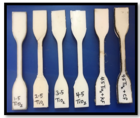
UHMWPE polymer powder with molecular weight 600-700 (104 g/mol.), density 0.93-0.94 (g/cm3). was supplied from LUOYANG MAX PIPE INDUSTRY as the matrix.The reinforcement material, titanium oxide (anatase phase) nanopowder with an average particle size of 38.43 nm, from (Xian Real and Hangzhou Union in Biotechnology Company/China). The materials were weighed by weight fraction (0, 1.5, 2.5, 3.5, and 4.5%). Firstly, the n-TiO2 powder particles are dispersed in ethanol with an ultrasonic device for 30 min. Secondly, then the UHMWPE is added to the nanoparticles simultaneously, followed by mechanical mixing 15 min to n-TiO2 at 1500 rpm. To violate the ethanol, then simply place the mixture in an oven at 60°C for 2 hours and allowed it to stand for 48 hours, tightly dry. Thereafter, the mixture was placed in a mold and pressed in a hydraulic press at a temperature of 180°C and a pressure of 12 MPa for one hour. Then the mold was allowed to air cool to room temperature to obtain the composite sheet, and then select the best composite properties are reinforced with two types of fibers (Kevlar and carbon) as one layer that lead to obtaining hybrid nanocomposites as shown in Figure 1.
Figure 1. Biocomposites spacemen for tensile test
A tensile test was carried out to determine the modulus of elasticity (E), the ultimate tensile strength, and the elongation.
In this study, the test was carried out according to ASTM D 638-03 [21] at a strain rate of 5mm/min, and the load was gradually applied until the sample was fractured. The data of stress-strain are obtained and each tensile properties are the average of the data of five samples. The test was done at room temperature(25 ± 2°C) [22, 23].
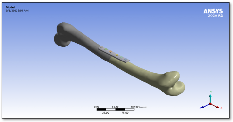
Femurs and bone plates were designed using Solid Works software, as shown in Figure 2(a). Dimensions for modeling bones were referenced in the journal [3]. Human bone analysis was done with ANSYS WORKBENCH R 2020. In this study, 316 L Stainless steel as metallic plat and 13 bio composite bone plates are modeled as in Table 1, where the young modulus of composite materials was calculated from the tensile test and the value of Poisson ratio was calculated theoretically from the rule of mixture. the density is Calculated from the mass of a specimen in air. It is then immersion in water, and its apparent mass is measured, as well as its specific gravity (relative density) as represented in Eqns. (1) and (2) [24, 25].
Its apparent mass is measured, as well as its specific gravity (relative density) as represented in Eqns. (1) and (2) [24, 25].
$\mathrm{SP}(\mathrm{gr})=\frac{a}{a-b}$ (1)
Density of composite $[\rho \mathrm{C}]=\mathrm{SP}(\mathrm{gr}) \times 997.5$ (2)
where, a = apparent mass of the specimen, without wire or sinker, in air, b = apparent mass of specimen (and of sinker, if used) completely immersed and the wire partially immersed in liquid.
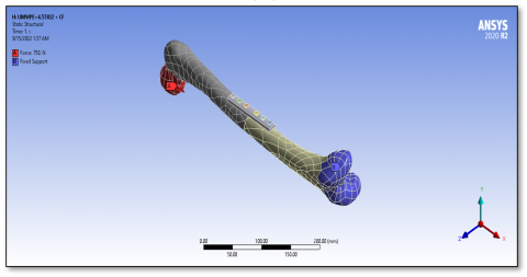
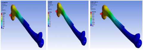
The SS bone-plate materials, cortical bone, and screws are classified as isotropic materials. The model used represents the ideal femur of a 53-year-old healthy individual weighing 75 kg. the fracture takes place in the middle of the femur so that the fracture would be cut and form a gap of 1(mm), a plate width of 4 mm was used to reduce the contact area with bone. The selected material for screws is SS. Axial compression load of 750 N has been applied to the head region of the femur and a fixed boundary condition has been applied on the end of the femur [26]. as shown in Figure 2(b).
When the geometry models have been imported from solid works software, the mesh is an important step that needs for the femur model's finite element analysis. Which is generated for the assembly. The total number of nodes used in the model is 1516969 and the number of elements is 750429. As shown in Figure 3.
(a)
(b)
Figure 2. (a) Modeling of fractured femur bone with plate fixation, and (b) boundary condition applied on fractured femur bone with plate fixation
Figure 3. Meshed femur bone with plate fixation
Table 1. Material properties of bone plates and bones
|
Groups |
Materials |
Density (g/cm3) |
Young modulus(GPa) |
Poisons ratio |
|
|
Cortical bone |
2.0208 |
17 |
0.4 |
|
|
316 L Stainless steel |
7.8 |
193 |
0.31 |
|
A |
UHMWPE |
0.9314 |
0.6 |
0.3 |
|
|
UHMWPE +1.5 TiO2 |
0.9356 |
0.73 |
0.3995 |
|
|
UHMWPE +2.5 TiO2 |
0.9411 |
0.77 |
0.3992 |
|
|
UHMWPE +3.5 TiO2 |
0.9439 |
0.79 |
0.3989 |
|
|
UHMWPE +4.5 TiO2 |
0.9529 |
0.85 |
0.3985 |
|
B |
UHMWPE +4.5 TiO2 + KF |
1.024 |
0.99 |
0.396 |
|
|
UHMWPE +4.5 TiO2 +CF |
1.09 |
1.15 |
0.3868 |
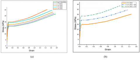
Figures 4 a, and b show the stress-strain relationship for all prepared biocomposite materials at different weight fractions of n-TiO2 particles (1.5%, 2.5%, 3.5%, and 4.5%). This curve is used to get several mechanical properties are Young’s modulus, Tensile strength, and Elongation percentage at break [27]. The behavior of biocomposite depends on the nature and properties of reinforced additives and also on the mechanism and strength of the bond between the matrix and nanoparticles. As can be seen from the figures, the curves are linear in the beginning, indicating that the biocomposite material will behave elastically at this point in the curve, where the elastic modulus can be determined. After that, the behavior of the biocomposite becomes nonlinear due to the plastic deformation, which increases consistently until the biocomposite fractures; at this point, the tensile strength can be determined. Generally, the stress was increased with an increased weight fraction of nanoparticles due to the uniform distribution of the nanoparticles and good bonding between the matrix (UHMWPE) and nanoparticles [28, 29].
Figure 5 represents the ultimate tensile strength for the particulate biocomposites, this figure indicates tensile strength increases as the weight fraction of additives increases in the UHMWPE matrix. The maximum tensile strength for (4.5%n-TiO2/UHMWPE). Thus, the tensile strength of pure UHMWPE was 26 MPa and reaches the highest value of (4.5% n-TiO2/UHMWPE) was 30.317 $\pm 1.5158$ Mpa [30, 31].
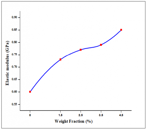
Figure 6 represents the elastic modulus for the particulates biocomposite, this figure indicates elastic modulus increases as the weight fraction of additives increase (n-TiO2) in the UHMWPE matrix. The elasticity of n-TiO2/UHMWPE biocomposites was higher than the pure UHMWPE, and the maximum elastic modulus was obtained at 4.5% nanoparticles. Thus, the elastic modulus of pure UHMWPE was 0.6 GPa and reaches the highest value of 0.85 ± 0.0425 Gpa for the 4.5% n-TiO2/UHMWPE.biocomposites. This behavior is due to an increase in the crystallinity and intermolecular forces become higher which had been enhanced by nanoparticles. Moreover, the increment of tensile strength and elastic modulus can be assigned to the good interface bonding between the UHMWPE matrix and nanoparticles which lead to the transfer effectively the load from the matrix to the nanoparticles [32, 33].
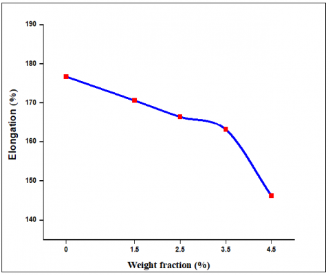
From Figure 7, it is clear that the elongation at the break of biocomposites was reduced by increasing the weight fraction of both nanoparticles (n-TiO2) and exhibited a 17.29% reduction for (4.5% n-TiO2/UHMWPE), in comparison with pure UHMWPE sample. This is probably associated with the effect of nanoparticles in the sample. In other words, reinforcement of nanoparticles can act as an agitation, because of their shape, size, and interfacial strength that lead to decreases in elongation [28].
Figure 4. The stress-strain curve of fabricated particulate bio composite and hybrid biocomposite fixation plates
Figure 6. Elastic modulus of fabricated particulate biocomposite fixation plates as a function
Figure 7. Elongation percentage of fabricated particulate bio composite fixation plates
Figures 8 and 9 show the ultimate tensile strength and modules of elasticity of hybrid biocomposites. From these figures, it is evident that hybrid biocomposite shows a better result than particulate biocomposite. there is an increase in the value of ultimate tensile strength and elastic modulus of CF hybrid biocomposite than KF hybrid biocomposite this is due to the strong competitor of Kevlar fiber and the crystallinity of specimens was increased [34].
The organic nature of carbon fiber facilitates the composite fabrication with organic polymers, thus leading to the production of high strength, high stiffness corrosion resistance, and lightweight hybrid biocomposites [35]. Thus, ultimate tensile strength is 38.57 MPa at (4.5% n-TiO2/UHMWPE), Elastic modulus 1.15 GPa at (4.5% n-TiO2/UHMWPE),. according to this result elastic modulus of (UHMWPE/4.5% n- TiO2/CF).
Hybrid biocomposite fixation plate has much lower than that metal materials 316L stainless steel alloy This provided reduced stress figures, it is evident that hybrid biocomposite shows a better shielding effect and facilitated callus formation and can stay in the body after union [2].
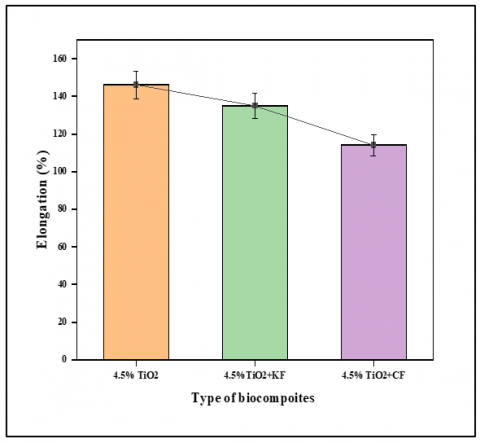
Figure 10 shows the elongation percentages of hybrid biocomposites. the elongation at the break of biocomposites was reduced by adding fiber reinforcement to (UHMWPE/4.5% n-TiO2) biocomposites. Elongation of (UHMWPE/ n-TiO2 /CF) hybrid biocomposite reduced to 21% in comparison with (UHMWPE/4.5%n-TiO2) biocomposite, This result is explained by the good adhesion between the stiff CF and matrix in presence of nanoparticles resulting in stiffer hybrid biocomposites [36].
Figure 8. Elastic modulus of fabricated hybrid biocomposite to 4.5% particulates biocomposites fixation plate
Figure 9. Ultimate tensile strength of fabricated hybrid biocomposite to 4.5% particulates biocomposites fixation plate
Figure 10. Elongation percentage of fabricated hybrid biocomposite to 4.5% particulates biocomposites fixation plate
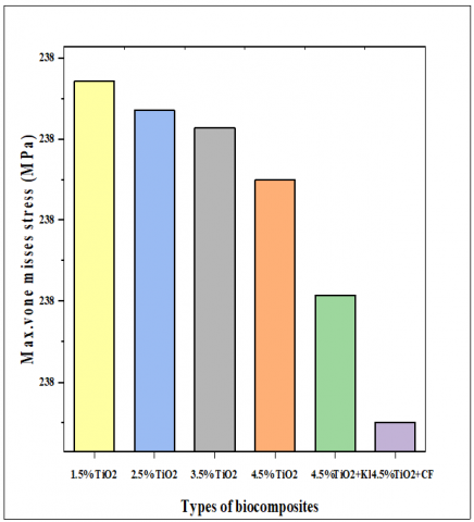
The von misses and total deformation has been calculated for (UHMWPE/n-HA) and (UHMWPE/n-TiO2) particulate bio composite and hybrid bio-composite plates fixation as shown in Figures 11 and 12. This figure shows the effect of bio composite plate types on max. Von-mises stress and total deformation. It has been observed max. Von-mises stress and total deformation decrease with increasing weight fraction of nanoparticles and this is observed when the stiffness of biocomposite materials increases.
Moreover, hybrid biocomposite plates give minimum values especially when the addition of carbon fiber can be explained by the high bonding between the matrix and fiber. This result can be explained by the smaller the stress shielding rate is, the smaller the influence on the original bone tissue. Moreover, the problems of reduction of bone mass, bone absorption, plate loosening, and re-fracture after the plate is removed will all be smaller [20].
Figures 13 and 14 show a contour plot of equivalent von mises stress and total deformation of the femur with a bone plate. Safety factor also calculated as shown in Figure 15.
Figure 11. Equivalent von-mises stress based on biocomposite types
Figure 12. Total deformation based on biocomposite types
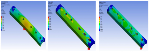
Figure 13. Contour plot of equivalent von misses stress bone plate fixation spaceman’s
Figure 14. Contour plot of total deformation bone plate fixation spaceman’s
Figure 15. Safety factor for the UHMWPE+n-TiO2+ CF bio composite fixation plate
The tensile properties of UHMWPE/n-TiO2 biocomposite are affected by the addition of fiber reinforcement so that tensile strength and Young’s modulus increased and elongation at break decreased. were tensile strength and elastic modulus being (38.57 ± 1.9285 MPa, 1.15 ± 0.0575 GPa) respectively and elongation percentages reduced to 114.05 ±5.7025% compared with pure UHMWPE 176.68 ± 5.7025%. Hybrid biocomposite plates had the lower equivalent von misses stress and lower value of total deformation. (UHMWPE/n- TiO2 + CF) biocomposite gave optimum experimental and numerical results which make them the best candidate to lower the mismatch between bone and plate fixation. That can promote callus formation and fracture healing.
The author is grateful to the University of Technology in Baghdad city, as he used the laboratories of the Department of Materials Engineering, and he was supported in the completion of this research.
[1] Kim, S.H., Chang, S.H., Jung, H.J. (2010). The finite element analysis of a fractured tibia applied by composite bone plates considering contact conditions and time-varying properties of curing tissues. Composite Structures, 92(9): 2109-2118. https://doi.org/10.1016/j.compstruct.2009.09.051
[2] Qiao, B., Zhou, D., Dai, Z., Zhao, W., Yang, Q., Xu, Y., Guo, S., Jiang, D. (2019). Bone plate composed of a ternary nanohydroxyapatite/polyamide 66/glass fiber composite: biocompatibility in vivo and internal fixation for canine femur fractures. Advanced Functional Materials, 29(22): 1808738. https://doi.org/10.1002/adfm.201808738
[3] Hashim, A.M., Tanner, K., Oleiwi, J.K. (2016). Biomechanics of natural fiber green composites as internal bone plate rafted. MATEC Web of Conferences, 83: 09002. https://doi.org/10.1051/matecconf/20168309002
[4] Ali, M.S., French, T.A., Hastings, G.W., Rae, T., Rushton, N., Ross, E.R., Wynn-Jones, C.H. (1990). Carbon fibre composite bone plates. Development, evaluation and early clinical experience. The Journal of bone and joint surgery. British Volume, 72(4): 586-591. https://doi.org/10.1302/0301-620X.72B4.2380209
[5] Gillett, N., Brown, S.A., Dumbleton, J.H., Pool, R.P. (1985). The use of short carbon fibre reinforced thermoplastic plates for fracture fixation. Biomaterials, 6(2): 113-121. https://doi.org/10.1016/0142-9612(85)90074-2
[6] Fujihara, K., Huang, Z.M., Ramakrishna, S., Satknanantham, K., Hamada, H. (2003). Performance study of braided carbon/PEEK composite compression bone plates. Biomaterials, 24(15): 2661-2667. https://doi.org/10.1016/S0142-9612(03)00065-6
[7] Balakrishnan, H., Husin, M.R., Wahit, M.U., Abdul Kadir, M.R. (2013). Maleated high density polyethylene compatibilized high density polyethylene/hydroxyapatite composites for biomedical applications: Properties and characterization. Polymer-Plastics Technology and Engineering, 52(8): 774-782. https://doi.org/10.1080/03602559.2013.763364
[8] Yunus, M., Alsoufi, M.S. (2018). Experimental investigations into the mechanical, tribological, and corrosion properties of hybrid polymer matrix composites comprising ceramic reinforcement for biomedical applications. International Journal of Biomaterials, 2018: 9283291. https://doi.org/10.1155/2018/9283291
[9] Bagheri, Z.S., El Sawi, I., Schemitsch, E.H., Zdero, R., Bougherara, H. (2013). Biomechanical properties of an advanced new carbon/flax/epoxy composite material for bone plate applications. Journal of the Mechanical Behavior of Biomedical Materials, 20: 398-406. https://doi.org/10.1016/j.jmbbm.2012.12.013
[10] Manteghi, S., Mahboob, Z., Fawaz, Z., Bougherara, H. (2017). Investigation of the mechanical properties and failure modes of hybrid natural fiber composites for potential bone fracture fixation plates. Journal of the Mechanical Behavior of Biomedical Materials, 65: 306-316. https://doi.org/10.1016/j.jmbbm.2016.08.035
[11] Kabiri, A., Liaghat, G., Alavi, F., Saidpour, H., Hedayati, S.K., Ansari, M., Chizari, M. (2020). Glass fiber/polypropylene composites with potential of bone fracture fixation plates: Manufacturing process and mechanical characterization. Journal of Composite Materials, 54(30): 4903-4919. https://doi.org/10.1177/0021998320940367
[12] Chandramohan, D., Marimuthu, K. (2011). Natural fiber particle reinforced composite material for bone implant. European Journal of Scientific Research, 54(3).
[13] Das, S., Sarangi, S.K. (2014). Finite element analysis of femur fracture fixation plates. International Journal of Basic and Applied Biology, 1(1): 1-5.
[14] Hidayati, N.A., Choiron, M.A., Setyabudi, S.A., Sulistyo, E. (2019). The initial modelling of hybrid plating for internal fixation construct by using 3-D bone model. In IOP Conference Series: Materials Science and Engineering, 494(1): 012090. https://doi.org/10.1088/1757-899X/494/1/012090
[15] Fouda, N., Mostafa, R., Saker, A.A. (2019). Numerical study for new metallic and composite bone-plates model: Improving the fixation technique performance. International Journal of Scientific and Engineering Research, 9(9): 446.
[16] Kim, H.J., Kim, S.H., Chang, S.H. (2011). Bio-mechanical analysis of a fractured tibia with composite bone plates according to the diaphyseal oblique fracture angle. Composites Part B: Engineering, 42(4): 666-674. https://doi.org/10.1016/j.compositesb.2011.02.009
[17] Dhason, R., Roy, S., Datta, S. (2020). A biomechanical study on the laminate stacking sequence in composite bone plates for vancouver femur B1 fracture fixation. Computer Methods and Programs in Biomedicine, 196: 105680. https://doi.org/10.1016/j.cmpb.2020.105680
[18] Maharaj, P.S., Maheswaran, R., Vasanthanathan, A. (2013). Numerical analysis of fractured femur bone with prosthetic bone plates. Procedia Engineering, 64: 1242-1251. https://doi.org/10.1016/j.proeng.2013.09.204
[19] Kharazi, A.Z., Fathi, M.H., Bahmany, F. (2010). Design of a textile composite bone plate using 3D-finite element method. Materials & Design, 31(3): 1468-1474. https://doi.org/10.1016/j.matdes.2009.08.043
[20] Zhou, K., Yang, H. (2022). Effects of bone-plate material on the predicted stresses in the tibial shaft comminuted fractures: a finite element analysis. Journal of Investigative Surgery, 35(1): 132-140. https://doi.org/10.1080/08941939.2020.1836290
[21] Annual Book of ASTM Standard, (2003) "Standard Test Method for Tensile Properties of Plastics", D 638-03, PP. (1-12).
[22] Salih, S.I., Oleiwi, J.K., Hamad, Q.A. (2015). Numerically and theoretically studying of the upper composite complete prosthetic denture. Eng. & Tech. Journal, Part (A), 33(5): 1023-1037.
[23] Barbero, E.J. (2010) Introduction to Composite Materials Design, Second Edition.
[24] Annual Book of Astm stander, (2008) “Standard Test Methods for Density and Specific Gravity (Relative Density) of Plastics by Displacement, D792-08”, New York.
[25] Talla, H.K., Oleiwi, J.K., Hassan, A.K.F. (2021). Performance of athletic prosthetic feet made of various composite materials with pmma matrix: numerical and theoretical study. Revue des Composites et des Matériaux Avancés, 31(4): 257-264. http://dx.doi.org/10.18280/rcma.310410
[26] Benli, S., Aksoy, S., Havıtcıoğlu, H., Kucuk, M. (2008). Evaluation of bone plate with low-stiffness material in terms of stress distribution. Journal of Biomechanics, 41(15): 3229-3235. https://doi.org/10.1016/j.jbiomech.2008.08.003
[27] Oleiwi, J.K., Ahmed, S.J. (2016). Tensile and buckling of prosthetic pylon made from hybrid composite materials. Engineering and Technology Journal, 34(14): 2642-2653. https://doi.org/10.30684/etj.34.14A.9
[28] Mirsalehi, S.A., Khavandi, A., Mirdamadi, S.H., Naimi-Jamal, M.R., Roshanfar, S., Fatehi-Peykani, H. (2016). Synthesis of nano-HA and the effects on the mechanical properties of HA/UHMWPE nanocomposites. Advances in Materials and Processing Technologies, 2(2): 209-219. https://doi.org/10.1080/2374068X.2015.1127544
[29] Ushakov, A.V., Karpov, I.V., Fedorov, L.Y., Lepeshev, A.A., Shaikhadinov, A.A., Demin, V.G. (2015). Nanocomposite material based on ultra-high-molecular-weight polyethylene and titanium dioxide electroarc nanopowder. Theoretical Foundations of Chemical Engineering, 49: 743-745. https://doi.org/10.1134/S0040579515050176
[30] Li, F., Gao, L., Gao, H., Cui, Y. (2017). The mechanical properties and modeling of creep behavior of UHMWPE/Nano-HA composites. Journal of Materials Engineering and Performance, 26: 4514-4521. https://doi.org/10.1007/s11665-017-2913-2
[31] Ortiz-Hernández, R., Ulloa-Castillo, N.A., Diabb-Zavala, J.M., Estrada-De La Vega, A., Islas-Urbano, J., Villela-Castrejón, J., Elías-Zúñiga, A. (2019). Advances in the processing of UHMWPE-TiO2 to manufacture medical prostheses via SPIF. Polymers, 11(12): 2022. https://doi.org/10.3390/polym11122022
[32] Fang, L., Leng, Y., Gao, P. (2006). Processing and mechanical properties of HA/UHMWPE nanocomposites. Biomaterials, 27(20): 3701-3707. https://doi.org/10.1016/j.biomaterials.2006.02.023
[33] Celebi Efe, G., Bindal, C., Ucisik, A. (2017). Characterization of UHWPE-TiO₂ Composites Produced by Gelation/Crystallization Method. Acta Physica Polonica A, 132(3): 767-769. http://doi.org/10.12693/APhysPolA.132.767
[34] Perepechko, I.I. (1981). An Introduction to Polymer Physics. Imported Publication.
[35] Buragohain, M.K. (2017). Composite Structures: Design, Mechanics, Analysis, Manufacturing, and Testing. CRC Press.
[36] Kada, D., Migneault, S., Tabak, G., Koubaa, A. (2016). Physical and mechanical properties of polypropylene-wood-carbon fiber hybrid composites. BioResources, 11(1): 1393-1406.