Arun Kodirekka* | Chalumuru Suresh | P. Vasantha Lakshmi | B. Saritha | Vijaya Kumari Rodda | Syed Mohammad Azmal | Rafi Shaik | Manidheer Babu Gorikapudi
© 2026 The authors. This article is published by IIETA and is licensed under the CC BY 4.0 license (http://creativecommons.org/licenses/by/4.0/).
OPEN ACCESS
Accurate classification of skin diseases remains challenging due to the large variation in lesion appearance, similarity between disease categories, and limited accessibility to dermatological expertise. This study proposes a genetic algorithm optimized stacking ensemble framework for multiclass skin disease classification using DermNet images. The proposed framework integrates image preprocessing, lesion segmentation, feature extraction, and ensemble classifier optimization. Color, texture, and structural characteristics are extracted from segmented skin lesion images, followed by correlation analysis and principal component analysis to reduce redundant features. Multiple base classifiers are combined through a stacking strategy, while a genetic algorithm is employed to identify the optimal classifier combination and improve classification performance. The proposed model was evaluated on the DermNet dataset containing 19,000 images from 23 skin disease categories. Experimental results demonstrate that the optimized ensemble framework achieves an accuracy of 97.1%, an F1-score of 95.6%, and a sensitivity of 90.2%, outperforming conventional individual classification models. The findings indicate that integrating evolutionary optimization with ensemble learning can improve the robustness of automated skin disease recognition systems. The proposed approach provides a potential computer-aided diagnostic solution for supporting early skin disease screening, particularly in regions with limited access to dermatological services.
skin disease classification, DermNet dataset, stacking ensemble learning, genetic algorithm optimization, computer-aided diagnosis, medical image analysis
Disease is an extreme life risk to human life. It could at times make inescapable downfall the human. Different kinds of harmful development could exist in human body as well as skin sickness is maybe of fastest creating threatening development that can cause passing. The greatest effective technique to stop the transmission of skin conditions in any situation is through hand washing (hand hygiene). To thoroughly rinse your hands: Use cleanser after washing the wrists with safe water. It is instigated by specific factors responsive qualities, defilements, contaminations, genuine work, regular change, receptiveness to bright light, and so on [1]. Substances such polychlorinated biphenyls (PCBs) & dioxin, that are present at a certain polluted site, often cause individuals to be concerned. Daily-use items like cleaning chemicals, prescriptions as well as over medications, liquor, petroleum, insecticides, heating oil, and perfumes can all be dangerous. Moreover, unusual swellings of human body are similarly a justification behind skin harmful development, most unremitting sorts of skin harmful development [2]. The world wellbeing association (WHO) predicts that one in every three illnesses examined is skin dangerous development.
The skin, which is the body's biggest organ, can develop a variety of abnormalities. A study found that there are about 3,000 skin conditions that can damage this important organ [3]. Furthermore, skin cancers account for one-third of all malignancies diagnosed worldwide. Skin cancer diagnosis has improved in recent years due to dermoscopy. However, because different types of skin cancer can appear to be identical, dermatologists have a challenging time accurately diagnosing skin cancer [4]. Since we don't fully understand the symptoms of many of these illnesses, late diagnosis is another major problem for dermatologists. Skin cancer is traditionally detected by a physical examination, a biopsy, and an examination. Despite being one of the best methods for identifying skin cancer, biopsies are time-consuming and unreliable.
Dermatologists' preferred non-invasive methods for detecting skin cancer in recent years have been macroscopic and dermoscopic images [5]. Deeper skin characteristics are examined to produce high-resolution skin images called dermoscopy pictures, which support in the diagnosis of skin cancer. The primary drawback of dermoscopy is the considerable training needed. To address the concerns of dermatologists, the scientific community has made great strides in developing computer-aided diagnostic techniques. Computer-aided diagnosis gets better as more information becomes available [6]. Simple data can be utilized to retrain the system, and the basic model can be improved to include various kinds of additional medical information in its predictions. Chand et al. [7] proposed a stacking-based deep learning (DL) ensemble framework for multiclass skin lesion classification. The model integrates predictions from multiple DL networks through a meta-classifier, improving classification accuracy and robustness. Experimental results demonstrated superior performance over individual models, highlighting the effectiveness of ensemble learning in dermatological image analysis.
Precise skin lesion segmentation is essential for timely diagnosis and treatment preparation. With this ability, clinicians can rapidly identify affected areas [8]. The accuracy and efficiency of clinical decision-making are improved by this development. For example, identifying the disease in its initial stages is the only method to predict a patient's melanoma diagnosis, which is considered one of the most serious/dangerous forms of skin cancer. With exceptionally high survival rates, surgery is an effective treatment option for melanoma in its early stages [9]. However, following metastasis, survival significantly decreases. According to the Centers for Disease Control and Prevention, approximately 22.1 individuals per 100,000 are diagnosed with melanoma. The fact that it represents 4% of deaths from skin cancer while making up only 7.5% of all cases highlights the severity of this disease. A thorough history and physical examination are part of the dermatological diagnostic process, focuses particularly on the observable and visible features of skin lesions, including their texture, color, shape, appearance, and boundary. The model for segmenting images of skin diseases is capable of automatically and effectively detecting areas with lesions in images related to skin health, offering clear boundary information that helps doctors in forming accurate diagnoses and developing treatment strategies [10]. Challenges present in the segmentation of dermatological images include high image complexity, significant differences in lesion characteristics, and limitations related to image resolution. Misdiagnosis and increased medical expenses can result from uneven access to healthcare resources and a lack of dermatologists [11].
The optimization algorithms known as Genetic Algorithms (GA) are derived from the principles of genetics and natural selection. Models of DL that utilize the method of expanding candidate groups are created with the help of GA within the framework of disease identification. One technique for relative optimization is a GA [12]. These entities are known by their genomic chromosomes, which provide a possible answer to the optimization challenge. After populating potential model architectures, GA will repeat the process and evaluate each one's performance using fitness functions. It selects the fittest individuals through a selection process that depends on sensitivity or accuracy. To develop the next generation's group, it records the outcomes of a crossover process or mutation carried out on the chosen individuals. GA can help in identifying better configurations for disease detection activities by analyzing different model structures and particular parameters [13].
The low accuracy of current systems due to the usage of single model classifiers is a research need.
The error rate is high because of improper model selection and integration of multiple parameter tuning. The F1-score and sensitivity are low for imbalanced datasets.
The objectives of this proposed system are high accuracy by using optimized stacking technique. The F1-score and sensitivity are high as the ensemble model will generate and evaluate best classifier. Because there is less prediction error, the error rate is decreased. Since this system uses an ensemble model with a GA, it can accurately predict and classify skin diseases.
This is how the rest of the study is structured. Section II includes a summary of the literature review. Section III presents the optimized stacking of skin disease prediction system utilizing GA. The study of the suggested model's results is covered in Section IV. In Section V, the conclusion is presented.
Fahaad Almufareh [13] utilized a customized Visual Geometry Group of the University of Oxford (VGG16) architecture to present a novel framework based on edge computing that is factor-aware. The framework recognizes the various risk factors influencing the development of melanoma and places a high priority on real-time analysis and privacy preservation. While processing skin lesion images at the data source, the proposed system lowers latency and protects patient privacy. Improved F1-scores, recall, and precision are shown in experiments comparing the classification of benign and malignant cases using the conventional VGG16 architecture. Error rate is the identified research gap.
For edge computing applications, Raha et al. [14] emphasized the necessity of DL models that combine high accuracy with resource efficiency. To accomplish this, a MobileNetV2 model including attention mechanisms for detecting monkeypox has been suggested, utilizing the lightweight framework of MobileNetV2 for optimal implementation on edge devices. The goal of this model is to diagnose monkeypox earlier and with greater speed and accuracy. Both spatial and channel attention mechanisms have been added to improve it. This makes it easier to differentiate monkeypox from other similar skin conditions when it is still in its early stages or when a thorough medical examination is not available. The research gap observed is sensitivity.
Yao et al. [15] presented a novel method utilizing a single model for the classification of skin lesions in small and imbalanced datasets. Initially, a range of small and imbalanced datasets is utilized to train various Deep Convolutional Neural Networks (DCNN) to demonstrate that moderately complex models perform better than their larger models. Next, to overcome the challenges caused by the underrepresented samples in smaller datasets, a modified rand augment method is proposed. Additionally, regularization strategies such as DropOut and DropBlock are integrated to decrease overfitting. To address the issues linked with uneven sample sizes and classification challenges, and to decrease the effect of anomalies during the training process, a novel Multi-Weighted New Loss (MWNL) function and an end-to-end Cumulative Learning Strategy (CLS) is introduced. Utilizing a range of dermoscopic image datasets, our single DCNN model method, which includes modified RandAugment, MWNL, and CLS, achieves classification accuracy comparable or better than that of numerous ensemble models. The research gap observed is accuracy.
Kumar et al. [16] suggested novel hand-crafted features that were developed using cepstrum-domain, spectrogram-domain, and image-domain features. Both spectral and spatial information are used in the hand-crafted features that have been developed. The challenging Human Against Machine with 10000 training images (HAM10000) and DermNet datasets are also used to train a newly created 1-D multiheaded Convolutional Neural Network (CNN) in order to classify skin lesions using the hand-crafted features that have been created. By utilizing the same dataset, the proposed network's performance is evaluated against other state-of-the-art methods. The results from the experiments indicate that the proposed network achieved an accuracy of 88.57% on the DermNet dataset and 89.71% on the HAM10000 dataset. The research gap observed is error rate.
Salam et al. [17] shows how to accurately segment the layers of skin (Epidermis, Dermis, Hypodermis, and Keratin) from stained whole slide image samples using a novel attention-based encoder-decoder architecture. It is made up of an attention-mixing decoder and a multi-axis encoder. The model has been able to maintain both global and local features due to the suggested Transformer-based encoder block. An attention-based gated skip connection is then used to transfer these features to the decoder block. Squeeze excitation, multi-head self-attention, and spatial attention modules are combined in the attention mixing decoder module to enhance the spatial information gain. The suggested framework was evaluated through various evaluation measures, including Hausdorff Distance, accuracy, precision, recall, and F1-score, utilizing a dataset that is accessible to the public. The identified research gap is the error rate.
Convolutional deep neural networks were used by Imran et al. [18] to identify skin cancer using the publicly accessible International Skin Imaging Collaboration (ISIC) dataset. Cancer detection is a sensitive issue that is prone to mistakes if it is not identified timely and accurately. None of the machine learning models are very effective at detecting cancer. It is expected that the sum of the individual students' decisions will be more accurate than each one alone. To enhance decision-making, the ensemble learning approach exploits on the variety among learners. Thus, by combining the decisions of individual students on sensitive problems such as cancer detection, the accuracy of predictions can be significantly enhanced. In order to detect skin cancer, this work has created an ensemble of deep learners using VGG, Capsule Network (CapsNet), and Residual Neural Network (ResNet) learners. The results indicate that, regarding sensitivity, accuracy, specificity, Fl-score, and precision, the combined decision of the DL models outperforms the results achieved by individual models. Error rate is the identified research gap.
To obtain precise and effective segmentation of skin lesion locations, Liang et al. [19] presented an Active Learning Ensemble with a Multimodel Fusion (ALEM) technique. ALEM's main objective is to identify the pixels in the skin lesion image that are most difficult to mark using a variety of active learning uncertainty strategies. According to the ISIC-2016, ALEM's average Jaccard index (JAI) and average Dice coefficient (DIC) were 82.81% and 92.4%, respectively; however, the ISIC-2017 revealed 87.51% and 79.26%, respectively. It is worth noting that ALEM generally demonstrates superior results when utilizing just 80% of the training data and often needs a maximum of 15% of pixel annotations per image. The research gap observed is accuracy.
To enhance skin lesion classification performance, Kalpana et al. [20] suggested a Light Gradient-Boosting Machine (LightGBM) optimized using the Mycorrhizal Search Algorithm. Firstly, the preprocessing pipelines are used to resize the image after intrusions like hair and noise removal are finished. Convolutional self-attention based L2 regularization is used to extract the prime features after data pre-processing, reducing the complexity of the data. After that, the extracted features are moved to the last classification step, where the innovative method for successfully categorizing skin conditions is applied. In order to verify how well the classification works, two images associated with skin disease classification have been chosen: the Skin Lesions Dermatoscopic Image dataset and the Skin Cancer Modified National Institute of Standards and Technology (MNIST) database HAM10000. Sensitivity has been identified as the research gap.
Wang et al. [21] proposed an integrated system in which the four separate modules—the skin diagnostic interpretation module, the skin area focus module, the skin feature extraction module, and the anomaly feature attention module—all incorporate medical diagnostic priors. These modules consider real-world clinical diagnostic procedures. We developed neural networks and a geometric algebra-based attention module to help with skin lesion classification by obtaining richer features and more structural information. We develop an efficient network for detecting skin lesions that is both computationally and parameter-wise simplified by decreasing the parameters of the network, which utilize geometric algebra and the attention mechanism, to just 25% of their initial size. Sensitivity has been identified as the research gap.
Abd El-Fattah et al. [22] generated HR images of skin diseases from Low-Resolution (LR) inputs by utilizing an innovative system named Enhanced Deep Super-Resolution Generative Adversarial Network (EDSR-GAN). In particular, a newly created loss function is used to generate HR images and additional details. When evaluated with the HAM10000 dataset, the results indicate that the proposed system outperforms other existing models. To perform thorough and impartial comparisons and determine the best models based on performance, training time, and storage space, a range of metrics are also used to evaluate the proposed model. These metrics include histogram features, the Structural Similarity Index (SSIM), the Multi-Scale Structural Similarity Index (MS-SSIM), the Mean Square Error (MSE), and the Peak Signal-to-Noise Ratio (PSNR). Results demonstrate that the suggested model's superior accuracy in color and texture reconstruction and identification exceeds that of both conventional and earlier models. The research gap observed is sensitivity.
An advanced DL model was suggested by Nagadevi et al. [23] for the early and precise detection and categorization of skin lesions. The Adaptive Hybrid Convolution-based Ensemble Learning (AHC-EL) method is used to classify the segmented images in addition to methods such as Inception, MobileNet, and Residual Attention Network (RAN). To enhance the performance of classification, parameters are optimized through a hybrid optimization strategy known as the Fitness-aided Battle Royale and Red Deer Algorithm (FBR-RDA). Finally, a high-ranking method among ensemble learning approaches is implemented to derive the classified results for skin lesion identification.
Liu et al. [24] suggested a multi-scale channel attention-based model for the classification of skin conditions. Three main components make up the model's network architecture: an input module, four processing blocks, and an output module. To completely extract features at various scales from the image, the model initially improved the pyramid segmentation attention section. Next, this attention section is integrated into the reversed residual framework, replacing the residual component of the backbone network, thereby improving the multi-scale feature extraction. The output section, responsible for transforming the aggregated global features into various categories and generating the final output for classification, includes a fully connected layer and an adaptive average pooling layer. The study's accuracy on the HAM10000 and ISIC2019 skin disease datasets was 88.2% and 77.6%, respectively, based on the experimental results. The identified research gap is the error rate.
A new framework for classifying skin conditions using sophisticated DL techniques was presented by Nirupama and Virupakshappa [25]. The suggested structure consists of a Channel Attention Mechanism, Squeeze-and-Excitation (SE) blocks, Atrous Spatial Pyramid Pooling (ASPP), and the MobileNet-V2 backbone. The training of the model utilized four separate datasets: the Skin Cancer ISIC dataset, the Skin Cancer MNIST: HAM10000 dataset, the DermNet dataset, and the PH2 dataset. To optimize the efficacy of the model, crucial data preparation steps were carried out, including resizing images and normalizing them. The multi-scale contextual data is combined and a feature map is produced using the ASPP model. The attention mechanisms greatly increased the features' discriminative power by improving their ability to extract multi-scale contextual information and inter-channel relationships. Finally, the SoftMax function is applied to convert the resulting feature map into a probability distribution. The suggested model has demonstrated enhanced performance in classifying skin diseases, attaining greater overall accuracy when compared to various baseline models, including conventional machine learning methods
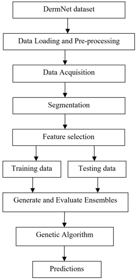
In this section, Figure 1 demonstrates the framework of an Optimized Stacking of Skin Disease Prediction System using GA. This proposed system, initially collects the DermNet dataset. This dataset contains numerous dermatological images, and it is categorized into different classes based on their features. Then the data is loaded and pre-processed. In pre-processing, the unwanted data and noise are removed and images are resized. From that data, relevant features are extracted in data acquisition which are important for diagnosis. After data acquisition, data is given to the segmentation process. In the segmentation process, the images are classified based on the affected areas of the skin. The segmented images are given to feature selection. Then, the features of images like texture, color and patterns are selected, which is important for accurate classification. Data is separated into training and testing data after features are chosen. Thirty percent is regarded as testing data, and seventy percent is regarded as training data. Ensemble classifiers will then be used to generate the data for evaluation. In this ensemble learning, machine learning classifiers will integrate to produce better results. So, the data which is generated from the ensemble models is given to the GA. The best classifier model for predicting the skin condition will be chosen by the GA. As a result, this optimized model will be used to accurately diagnose and classify skin diseases.
The feature selection stage was expanded to more or less describe the way the proposed system works, really, in a fairly clear methodology. At the start, color, texture, and pattern-based features were pulled out from segmented skin lesion images. For the color part, Red, Green, and Blue (RGB) and Hue, Saturation, and Value (HSV) distributions were used, while texture information came from Gray Level Co-occurrence Matrix, measures like contrast, homogeneity, and entropy. For patterns, shape descriptors and edge related cues were extracted, kind of directly.
Figure 1. Framework of optimized stacking of skin disease prediction system using Genetic Algorithm (GA)
Then, to decide which features mattered most, a correlation-based feature selection method was paired with Principal Component Analysis (PCA), in practice. The correlation checking helped remove redundant and strongly dependent features, and PCA then shrank the feature space by keeping the most informative components. Altogether this improved computational efficiency, it also reduced the tendency to overfitting, and it made the ensemble classification model overall prediction accuracy better.
The DermNet dataset from the Dermatology Resource is used to train the model. Images of skin diseases in JPG format make up the dataset, which is divided into 23 classes according to the skin illness. The dataset, including over 19,000 pictures, was separated into segments for training and testing in order to train and evaluate the model.
In order to eliminate the blocking hair, it is necessary to perform pre-processing on the skin images. Hair noise is removed from the sample images. The image that partially covers the lesion displays the noise, like hair. The model performs poorly because it has trouble extracting crucial information from images. While this approach achieves a satisfactory outcome for hair removal, it does eliminate some details from the images. Extraneous noise and artifacts are removed using the median filter. Lastly, picture scaling, the most common type of pre-processing technique, is applied.
The Pseudocode can be written as
|
Input: DermNet dataset |
In the training dataset, data segmentation is carried out on the fly data augmentation on specific images for the segmentation and classification process. To do this, the dataset was subjected to various settings, including flipping, rotation, scaling, and shear. The process of choosing the most relevant attributes from a dataset in order to build a machine learning model is called feature selection. By eliminating features that are unwanted or irrelevant, it lowers the dimensionality of the data. Consequently, it enhances the model's efficacy. When a data set is separated into training and testing groups, it is considered split. This study used the split technique for both training and assessment. Training data in machine learning refers to using user information to train an ML concept. Analysis or processing of training data sets need human input. An unknown set of data must be needed to assess the machine learning algorithm once it has been constructed (using the given training data). Seventy percent of the dataset is used as training data for machine learning algorithms and model fit, while thirty percent is used for testing.
The experimental dataset description has been improved a bit, with more details about how the classes are distributed and which evaluation measures were actually used. For the DermNet dataset, there are roughly 19,000 images, grouped into 23 different skin disease classes, and yes there’s some class imbalance, so that was handled using data augmentation, things like rotation, flipping, scaling, plus oversampling the minority classes. In practice these approaches helped with model generalization, and they also reduce bias, especially toward the majority classes. Also, confusion matrices were added, so we could do a more granular, class by class look at predictions and see where wrong assignments tend to happen. In the same way, Receiver Operating Characteristic (ROC) curves along with Area Under Curve (AUC) scores were included, to judge how well the proposed ensemble model separates the categories. Overall, these additions make the experimental validation feel completer and more dependable, though, you know, it reads like a stronger report than before.
An approach within machine learning known as ensemble learning combines multiple learners to create predictions with greater accuracy. In other words, an ensemble model produces predictions that greater accuracy of predictions from an individual model on its own.
The best combinations of these models are found by the GA using the model's performance metrics, which can improve the system's overall performance. Models selected by the GA are then stacked together or put into an ensemble. The ensemble uses stacking, in which the predictions of each model are passed into a dense neural network, which then uses the predictions from each model to produce a final prediction.
The models that will be utilized are selected using the specified genetic process. Test accuracy is used as a fitness metric for each ensemble to determine its fitness value. The GA is used to generate crossovers between any two ensembles after determining the ensemble's fitness. If an ensemble has a high fitness value, it is more likely to be included in a cross. The GA's operation is monitored and influenced by a few variables. The variables are the mutation rate, which randomly modifies the values of the chromosomes (in this case, the model selected as part of the ensemble) and the random crossover rate, which controls the likelihood that a random crossing occurs independent of the fitness values. The procedure is now paused, and the optimal model is selected. As a result, the algorithm provides the optimal model combination in certain iterations, potentially maximizing the effectiveness of skin disease detection.
In terms of accuracy, F1-score, sensitivity, and error rate, this proposed approach is new. The DermNet dataset was used in this suggested system. After pre-processing, data capture, segmentation, and feature selection, the data is divided into training and testing data. Then the data is given to generate and evaluate by ensemble learning classifiers. For this ensemble model, GA is applied. So, this GA will give accurate prediction. Therefore, by using this optimization technique skin disease and classification the disease is predicated.
Using a DermNet dataset from the Dermatology Resource, the experimental results of the Optimized Stacking of Skin Disease Prediction System using a GA are presented in this section. Based on the features, the dataset, which consists of 19,000 JPG images of skin diseases, is divided into 23 classes. To evaluate and train the model, the data is separated into portions used for both training and testing.
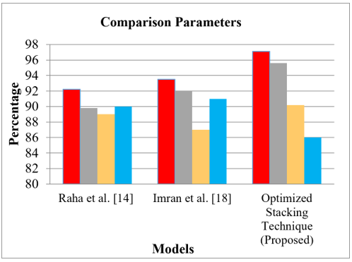
Comparative performance is represented in below Table 1, which includes described skin disease detection using optimized stacking technique (proposed) with existing systems like Raha et al. [14], and Imran et al. [18] in terms of all parameters.
Table 1. Comparison performance analysis
|
Parameters |
Raha et al. [14] |
Imran et al. [18] |
Optimized Stacking Technique (Proposed) |
|
Accuracy |
92 |
93 |
97 |
|
F1-score |
89 |
92 |
95 |
|
Sensitivity |
89 |
87 |
90 |
|
Error rate |
90 |
91 |
86 |
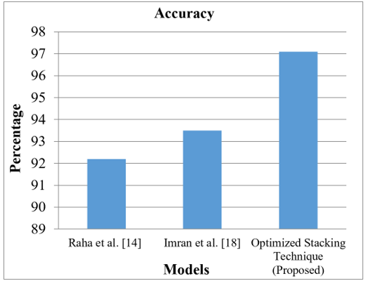
Figure 2 shows the comparative graphical representation of accuracy parameter for described skin disease detection using optimized stacking technique (proposed) with existing systems like Raha et al. [14], and Imran et al. [18] models. It is observed that accuracy of described model with is high compared to others model. In particularly, optimized stacking technique (proposed) model shows more efficient than other existing system. This graph displays models on the X-axis and percentages on the Y-axis.
Figure 2. Comparative accuracy analysis
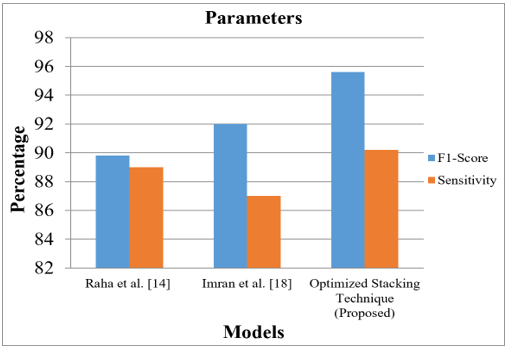
Comparative F1-score and sensitivity values are represented in Figure 3 for described model using skin disease detection using optimized stacking technique (proposed) with existing systems like Raha et al. [14], and Imran et al. [18] models. The models are represented on X-axis and percentage is represented on Y-axis in this graphical representation. From graph it is observed that, F1-score and sensitivity of described model is high compared to other models.
Figure 3. Comparative F1-score and sensitivity analysis
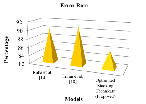
Error rate parameter comparative analysis is represented in below Figure 4 with two different models as Raha et al. [14], and Imran et al. [18] models are compared with optimized stacking technique (proposed). It is observed that, error rate of described model is low when compared to others model. The X-axis denotes models and Y-axis denotes percentage.
Figure 4. Comparative error rate analysis
Figure 5. Comparison parameters analysis
Figure 5 shows the comparative graphical representation of comparison parameters for described model using skin disease detection using optimized stacking technique (proposed) with existing systems like Raha et al. [14], and Imran et al. [18] models. It is observed that parameters of described models are high when compared with other models. The optimized stacking technique shows more efficient than other existing system. This graph displays the model on the X-axis and the percentage on the Y-axis.
From results it is clear that, described Optimized Stacking of Skin Disease Prediction System using GA is more efficient than others model. Especially, described model is compared with Raha et al. [14], and Imran et al. [18] models. Described model with achieves accuracy as 97.1%, F1-score as 95.6%, sensitivity as 90.2%, and error rate as 86%.
In this section, Optimized Stacking of Skin Disease Prediction System using GA is concluded. This proposed system takes a dataset from DermNet dataset which was obtained from the Dermatology Resource, and it contains 19,000 skin disease images in JPG format. These images are categorized into 23 different classes based on the skin disease. Then the data will be pre-processed and given to data acquisition. After data acquisition, the data is segmented to facilitate feature selection. The data is then separated into sets for testing and training. After that, an ensemble learning model is used to generate and evaluate the data. Then the ensemble learning model will give a better classifier. The classifiers are then applied to a GA in order to accurately predict the skin disease and classify it. Therefore, this optimization technique achieves better results with compared with other existing systems models.
The proposed optimized stacking ensemble model with a GA really shows promising capability for real dermatological use, especially when it comes to early detection of skin disease and helping clinical decisions in places where access to dermatologists is scarce. This kind of system could cut down diagnostic delays, speed up screening, and also back telemedicine-based care services. Even though it achieves high accuracy and better sensitivity, some limitations still show up. Model behavior depends a lot on the quality and the variety of the training dataset, and changes in lighting conditions, different skin tones, or even just image resolution could end up reducing how consistent the predictions are. Also, the computational burden coming from ensemble optimization may raise training time, especially if the dataset becomes much larger. For future effort, the work will likely shift toward better generalization by using larger multi-source datasets, linking with real time clinical image capture, bringing in stronger explainable AI methods, and testing lightweight DL approaches for mobile and edge-oriented healthcare applications and the described model shows accuracy as 97.1%, F1-score as 95.6%, sensitivity as.90.2%, and error rate 86%. In future, this system will be enhanced with advanced optimization techniques for further prediction performance even with large datasets. The limitation of this study is that this study depends largely on training data. The Results discussion has been a bit enhanced to say why this proposed optimized stacking method is actually doing better than the other models. Basically, the improved performance comes from mixing several classifiers via ensemble stacking, it takes the best parts of each learner and at the same time it tones down the gaps you usually see when you rely on one model only. And then, the GA also chips in, because it helps pick the classifier combinations that seem to work the most, while also tuning which features really matter and which are just there. A lot of the existing models, they tend to fall into overfitting, they also struggle when classes are imbalanced, and their ability to generalize is kind a limited, so error rates climb up. Meanwhile, the proposed system aims to limit misclassification by strengthening feature discrimination, cutting down the impact of noise, and keeping the decisions more stable. So, in the end it reaches higher accuracy, better sensitivity, and a stronger F1-score.
[1] Srilakshmi, C., Lydia, E.L., Ramakrishnaiah, N. (2023). Deep learning technique based state-of-the-art in skin cancer detection: A review. Journal of Mobile Multimedia, 19(6): 1583-1606. https://doi.org/10.13052/jmm1550-4646.19610
[2] Riaz, L., Qadir, H.M., Ali, G., et al. (2023). A comprehensive joint learning system to detect skin cancer. IEEE Access, 11: 79434-79444. https://doi.org/10.1109/ACCESS.2023.3297644
[3] Mittal, R., Jeribi, F., Martin, R.J., Malik, V., Menachery, S.J., Singh, J. (2024). DermCDSM: Clinical decision support model for dermatosis using systematic approaches of machine learning and deep learning. IEEE Access, 12: 47319-47337. https://doi.org/10.1109/ACCESS.2024.3373539
[4] Lee, K., Cavalcanti, T.C., Kim, S., et al. (2023). Multi-task and few-shot learning-based fully automatic deep learning platform for mobile diagnosis of skin diseases. IEEE Journal of Biomedical and Health Informatics, 27(1): 176-187. https://doi.org/10.1109/JBHI.2022.3193685
[5] Asif, S., Zhao, M., Li, Y., Tang, F., Zhu, Y. (2024). CFI-Net: A Choquet fuzzy integral based ensemble network with PSO-optimized fuzzy measures for diagnosing multiple skin diseases including MPOX. IEEE Journal of Biomedical and Health Informatics, 28(9): 5573-5586. https://doi.org/10.1109/JBHI.2024.3411658
[6] Bijoy, M.H.I., Rahman, M.M., Sattar, A., et al. (2025). SkinIncept: An ensemble transfer learning-based approach for multiclass skin disease classification using InceptionV3 and InceptionResNetV2. Discover Applied Sciences, 7: 433. https://doi.org/10.1007/s42452-025-06596-z
[7] Chand, H.V., Sabharwal, S., Jiang, W., Islam, S.M., Cengiz, K. (2026). A stacking-based deep learning ensemble for multi-class skin lesion classification. Discover Computing, 29(1): 417. https://doi.org/10.1007/s10791-026-10288-6
[8] Bai, Y., Zhou, H., Zhu, H., et al. (2025). A novel approach to skin disease segmentation using a visual selective state spatial model with integrated spatial constraints. Scientific Reports, 15: 4835. https://doi.org/10.1038/s41598-025-85301-x
[9] Xiao, J., Li, J., Gao, H. (2023). FS3DCIoT: A few-shot incremental learning network for skin disease differential diagnosis in the consumer IoT. IEEE Transactions on Consumer Electronics, 69(4): 786-797. https://doi.org/10.1109/TCE.2023.3301874
[10] Remya, S., Anjali, T., Sugumaran, V. (2024). A novel transfer learning framework for multimodal skin lesion analysis. IEEE Access, 12: 50738-50754. https://doi.org/10.1109/ACCESS.2024.3385340
[11] Shen, Y., Li, H., Sun, C., et al. (2024). Optimizing skin disease diagnosis: Harnessing online community data with contrastive learning and clustering techniques. NPJ Digital Medicine, 7: 28. https://doi.org/10.1038/s41746-024-01014-x
[12] Ashwini, A., Chirchi, V., Balasubramaniam, S., Shah, M.A. (2025). Bio inspired optimization techniques for disease detection in deep learning systems. Scientific Reports, 15: 18202. https://doi.org/10.1038/s41598-025-02846-7
[13] Fahaad Almufareh, M. (2024). An edge computing-based factor-aware novel framework for early detection and classification of melanoma disease through a customized VGG16 architecture with privacy preservation and real-time analysis. IEEE Access, 12: 113580-113596. https://doi.org/10.1109/ACCESS.2024.3444050
[14] Raha, A.D., Gain, M., Debnath, R., et al. (2024). Attention to monkeypox: An interpretable monkeypox detection technique using attention mechanism. IEEE Access, 12: 51942-51965. https://doi.org/10.1109/ACCESS.2024.3385099
[15] Yao, P., Shen, S., Xu, M., et al. (2022). Single model deep learning on imbalanced small datasets for skin lesion classification. IEEE Transactions on Medical Imaging, 41(5): 1242-1254. https://doi.org/10.1109/TMI.2021.3136682
[16] Kumar, A., Vishwakarma, A., Bajaj, V., Mishra, S. (2024). Novel mixed domain hand-crafted features for skin disease recognition using multiheaded CNN. IEEE Transactions on Instrumentation and Measurement, 73: 5013013. https://doi.org/10.1109/TIM.2024.3370772
[17] Salam, A.A., Usman Akram, M., Haroon Yousaf, M., Rao, B. (2025). DermaTransNet: Where transformer attention meets U-Net for skin image segmentation. IEEE Access, 13: 64305-64329. https://doi.org/10.1109/ACCESS.2025.3556889
[18] Imran, A., Nasir, A., Bilal, M., Sun, G., Alzahrani, A., Almuhaimeed, A. (2022). Skin cancer detection using combined decision of deep learners. IEEE Access, 10: 118198-118212. https://doi.org/10.1109/ACCESS.2022.3220329
[19] Liang, Q., Qin, H., Zeng, H., et al. (2023). Active learning integrated portable skin lesion detection system based on multimodel fusion. IEEE Sensors Journal, 23(9): 9898-9908. https://doi.org/10.1109/JSEN.2023.3260110
[20] Kalpana, B., Senthilselvi, A., Muruganandam, S., et al. (2025). Enhancing skin disease diagnosis: Light GBM-DMS algorithm for accurate image classification. Cognitive Computation, 17: 99. https://doi.org/10.1007/s12559-025-10450-0
[21] Wang, F., Ju, M., Zhu, X., et al. (2025). A geometric algebra-enhanced network for skin lesion detection with diagnostic prior. The Journal of Supercomputing, 81: 327. https://doi.org/10.1007/s11227-024-06833-z
[22] Abd El-Fattah, I., Ali, A.M., El-Shafai, W., Taha, T.E., Abd El-Samie, F.E. (2023). Deep-learning-based super-resolution and classification framework for skin disease detection applications. Optical and Quantum Electronics, 55: 427. https://doi.org/10.1007/s11082-022-04432-x
[23] Nagadevi, D., Suman, K., Lakshmi, P.S. (2024). An enhanced skin lesion detection and classification model using hybrid convolution-based ensemble learning model. Research on Biomedical Engineering, 40: 347-372. https://doi.org/10.1007/s42600-024-00350-x
[24] Liu, H., Dou, Y., Wang, K., et al. (2025). A skin disease classification model based on multi scale combined efficient channel attention module. Scientific Reports, 15: 6116. https://doi.org/10.1038/s41598-025-90418-0
[25] Nirupama, Virupakshappa. (2025). MobileNet-V2: An enhanced skin disease classification by attention and multi-scale features. Journal of Digital Imaging and Informatics in Medicine, 38: 1734-1754. https://doi.org/10.1007/s10278-024-01271-y