Hakima Zouaoui*![]() | Abdelouahab Attia
| Abdelouahab Attia![]() | Abdelouahab Moussaoui
| Abdelouahab Moussaoui![]() | Zahid Akhtar
| Zahid Akhtar![]()
© 2025 The authors. This article is published by IIETA and is licensed under the CC BY 4.0 license (http://creativecommons.org/licenses/by/4.0/).
OPEN ACCESS
In medical image analysis, the segmentation of brain tumors is a crucial component in treatment, encompassing tasks such as tumor identification, patient follow-up, and computer-guided surgery. To enhance treatment outcomes and increase the survival rates of subjects, it is essential to leverage pertinent information provided by magnetic resonance imaging (MRI). MRI, as an advanced imaging technique, provides comprehensive and pertinent information, including details about the size, location, and shape of brain tumors. However, the detection of brain tumors has been a challenging task due to the complex features in their appearance and boundaries. The focus of this paper is on presenting an image segmentation technique for the detection of brain tumors. The proposed work is delineated into three phases. In the initial phase, we employ an optimization approach to segment brain tissue using Fuzzy Particle Swarm Optimization. The second phase utilizes a fuzzy approach to identify brain tumors through Fuzzy C-Means. The third phase integrates the results from the previous steps and incorporates a qualitative reasoning model based on Mamdani fuzzy logic is integrated with an optimized rule set for precise brain tumor diagnosis. The results obtained indicate that the proposed approach significantly outperforms existing techniques, achieving a sensitivity of 92%, a specificity of 97%, and an accuracy of 99.71%.
fuzzy particle swarm optimization, Fuzzy C-Means, brain tumor, magnetic resonance imaging, segmentation, Mamdani fuzzy logic
The brain serves as the central hub of the nervous system. It is composed of spongy and non-replaceable soft tissues [1, 2]. Various diseases, including brain tumors, can impact the brain. The brain tumor is a collection of atypical cells which proliferate rapidly within the brain. Several types of brain tumors exist, classified into two primary groups: noncancerous (benign) brain tumors, which are less aggressive, develop slowly, and typically remain confined to the surrounding normal brain tissues; and malignant brain tumors (cancerous), which pose a challenge in distinguishing them from the adjacent normal tissues [3]. Different malignant tumors (e.g., astrocytoma, glioma, meningioma, metastatic, and medulloblastoma tumors) exhibit significant differences in their appearance, size, and location [4]. Detecting brain tumors remains an unresolved challenge due to the intricate nature of their structure [5].
In medical imaging, the MRI method is employed to retrieve comprehensive information about the internal tissues of a specific body part [6]. MRI provides a variety of series, comprising T2-weighted, T1-weighted, and proton density images (PD), offering a comprehensive view of internal structures [7]. In the investigation of brain tumors, precise localization is vital as it plays a significant role in discovering the shape and tumor size. Hence, numerous segmentation approaches and techniques have been introduced in the literature to achieve accurate tumor extraction.
Image segmentation is a vital and pivotal step in comprehending images, extracting features, and examining and interpreting them for diverse applications. It finds common applications in medical science, including tissue classification, tumor identification, and estimation of tumor volume, surgical planning, and delineation of blood cells, image registration, and atlas matching [8]. For instance, it is instrumental in the pathological identification of brain abnormalities and the uncovering of tumors from MRI images [9]. Brain tumor segmentation involves distinguishing different tumor tissues from typical brain tissues, including gray matter (GM), cerebrospinal fluid (CSF), and white matter (WM) [10, 11]. Image segmentation has a pivotal role in various phases, including treatment planning, surgical navigation, and disease diagnosis, highlighting its significant impact in the medical field. Planning an efficient and robust segmentation algorithm for brain tumor has become an essential step in the process [12]. This essential role of image segmentation has become increasingly prominent, serving as a cornerstone in image processing that significantly enhances the efficiency of clinicians during the medical diagnosis process [13]. Automated brain tumor detection, classification, and segmentation using MRI information primarily rely on a fully automated tumor segmentation method for extracting areas, providing an improved classification of brain tumors, and ultimately aiding in the early and precise discovery of brain tumors [5].
This paper presents a novel approach to enhance and refine the process of brain tumor detection and segmentation, focusing on improving both efficiency and accuracy. The proposed framework removes the necessity of expert intervention and combines the FPSO and FCM algorithms, leveraging the strengths of both methods and mitigating their respective drawbacks. In the final phase, when the FCM algorithm detects lesions as outlier tissues, a decision-making system based on a Mamdani fuzzy inference model is applied.
The paper is structured as follows: Section II provides a review of prior research on tumor segmentation in MRI. Section III describes the methodology proposed in this work. Section IV provides and analyzes the experimental results. Finally, Section V offers the conclusions of the study.
In recent years, various research approaches have been explored and are still evolving, aiming to propose an effective, robust, and accurate automatic segmentation tool for brain tumors [13]. However, this observation not only underscores the demand for automated tools in brain tumor segmentation but also emphasizes that research in this area is an ongoing work in progress [13]. Segmentation methods for brain tumors, particularly those focused on MRI, can be broadly categorized into three classes: (i) manual methods, (ii) interactive (semi-automatic) approaches, and (iii) fully-automatic techniques [14-16].
Brain tumors manual segmentation demands the expertise and knowledge of multiple clinicians to manually define the tumors. Consequently, this task is deemed tedious and time-consuming. When conducted by experts, manual segmentation is subject to intra and inter-rater inaccuracies. Therefore, fuzzy ranges are established when estimating the segmentation to account for such variability [15]. Manual segmentations are typically used to assess the accuracy of semi-automatic and fully automatic methods [17].
Human participation in semi-automated brain tumor segmentation is frequently needed to initiate the segmentation process, assess the accuracy of the segmentation result, or manually guide the segmentation outcome [13]. Generally, existing research focuses on semi-automatic-based segmentation of brain tumors with the objective of minimizing human interaction to the greatest extent possible [10]. The level-sets method for semi-automatic brain tumor segmentation involves operator-selected tumor regions as initialization points, followed by iterative parameter adjustments and segmentation refinement through visual evaluation [18]. A neural network-based deformable model approach has been developed for enhanced segmentation performance [19]. It's important to note that the system is not a true 3D method, as the algorithm processes each slice separately [20]. Recent work has developed semi-automatic systems for brain tumor analysis, combining interactive extraction with classification capabilities while utilizing training and generalization processes for segmentation [21]. It's noteworthy that the inclusion of spatial features enhances the accuracy of classifiers such as KNN, SVM, or random forests [22]. A semi-automatic segmentation and classification approach has been developed using post-contrast T1-weighted MRI data [23]. Therefore, to facilitate the multiclass cancer tumors classification. The designed system comprises four main modules. For more detailed information, a comprehensive survey of various classification process employed in MRI images is elaborated [24].
While semi-automatic segmentation of brain tumors has proven to be less time-consuming compared to manual methods and can yield efficient results, it still remains susceptible to inter-and intra-rater or user variability [25]. Thus, recent investigations in brain tumor segmentation are predominantly focused on developing fully automatic methods [17]. In entirely automatic segmentation methods for brain tumors, there is no need for expert involvement. Typically, artificial intelligence (AI) and prior knowledge are amalgamated to address the segmentation challenge [17]. An automated brain tumor segmentation method incorporating Enhanced Darwinian Particle Swarm Optimization (EDPSO) has been developed to address limitations in conventional PSO approaches [26]. This novel method comprises three steps. The first step involves the removal of film artifacts and unwanted portions of MRI images utilizing a tracking procedure, along with the elimination of noise and high-frequency components through a Gaussian filter. In the second step, segmentation is performed using the Darwinian PSO method. The final step involves classification that is achieved through the Adaptive Neuro Fuzzy Inference System. Sehgal et al. [27] have proposed a fully automatic scheme for brain tumor segmentation, demonstrating high accuracy in tumor identification. The method employs the Fuzzy C-means clustering algorithm for segmentation. This approach involves a preprocessing step, and area features along with circularity are utilized for the extraction of tumors from the segmented images.
Tejashwini et al. [28] propose SLCA-UNet, a modified UNet architecture for automated brain tumor segmentation in MRI images. The model integrates residual dense blocks, layered and channel attention, and stacked convolution to enhance feature extraction and reduce complexity. The model is tested on the BraTS 2020 dataset, it achieved good results in terms of sensitivity and specificity.
A hybrid algorithm combining SVM and FCM with contrast-enhanced preprocessing has been developed for brain tumor prediction [29]. Skull stripping is performed through both thresholding and morphological actions. The image segmentation step utilizes the FCM clustering algorithm. For feature extraction, the Grey Level Run Length Matrix (GLRLM) is employed. The classification of brain MRI images is then carried out using the Linear Quadratic with Polynomial SVM technique.
An effective segmentation method (AMSOMFKM) has been developed to address the challenges of tumor detection and extraction across diverse MR brain image datasets [30]. When it came to solving the tumor area segmentation problem, the proposed solution outperformed the AMSOM and FKM algorithms. By combining features taken from brain magnetic resonance imaging with previously acquired data. A brief overview of some existing methods for brain tumor recognition and segmentation is presented in Table 1.
Table 1. Representative existing studies on automated segmentation in brain tumor identification
|
Reference |
Methods |
Performance |
|
Lefohn et al. [18] |
level-sets+Graphics Processing Units |
Pre=94.04% |
|
Khan et al. [22] |
marker-based watershed algorithm+SVM |
Acc=92.26% Sen=91.01% |
|
Sachdeva et al. [23] |
SROI+PCA |
Over=95.85% |
|
Vijay et al. [26] |
EDPSO+ANFIS |
Pre=Acc=95% |
|
Sehgal et al. [27] |
Fuzzy C-Means+Labeling of segmented Images |
Dice=72.9 % |
|
Tejashwini et al. [28] |
SLCA+UNet |
Acc=98.95 |
|
Parveen and Singh [29] |
FCM+GLRLM+SVM |
Spe=100% Sen=83.33% |
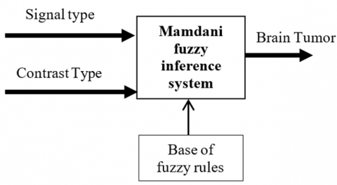
The proposed framework consists of three principal steps: segmentation, detection, and decision-making. Segmentation is performed using the FPSO algorithm, while detection is performed employing FCM. Decision-making is performed using a Mamdani-based fuzzy inference model. Figure 1 shows the phases of the proposed brain tumor segmentation technique using MRI images.
Figure 1. Proposed method diagram for brain tumor detection
3.1 Dataset
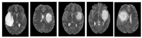
In this work, the objective is to generate a segmentation of the heterogeneous tumor region. To achieve this, two datasets were utilized, including the publicly available BRATS 2015 [31]. Furthermore, a private dataset was collected from the Civil Hospital of Setif, comprising 18 healthy and 18 unhealthy subjects. In the BRATS 2015 dataset, we specifically chose malignant tumors for segmentation and classification. A sample of the selected datasets is illustrated in Figure 2.
Figure 2. Brain tumor samples from the image dataset used in this study
3.2 Preprocessing
The preprocessing stage plays a significant role in enhancing the quality of the image that leads to attaining good effects in segmentation processes.
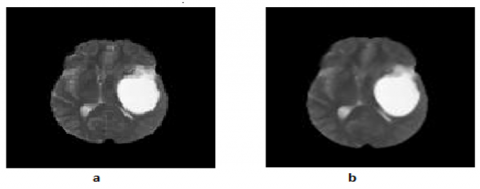
Figure 3. Preprocessing step: (a) Original MRI image and (b) Enhanced image after preprocessing
It consists of basic pre-processing techniques including adjusting image size, image normalization [32], image enhancement [31], image binarization and more [33]. Figure 3 shows the enhanced image after preprocessing
3.3 Fuzzy particle swarm optimization for segmentation
The segmentation process involves the method of partitioning an image into various homogeneous parts. Over the years, remarkable research progress in the field of brain tumor segmentation has been made. In this study, the FPSO is employed to segment the tumor zone from MRI brain. The selection of the FPSO is based on its simplicity, efficiency in segmentation applications, and its capability to handle information characterized by high-dimensional data [34].
The Particle Swarm Optimization (PSO) algorithm, developed in 1995, is an evolutionary computation technique inspired by natural swarm behavior that operates through iterative refinement [35]. In the PSO framework, a swarm consists of a set number of particles. In each iteration, these particles explore the N-dimensional problem space to evaluate and identify the global optimum. The velocity and position of each particle are updated according to the following equations:
$V(t+1)=w \cdot V(t)+c_1 \cdot \operatorname{rand}_1 \cdot(\operatorname{Pbest}(t)-X(t))+c_2 \cdot \operatorname{rand}_2 \cdot(\operatorname{Gbest}(t)-X(t))$ (1)
$X(t+1)=X(t)+V(t+1)$ (2)
where,
The PSO algorithm faces a common challenge where particles can be trapped in local minima during convergence, leading to suboptimal solutions. This issue affects its ability to find the global minimum [37].
An enhanced fuzzy particle swarm optimization (FPSO) method has been developed to effectively solve the Traveling Salesman Problem [30]. This FPSO approach has since been applied to tackle fuzzy clustering challenges in image analysis [36, 38]. In this framework, the position of each particle (denoted by X) corresponds to the fuzzy membership values {p1, p2, ... pN} of pixels to a set of cluster centers {C1, C2, ..., Cc}. The representation of X is as follows:
$X=\left[\begin{array}{ccc}\mu_{11} & \cdots & \mu_{1 c} \\ \vdots & \ddots & \vdots \\ \mu_{n 1} & \cdots & \mu_{n c}\end{array}\right]$ (3)
uij indicates the degree of membership of the i-th pixel to the j-th cluster, based on certain constraints. The particle position matrix is structured similarly to the fuzzy matrix in the FCM. The particles' velocities are represented by a matrix V with dimensions (N, c), where N and c correspond to the number of rows and columns, respectively. The position and velocity updates in this process are determined by Eqs. (4) and (5) [39].
$\begin{gathered}V(t+1)=w \otimes V(t) \oplus(c 1 r 1) \otimes p b e s t(t) \ominus \\ X(t)) \oplus(c 2 r 2) \otimes(g b e s t(t) \ominus X(t))\end{gathered}$ (4)
$X(t+1)=X(t) \oplus \mathrm{X}(\mathrm{t}+1)$ (5)
The novel matrix by normalized values is supposed as follows [40]:
$X_{\text {normal }}=\left[\begin{array}{ccc}\frac{\mu_{11}}{\sum_{j=1}^c \mu_{1 j}} & \cdots & \frac{\mu_{1 c}}{\sum_{j=1}^c \mu_{1 j}} \\ \vdots & \ddots & \vdots \\ \frac{\mu_{n 1}}{\sum_{j=1}^c \mu_{n j}} & \cdots & \frac{\mu_{n c}}{\sum_{j=1}^c \mu_{n j}}\end{array}\right]$ (6)
The FPSO method's fitness function evaluates generalized solutions, as defined in Eq. (7).
$f(X)=\frac{K}{J_m}$ (7)
where, K is a constant, $J_m$ is the objective function of FCM method. The pseudo algorithm FPSO for fuzzy clustering problem is concise by Algorithm 1.
|
Algorithm 1. FPSO |
|
Input: MRI Image.
4.1 Determine the cluster centers for each particle based on Eq. (11). 4.2 Evaluate the fitness function for each particle based on Eq. (7). 4.3 Update Pbest for each particle. 4.4 Update Gbest for the swarm. 4.5 Update V for each particle based on Eq. (4). 4.6 Update X for each particle based on Eq. (5). 4.7 If the stopping condition has not been met, repeat from step 4.1.
Output: Segmented image. |
3.4 Tumor detection using Fuzzy C-Means
The subsequent steps involve the separation of outlier data, as previously mentioned, with the segmented image aiming to highlight the tumor. A non-supervised Fuzzy C-Means algorithm approach is employed. This choice is because of its success in different domains, such as image analysis and medical diagnosis. Despite its low complexity and simple implementation, especially for large datasets [41], the FCM algorithm has proven to be fruitful for segmenting brain tumors. FCM is an unsupervised fuzzy clustering algorithm, classified as a constrained soft clustering algorithm. The approach is based on minimizing a quadratic criterion [40]. In the FCM algorithm, data point membership to clusters is based on their proximity to cluster centers, aiming to minimize an objective function related to the fuzzy membership set U and centroids V [41].
$J=\sum_{i=1}^C \sum_{j=1}^N\left(u_{i j}\right)^m d^2\left(x_j, c_i\right)$ (8)
With the following constraint:
$\begin{gathered}\forall j \in[1, N]: \sum_{i=1}^C u_{i j}=1 \\ \forall i \in[1, C], \forall j \in[1, N]: u_{i j} \in[0,1]\end{gathered}$ (9)
In these equations, uij represents the membership of pixel xj for the j-th cluster, where ci is the center of the i-th cluster. Additionally, d(xj, ci)2 is the Euclidean distance between xj and ci. The parameter m, which is greater than 1, directs the resulting partition’ fuzziness. The cluster centers and membership functions are updated using Eq. (10) and Eq. (11), respectively.
$u_{i j}=\left[\sum_{k=1}^c\left(\frac{d\left(x_j, b_i\right)}{d\left(x_j, b_k\right)}\right)^{\frac{2}{(m-1)}}\right]^{-1}$ (10)
$b_i=\frac{\sum_{k=1}^N\left(u_{i k}^m+t_{i k}^\lambda\right) x_k}{\sum_{k=1}^N\left(u_{i k}^m+t_{i k}^\lambda\right)}$ (11)
The FCM algorithm iteratively repeats two basic conditions till a solution is obtained. The final phase of segmentation is achieved by allocating data points to the cluster with the most significant membership value [42]. The steps of this algorithm are considered as follows:
|
Algorithm 2. FCM |
|
Input: A set of image segments segmented by FPSO. 1). Ensemble of values of the number of clusters c, the fuzziness degree m and the error epsilon (ε). 2). For i=1, 2, ..., c: Compute the center of the fuzzy cluster ci, using Eq. (11). 3). Update the fuzzy membership uij using Eq. (10). 4). If the value of J is smaller than the predefined threshold ε, stop; otherwise, return to step 2. Output: Tumor detection. |
3.5 Decision-making
At the final stage, the objective is to determine if a given pixel corresponds to a brain tumor or not. To accomplish this, a Mamdani model based fuzzy inference system has been implemented. The Mamdani Fuzzy Inference System offers a robust framework for integrating expert knowledge into the decision-making process. It is composed of rules structured as "IF... THEN..." statements, and the relationships between rules are expressed using "AND" and "OR" connectors [43]. In the proposed algorithm, a fuzzy inference system of the Mamdani type is employed based on expert knowledge to generate a knowledge base. This imparts the system with the ability to make decisions using the rules created in the knowledge base for the system's output. Brain tumors typically appear either hypointense in the T1-w sequence or hyperintense in T2-w and PD-w sequences [44]. The proposed Mamdani fuzzy inference system for the evaluation of brain tumor risk comprises two inputs: the contrast of MR Image and the signal of tumor detection. The system has one output that indicates the risk of brain tumor (refer to Figure 4).
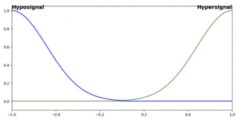
Depending on the characteristics of the input signal, two membership functions are defined: 'hyposignal' and 'hypersignal', as illustrated in Figure 5.
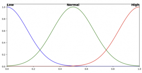
Based on the output for brain tumor classification, three Gaussian membership functions are utilized to represent the tumor states: high, normal, and low, as illustrated in Figure 6.
Figure 4. Diagram of the fuzzy inference system implemented in our approach
Figure 5. Membership functions for different signal types
Figure 6. Gaussian membership functions for brain tumor classification
The fuzzy rules set for MIN-MAX Mamdani fuzzy inference system are defined as follows:
In this work, we opted for the MIN-MAX Mamdani inference system. This model employs the connective operators AND and OR to implement min and max, respectively. We used Tanaka's proposed Center of Gravity strategy as the defuzzification method [45] for our case studies, which turns the fuzzy set produced by inference into a numerical value by taking into account all possible outputs.
3.6 Evaluation metrics
To assess the capability of the proposed brain tumor segmentation, three metrics are computed: sensitivity, Jaccard index, Accuracy and specificity. These metrics are defined by the following equations [46]:
Jaccard $=\frac{\mathrm{TP}}{\mathrm{TP}+\mathrm{FN}+\mathrm{FP}}$ (12)
Sensitivity $=\frac{\mathrm{TP}}{\mathrm{TP}+\mathrm{FN}}$ (13)
Specificity $=\frac{\mathrm{TN}}{\mathrm{TN}+\mathrm{FP}}$ (14)
Accuracy $=\frac{\mathrm{TP}+\mathrm{TN}}{\mathrm{TP}+\mathrm{TN}+\mathrm{FN}+\mathrm{FP}}$ (15)
where,
4.1 Brain tumor segmentation and detection using FPSO and FCM algorithms
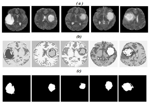
To assess the performance of each employed segmentation algorithm (FPSO, FCM, and FPSOFCM), corresponding to specific stages in the proposed pipeline for brain tumor segmentation, axial slices of the original MRI images are presented in Figure 7. The rows in the figure depict: (a) Original MRI images before segmentation, (b) Segmented images using the FPSO algorithm showing different tissues, and (c) Tumor extraction using the FCM algorithm in the final stage, where the number of clusters (c) was set to 2 and the fuzziness parameter (m) was set to 2.0 to ensure reproducibility.
The impact of each result on the proposed approach is assessed by analyzing the improvements in sensitivity, specificity, and Jaccard index performance metrics. Table 2 presents the quantitative results of these evaluations.
Table 2 presents the performance evaluation measures, including the Jaccard index, sensitivity, and specificity, for MRI brain tumor images using the proposed segmentation technique for brain tumor diagnosis. The results indicate that the proposed approach (FPSOFCM) is both effective and clinically acceptable. Specifically, the Jaccard index shows promising results, with an average of 0.85 across the datasets. The sensitivity values have an average of 0.92, while the specificity values show an average of 0.97.
Figure 7. Scheme of the complete brain tumor segmentation process: (a) Original MR images, (b) Segmented images using FPSO, and (c) Brain tumor detection using FCM
Table 2. Value of Jaccard, sensitivity and specificity for brain tumor segmentation
|
Performances (Tumors Detected) |
Jaccard |
Sensitivity |
Specificity |
|
Brain tumors |
0.85 |
0.92 |
0.97 |
4.2 Decision-making process
In this step, the output result aids in making decisions for brain tumor detection. The fuzzy inference system requires concepts characterized in membership functions, logical operations, and rule-based structures. The fuzzy inference process starts with fuzzification and ends with defuzzification. In our proposed Mamdani model based fuzzy inference, MIN and MAX operators are implemented. The potential brain tumors detected are progressed via this inference system that acts as an expert in determining whether a pixel belongs to the brain tumor cluster or not. Table 3 reports the achieved results considering the three MRI sequences.
Table 3. Defuzzification results on different MRI sequences.
|
MRI Sequences/ Tumors Detected |
T1-w (%) |
T2-w (%) |
PD-w (%) |
|
Brain Tumor |
53.53 |
88.17 |
77.04 |
Proactive identification and treatment of brain tumors are vital for successful management. Decision-making in clinical applications heavily relies on expertise. From Table 3, it is evident that the patient has a brain tumor. The tumors are identified in all sequences of MRI images with a high level of description.
4.3 Comparative study
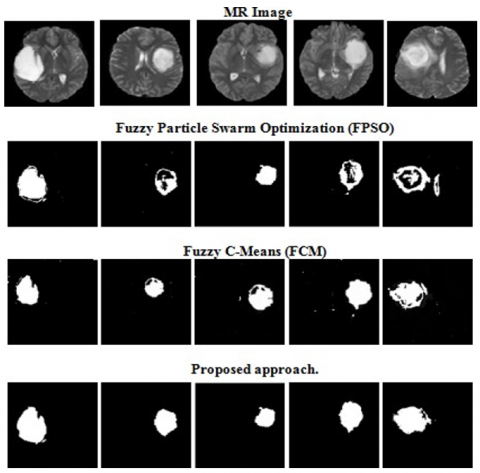
Here, we illustrate comparative study of the offered algorithm with the FPSO and FCM algorithms by employing an ensemble of MRI brain images for brain tumor detection. The segmentation results of a sample of selected datasets are illustrated in Figure 8.
Figure 8. Segmentation results obtained using various methods
In this proposed technique we got 99.71% accuracy that is good than the state of the art results obtained by the studies [22, 28, 25, 30] as shown in the Table 4.
Table 4. Comparison of segmentation performance
|
References |
Techniques |
Accuracy % |
|
[30] |
AMSOM+FKM |
99.8 |
|
[22] |
MARKER-BASED WATERSHED ALGORITHM + SVM |
93.29 |
|
[28] |
SLCA+UNet |
98.95 |
|
[25] |
SVM+CNN |
99.74 |
|
Proposed approach |
FPSO+FCM |
99.71 |
Detecting and identifying tumors from MRI images quickly, accurately, and reproducibly remains a challenging problem. Brain tumor segmentation has shown immense potential in identifying and examining tumors in medical imaging, and this trend is expected to persist in the future. Therefore, this paper presents a fully automated method for brain tumor detection. The combination of FCM algorithms and FPSO was utilized for brain tumor segmentation. Experimental results demonstrated that the segments extracted by the FPSOFCM algorithm from MRI images outperformed those obtained by classical methods (FPSO and FCM). Furthermore, in a comparative study with previous existing works, all three presented approaches achieved higher performances. In conclusion, the high accuracy of the proposed FPSOFCM method makes it a promising system for brain tumor diagnosis in a clinical setting based on MRI images. In future work, we plan to extend this system by incorporating other hybrid techniques, including combining FPSO/FCM with deep learning models such as Convolutional Neural Networks (CNNs) or Transformer-based architectures, to further enhance brain tumor detection performance. Additionally, the proposed method will be further analyzed for its applicability to other areas of biomedical image processing.
[1] Amin, J., Sharif, M., Yasmin, M., Fernandes, S.L. (2020). A distinctive approach in brain tumor detection and classification using MRI. Pattern Recognition Letters, 139: 118-127. https://doi.org/10.1016/j.patrec.2017.10.036
[2] Amin, J., Sharif, M., Gul, N., Yasmin, M., Shad, S.A. (2020). Brain tumor classification based on DWT fusion of MRI sequences using convolutional neural network. Pattern Recognition Letters, 129: 115-122. https://doi.org/10.1016/j.patrec.2019.11.016
[3] Bousselham, A., Bouattane, O., Youssfi, M., Raihani, A. (2019). Towards reinforced brain tumor segmentation on MRI images based on temperature changes on pathologic area. International Journal of Biomedical Imaging, 2019(1): 1758948. https://doi.org/10.1155/2019/1758948
[4] Rana, R., Bhadauria, H.S., Singh, A. (2013). Study of various methods for brain tumour segmentation from MRI images. International Journal of Emerging Technology and Advanced Engineering, 3(2): 338-342. https://api.semanticscholar.org/CorpusID:9226087.
[5] Batool, A., Byun, Y.C. (2024). Brain tumor detection with integrating traditional and computational intelligence approaches across diverse imaging modalities—Challenges and future directions. Computers in Biology and Medicine, 108412. https://doi.org/10.1016/j.compbiomed.2024.108412
[6] Lobo, M.D., Tavares, S.M., de Almeida, R.P.P., Garcia, M.B. (2025). Advancing precision in physical education and sports science: A review of medical imaging methods for assessing body composition. Global Innovations in Physical Education and Health, 293-326. https://doi.org/10.4018/979-8-3693-3952-7.ch011
[7] Hwang, J.H., Park, C.K., Kang, S.B., Choi, M.K., Lee, W.H. (2024). Deep learning super-resolution technique based on magnetic resonance imaging for application of image-guided diagnosis and surgery of trigeminal neuralgia. Life, 14(3): 355. https://doi.org/10.3390/life14030355
[8] Ben Rabeh, A., Benzarti, F., Amiri, H. (2017). Segmentation of brain MRI using active contour model. International Journal of Imaging Systems and Technology, 27(1): 3-11. https://doi.org/10.1002/ima.22205
[9] Hill, J.E., Nutter, B., Mitra, S. (2014). An information theoretic approach via IJM to segmenting MR images with MS lesions. In 2014 IEEE 27th International Symposium on Computer-Based Medical Systems, New York, NY, USA, pp. 189-192. https://doi.org/10.1109/CBMS.2014.130
[10] Gordillo, N., Montseny, E., Sobrevilla, P. (2013). State of the art survey on MRI brain tumor segmentation. Magnetic Resonance Imaging, 31(8): 1426-1438. https://doi.org/10.1016/j.mri.2013.05.002
[11] Wadhwa, A., Bhardwaj, A., Verma, V.S. (2019). A review on brain tumor segmentation of MRI images. Magnetic Resonance Imaging, 61: 247-259. https://doi.org/10.1016/j.mri.2019.05.043
[12] Tong, J., Zhao, Y., Zhang, P., Chen, L., Jiang, L. (2019). MRI brain tumor segmentation based on texture features and kernel sparse coding. Biomedical Signal Processing and Control, 47: 387-392. https://doi.org/10.1016/j.bspc.2018.06.001
[13] Alqazzaz, S., Sun, X., Yang, X., Nokes, L. (2019). Automated brain tumor segmentation on multi-modal MR image using SegNet. Computational Visual Media, 5: 209-219. https://doi.org/10.1007/s41095-019-0139-y
[14] Sharma, N., Aggarwal, L.M. (2010). Automated medical image segmentation techniques. Journal of Medical Physics, 35(1): 3-14. https://doi.org/10.4103/0971-6203.58777
[15] Bauer, S., Wiest, R., Nolte, L.P., Reyes, M. (2013). A survey of MRI-based medical image analysis for brain tumor studies. Physics in Medicine & Biology, 58(13): R97. https://doi.org/10.1088/0031-9155/58/13/R97
[16] Sharma, A., Kumar, S., Singh, S.N. (2019). Brain tumor segmentation using DE embedded OTSU method and neural network. Multidimensional Systems and Signal Processing, 30: 1263-1291. https://doi.org/10.1007/s11045-018-0603-3
[17] Işın, A., Direkoğlu, C., Şah, M. (2016). Review of MRI-based brain tumor image segmentation using deep learning methods. Procedia Computer Science, 102: 317-324. https://doi.org/10.1016/j.procs.2016.09.407
[18] Lefohn, A.E., Cates, J.E., Whitaker, R.T. (2003). Interactive, GPU-based level sets for 3D segmentation. In International Conference on Medical Image Computing and Computer-Assisted Intervention, Berlin, pp. 564-572. https://doi.org/10.1007/978-3-540-39899-8_70
[19] Zhu, Y., Yan, Z. (1997). Computerized tumor boundary detection using a Hopfield neural network. IEEE Transactions on Medical Imaging, 16(1): 55-67. https://doi.org/10.1109/42.552055
[20] Khotanlou, H., Colliot, O., Atif, J., Bloch, I. (2009). 3D brain tumor segmentation in MRI using fuzzy classification, symmetry analysis and spatially constrained deformable models. Fuzzy Sets and Systems, 160(10): 1457-1473. https://doi.org/10.1016/j.fss.2008.11.016
[21] Havaei, M., Larochelle, H., Poulin, P., Jodoin, P.M. (2016). Within-brain classification for brain tumor segmentation. International Journal of Computer Assisted Radiology and Surgery, 11: 777-788. https://doi.org/10.1007/s11548-015-1311-1
[22] Khan, M.A., Lali, I. U., Rehman, A., Ishaq, M., Sharif, M., Saba, T., Zahoor, S., Akram, T. (2019). Brain tumor detection and classification: A framework of marker‐based watershed algorithm and multilevel priority features selection. Microscopy Research and Technique, 82(6): 909-922. https://doi.org/10.1002/jemt.23238
[23] Sachdeva, J., Kumar, V., Gupta, I., Khandelwal, N., Ahuja, C.K. (2012). A dual neural network ensemble approach for multiclass brain tumor classification. International Journal for Numerical Methods in Biomedical Engineering, 28(11): 1107-1120. https://doi.org/10.1002/cnm.2481
[24] Iqbal, S., Khan, M.U.G., Saba, T., Rehman, A. (2018). Computer-assisted brain tumor type discrimination using magnetic resonance imaging features Biomedical Engineering Letters, 8(1): 5-28. https://doi.org/10.1007/s13534-017-0050-3
[25] Chattopadhyay, A., Maitra, M. (2022). MRI-based brain tumour image detection using CNN based deep learning method. Neuroscience Informatics, 2(4): 100060. https://doi.org/10.1016/j.neuri.2022.100060
[26] Vijay, V., Kavitha, A.R., Rebecca, S.R. (2016). Automated brain tumor segmentation and detection in MRI using enhanced Darwinian particle swarm optimization (EDPSO). Procedia Computer Science, 92: 475-480. https://doi.org/10.1016/j.procs.2016.07.370
[27] Sehgal, A., Goel, S., Mangipudi, P., Mehra, A., Tyagi, D. (2016). Automatic brain tumor segmentation and extraction in MR images. In 2016 Conference on Advances in Signal Processing (CASP), Pune, India, pp. 104-107. https://doi.org/10.1109/CASP.2016.7746146
[28] Tejashwini, P.S., Thriveni, J., Venugopal, K.R. (2025). A novel SLCA-UNet architecture for automatic MRI brain tumor segmentation. Biomedical Signal Processing and Control, 100: 107047. https://doi.org/10.1016/j.bspc.2024.107047
[29] Parveen, Singh, A. (2015). Detection of brain tumor in MRI images, using combination of fuzzy c-means and SVM. In 2015 2nd International Conference on Signal Processing and Integrated Networks (SPIN), Noida, India, pp. 98-102. https://doi.org/10.1109/SPIN.2015.7095308
[30] Dalal, S., Lilhore, U.K., Manoharan, P., Rani, U., Dahan, F., Hajjej, F., Keshta, I., Sharma, A., Simaiya, S., Raahemifar, K. (2023). An efficient brain tumor segmentation method based on adaptive moving self-organizing map and fuzzy k-mean clustering. Sensors, 23(18): 7816. https://doi.org/10.3390/s23187816
[31] Uka, A., Polisi, X., Barthes, J., Halili, A.N., Skuka, F., Vrana, N.E. (2020). Effect of preprocessing on performance of neural networks for microscopy image classification. In 2020 International Conference on Computing, Electronics & Communications Engineering (iCCECE), pp. 162-165. https://doi.org/10.1109/iCCECE49321.2020.9231071
[32] Pal, K.K., Sudeep, K.S. (2016). Preprocessing for image classification by convolutional neural networks. In 2016 IEEE International Conference on Recent Trends in Electronics, Information & Communication Technology (RTEICT), pp. 1778-1781. https://doi.org/10.1109/RTEICT.2016.7808140
[33] Pei, X., Zhao, Y.H., Chen, L., Guo, Q., Duan, Z., Pan, Y., Hou, H. (2023). Robustness of machine learning to color, size change, normalization, and image enhancement on micrograph datasets with large sample differences. Materials & Design, 232: 112086. https://doi.org/10.1016/j.matdes.2023.112086
[34] Gripsy, J.V., Divya, T. (2024). Lung cancer prediction using combination of oversampling with standard random forest algorithm. Smart Data Intelligence: Proceedings of ICSMDI 2024, 1. https://doi.org/10.1007/978-981-97-3191-6_1
[35] Kennedy, J., Eberhart, R. (1995). Particle swarm optimization. In Proceedings of ICNN'95-International Conference on Neural Networks. Perth, WA, 4: 1942-1948. http://dx.doi.org/10.1109/ICNN.1995.488968
[36] Izakian, H., Abraham, A. (2011). Fuzzy c-means and fuzzy swarm for fuzzy clustering problem. Expert Systems with Applications, 38(3): 1835-1838. https://doi.org/10.1016/j.eswa.2010.07.112
[37] Abdel-Kader, R.F. (2011). Fuzzy particle swarm optimization with simulated annealing and neighborhood information communication for solving TSP. International Journal of Advanced Computer Science and Applications, 2(5). https://doi.org/10.14569/IJACSA.2011.020503
[38] Zhang, H., Huang, S.L. (2025). Improved fuzzy c-means clustering algorithm based on fuzzy particle swarm optimization for solving data clustering problems. Mathematics and Computers in Simulation. https://doi.org/10.1016/j.matcom.2025.02.012
[39] Silva Filho, T.M., Pimentel, B.A., Souza, R.M., Oliveira, A.L. (2015). Hybrid methods for fuzzy clustering based on fuzzy c-means and improved particle swarm optimization. Expert Systems with Applications, 42(17-18): 6315-6328. https://doi.org/10.1016/j.eswa.2015.04.032
[40] Benaichouche, A.N., Oulhadj, H., Siarry, P. (2013). Improved spatial fuzzy c-means clustering for image segmentation using PSO initialization, Mahalanobis distance and post-Segmentation correction. Digital Signal Processing, 23(5): 1390-1400. https://doi.org/10.1016/j.dsp.2013.07.005
[41] Alam, M.S., Rahman, M.M., Hossain, M.A., Islam, M.K., Ahmed, K.M., Ahmed, K.T., Singh, B.C., Miah, M.S. (2019). Automatic human brain tumor detection in MRI image using template-based K means and improved fuzzy C means clustering algorithm. Big Data and Cognitive Computing, 3(2): 27. https://doi.org/10.3390/bdcc3020027
[42] Kouhi, A., Seyedarabi, H., Aghagolzadeh, A. (2020). Robust FCM clustering algorithm with combined spatial constraint and membership matrix local information for brain MRI segmentation. Expert Systems with Applications, 146: 113159. https://doi.org/10.1016/j.eswa.2019.113159
[43] Ghnemat, R. (2022). Hybrid framework for diabetic retinopathy stage measurement using convolutional neural network and a fuzzy rules inference system. Applied System Innovation, 5(5): 102. https://doi.org/10.3390/asi5050102
[44] Dou, W., Liao, Q., Ruan, S., Bloyet, D., Constans, J.M., Chen, Y. (2002). Automatic brain tumor extraction using fuzzy information fusion. In Second International Conference on Image and Graphics. SPIE, 4875: 604-609. https://doi.org/10.1117/12.477203
[45] Hernández-Julio, Y.F., Prieto-Guevara, M.J., Nieto-Bernal, W., Meriño-Fuentes, I., Guerrero-Avendaño, A. (2019). Framework for the development of data-driven Mamdani-type fuzzy clinical decision support systems. Diagnostics, 9(2): 52. https://doi.org/10.3390/diagnostics9020052
[46] Pereira, S., Pinto, A., Alves, V., Silva, C.A. (2016). Brain tumor segmentation using convolutional neural networks in MRI images. IEEE Transactions on Medical Imaging, 35(5): 1240-1251. https://doi.org/10.1109/TMI.2016.2538465