Ahmed J. Jabur![]() | Kian Raheem Qasim
| Kian Raheem Qasim![]() | Noor M. Naser*
| Noor M. Naser*![]()
© 2025 The authors. This article is published by IIETA and is licensed under the CC BY 4.0 license (http://creativecommons.org/licenses/by/4.0/).
OPEN ACCESS
This study proposed an AI system to diagnose cataracts using Neural Networks. The development of the system was carried out following the agile Scrum framework, including the development of artefacts defined by the Rational Unified Process software development process. 10 sprints were defined to complete the software development with the defined artefacts. Two methods were described to validate the model and the system in general. The precision, recall and F1-Score metrics were also determined to evaluate the performance and effectiveness of the model in diagnosing cataracts. Cataract classification yields 80% of positive cases found and 20% of positive cases not found. The proposed system uses fundus images to diagnose cataracts through the smartphone camera, obtain an automatic diagnosis and report, and assign an ophthalmologist to give the verdict. For the "Normal" classification, 93% of positive cases were found, while 7% were not. The average number of positive cases found is 86%, with 14% of positive cases not found. In all cases, we have a percentage of more than 80%. After obtaining the results using the established indicators, it is deduced that the preliminary diagnosis system can be considered support so that the doctor's activity is more optimal.
AI system, senile cataract, Convolutional Neural Network (CNN), Support Vector Machine (SVM), cataract diagnosis system and validation
Senile cataract is a public health problem that is becoming a determining agent in terms of the loss of visual capacity in older adults, caused by the deterioration of the transparency of the lens taking as risk factors that can complicate diabetes, hypertension, lack of prevention of UV rays, Glaucoma, etc. authors in studies [1-3] proposes using a smartphone to give the general public the ability to use a mobile application to perform a cataract examination without the assistance of a therapeutic professional. The authors aim to develop this software to detect cataracts in vivo using the camera automatically. The authors experimented with 50 people. 20 of these have cataracts, and 30 of these have normal eyes. Their evaluation results indicate that the system shows competitive retinal disease detection accuracy rates over 90%. The authors conclude that developing a mobile application for early detection is possible. The authors in studies [4, 5] propose a retinal fundus image segmentation algorithm based on a Fully Convolutional Network (FCN). This method is capable of automatic functional learning and identifies images of the vascular fundus of the cataract. In supervised methods, they find deficiencies inherent to the methods that prevent them from detecting small blood vessels. In unsupervised methods, they find that their segmentation is sensitive to noise in the images, and their performance is low [6]. A sparse learning algorithm for image classification was proposed inspired by doctors' manual classification process. Sparse learning is a representation learning method that aims to find a sparse representation of the input data in a linear combination of essential elements. An iris localisation algorithm that uses fuzzy logic-based edge estimation as input to the Hough transform algorithm proposed by Dixit et al. [7]. Pathak and Kumar [8] state that a robust, automated, and reliable iris localisation algorithm is paramount for automatic cataract detection. The authors use 2D-Gaussian filtering for image preprocessing to obtain a smoothed image [9]. In the study of Pratap and Kokil [10], the authors focus their research on automatic cataract classification from digital fundus images. Their methodology comprises four steps: image quality selection, preprocessing, feature extraction and classification. The authors use a module that filters good-quality background images for subsequent diagnosis, applying reference-free image quality evaluators such as naturalness (NIQE) and perception-based (PIQE). Raters when they have a low score, it means that the image is of good quality, and when they have a high score, it means that the image is of low quality. The authors only used the images that achieved a score less than or equal to 5 for the NIQE evaluator and less than or equal to 50 for the PIQE evaluator for the training and testing phase.
Artificial intelligence, specifically CNN, has increased significantly over the last five years, so it has been used for different purposes in biology, agriculture, and the field of health, as well as for detecting COVID-19 through X-ray images. This has motivated Li et al. [11] to recommend the adoption of artificial intelligence in ophthalmology, highlighting its practicality due to the use of a large number of images, both of the anterior and posterior segment of the eye, recognising at least six tests and two types of clinical data.
Based on the literature survey [12-18], the lack of a technological tool to support the diagnosis and classification of senile cataracts, the evaluation of the images obtained through ocular projection techniques carried out by specialised personnel, which is in this case, ophthalmologist implies effort and expertise, this being a manual analysis for the identification of particular visual characteristics that contribute to determining the presence or absence of the ocular disease describes how difficult it is to detect and classify senile cataract.
The development of this research implies clarifying the practicality of adopting the two convolutional neural networks with the highest performance in detection and diagnosis, according to the literature reviewed [12-18]. This allows us to know if the adoption of artificial intelligence in the detection of glaucoma or cataracts complies with the two fundamental aspects of the field of health, which are accuracy and the time it takes to make a diagnosis.
From the above, the absence of a support system for the diagnosis and classification of senile cataracts can be evident, as well as a long time [19], which is why the development of a preliminary diagnostic system for the classification of senile cataracts is proposed as a solution through convolutional neural networks, collaborating fully with the doctor, optimizing the diagnostic process and providing a reliable result with the minimum margin of error.
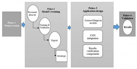
The research methodology for this work consists of 4 phases: Preprocessing, Model Training, Mobile Application Design and Validation (Figure 1).
2.1 Phases of the research methodology
· Phase 1 of preprocessing consists of a series of works on the original set of images to obtain better performance during model training and better results. First, we apply the augmentation technique to increase the volume of the image set. Next, we perform segmentation for the test and training images. Finally, a filtering technique is applied to the selected images.
· In Phase 2 of model training, the convolutional neural network model is designed based on what was analysed in the systematic literature review. Once the training and validation have been carried out, with the images already previously processed, the statistics of both steps are collected for subsequent analysis, and the model is exported to be used in the system.
· In Phase 3, the design of the mobile application is described in a series of steps and through a general diagram showing the components of the system, which are the cataract diagnosis component, which will use the model exported in Phase 2, the ophthalmic surveillance component, and the Results Verification Component. In addition, the methodology for software development is defined as the artefacts that will be generated and a description of the use of the system.
· In phase 4, the population, sample and unit of analysis are defined; the validation process for the model and the Cataract Diagnostic System is explained through a flow chart; the quality metrics, with their respective formulas, to evaluate the performance of the convolutional neural network model. The results of the quality metrics are also analysed, and the results are compared and averages are calculated. Finally, a discussion of the results commenting on the reasons or comments on the results obtained from the two case studies.
The development of this research implies clarifying the practicality of adopting the two convolutional neural networks with the highest performance in detection and diagnosis, according to the literature reviewed. This allows us to know if the adoption of artificial intelligence in the detection of glaucoma or cataracts complies with the two fundamental aspects of the field of health, which are accuracy and the time it takes to make a diagnosis. By relating the use of convolutional neural networks together with the needs of an ophthalmological centre for the diagnosis of eye diseases, it is possible to know if artificial intelligence has an adequate level of efficiency to use in the detection of cataracts and glaucoma and the practicality implicit in the time factor that could allow the replacement of labour, increase a diagnostic unit, etc.
Figure 1. Research methodology
3.1 Description of the convolutional neural network model
Data set: The dataset used for training (Retina Dataset, 2016) consists of 300 healthy fundus images and 100 cataract fundus images from the “Retina Blood Vessel Segmentation dataset”. A valuable resource for advancing medical image analysis and improving the diagnosis of retinal vascular diseases. For this project, only 100 images were extracted from each class. However, given the reduced data set, a data augmentation technique was used to train the model. The Python library KERAS generated more images from the data set. KERAS performs operations to rotate the image randomly from 0 to 40 degrees and flip the images horizontally and randomly. As a result, 400 images of each class were generated, finally having 1000 images as the training set. Finally, it is divided into two sets of images for the training and validation stages, with 800 and 200 images for each stage, respectively.
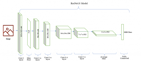
Architecture: The proposed model is based on the work of Xiong et al. [20], in which they propose using the Resnet-18 convolutional neural network (CNN) and the SVM Machine Learning algorithm as a classifier.
Figure 2 shows the architecture of Resnet-18, proposed by Kaur et al. [21], which is trained with the ImageNet data set containing 4, 217 image classes. According to Xiong et al. [20], this pre-trained model applies transfer learning in image recognition, allowing less effort when training with images from another domain.
Figure 2. The architecture of the proposed model and the architecture of the Resnet-18 convolutional neural network [21]
For the proposed model (Figure 2), the output of the layer before the Resnet-18 classification layer (512 features) is extracted to use as training data for the SVM. This model is implemented in the Python programming language with the Pytorch library. High-level features of cataract degrees were extracted using the ResNet18 network, and texture features were extracted using grey-level co-occurrence matrices (GLCM). Support vector machines (SVM) and a fully convolutional neural network (FCNN) were used to classify fundus images. There are many studies using deep learning in the literature [12-18].
3.2 Training steps
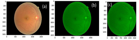
Preprocessing: The first step for training is to apply some transformations to the fundus image (Figure 3(a)) with the help of the Opencv Python library. The first transformation is to use a green filter. As the state-of-the-art filter shows, applying this filter can better visualise the veins (Figure 3(b)). The second transformation is to find the region of interest. Since the original image has a black background covering most of the image, only the eye features must be found. Figure 3(c) shows the region of interest for training.
Figure 3. (a) Original background image; (b) Background image with green filter; (c) Background image with green filter and with the region of interest
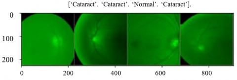
Finally, the transformations that the pre-trained Resnet-18 model requires are applied, such as modifying the image size to 256 and normalising it with the average parameters [0.485, 0.456, 0.406] and the standard deviation [0.229, 0.224, 0.225] for each color channel. Also, a random zoom to have different angles and focuses of the image. Figure 4 shows the final result.
Figure 4. Background image that will go to training
Resnet-18 model training: When training the model for each iteration, training and validation are performed. In this way, the parameters are adjusted and refined, avoiding overfitting, which occurs when the model only memorizes the training images. In each iteration, statistics of the loss and precision of the model are performed. A history is made so that it can be displayed in a graph in order to analyse the model's training performance later. As we already mentioned, 512 features are extracted from the image from a layer prior to the classifier. These features are chosen from the iteration that resulted with the best accuracy.
SVM classifier training: The classification Machine Learning algorithm known as SVM will be the one that learns the features extracted by the Resnet-18 CNN. This classifier has some parameters that must be chosen to obtain better results. These are the kernel, the coefficient for the kernel, and the regularization parameter.
For the kernel, the choice of job from which we are basing is "rbf". In order to find the value that provides the most outstanding performance to the algorithm, the GridSearch technique will be used. This technique makes a combination of all the parameters that are provided and looks for the one that has the best result (Figure 5).
Figure 5. Best parameters for the SVM classifier
In each combination, the parameters of the classifier will be adjusted. Once the parameters are found, they are saved using the Joblib library.
3.3 Statistics and export
Finally, with the support of a library, we extract the confusion matrix. These data will be used in the validation stage to calculate quality metrics and evaluate the performance of the proposed model. Then, we export the model in a ".pt" file that will be used in a system component.
3.4 Description of the cataract diagnostic system
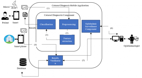
Figure 6 shows the general scheme of the system for diagnosing cataracts. The parts of the contribution are described below, which satisfies the need for portability as well as a remote monitoring scheme.
Figure 6. Proposed cataract diagnosis system
(1) Using a smartphone that integrates a micro lens as an accessory, an image of the patient's fundus is captured.
(2) The Cataract Diagnostic Component preprocesses the image by extracting the green channel of the image, which provides more luminance detail and the region of interest.
(3) The Cataract Diagnosis Component, with the pre-processed image, extracts the features necessary for the classification of cataract images using the pre-trained ResNet-18 model.
(4) The Cataract Diagnostic Component classifies the grade of the cataract using the SVM classifier. Once the classification is obtained, the diagnosis result is given to the smartphone.
(5) The medical entity, with access to the internet, may send the cataract results through the Results Verification Component. This result will be stored in a database to create a patient history.
(6) In turn, the Ophthalmologist is remotely notified of a new examination that resulted in a cataract. The Result Verification Component will allow you to review the image and verify the result. The Ophthalmologist is able to notify his verification made to the diagnosis.
(7) The Ophthalmic Surveillance Component allows the Ophthalmologist to monitor the patient with cataracts by having a history of images extracted from the database.
For this work, the good practices of the agile SCRUM Framework will be applied, which allows us to work in sprints or iterations and, in each iteration, have an increment of the product. Below is a brief summary of a sprint in SCRUM [22]:
⮚ Sprint Planning [22]
⮚ Daily SCRUM [22]
⮚ Sprint review meeting [22]
⮚ Sprint Retrospective Meeting [23]
In turn, artifacts defined by the RUP software development process will also be developed, as shown in Table 1.
Each artefact will be versioned and stored in a Version Control System repository.
Table 1. SCRUM
|
Sprint |
Artifact |
|
Sprint 1 |
Requirements Specification Document |
|
Sprint 2.3 |
System Analysis Document |
|
Design and Architecture Document |
|
|
Sprint 4 |
Testing Specification Document |
|
Sprint 5,6,7,8,9 |
Source Code |
|
Sprint 10 |
Specification Document and Test Results |
4.1 Requirements specification document
This artifact will specify the functional and non-functional requirements. These will be refined in iterations so that they are complete, consistent, verifiable correct, modifiable, traceable, understandable, unambiguous and prioritized.
Based on an evaluation of the cataract diagnosis process, the requirements for the present work are extracted. In total there were 12 functional requirements and 8 non-functional requirements. It is assigned an identifier that we can trace throughout the software development process. Among the most notable functional requirements would be the diagnosis of the cataract, the verification of the result by the Ophthalmologist, being alerted by a notification. The most notable non-functional requirements would be the use of a convolutional neural network for automatic diagnosis and to be implemented for a smartphone with the Android operating system. In addition, 6 prototypes of the main screens of the system were developed.
4.2 System analysis document
This artifact develops the system analysis based on the requirements. In this, we find the validation of the requirements, the use cases of the system the relationship with the requirements, and their specification. In the validation, a requirement vs requirements matrix was created to evaluate whether a requirement conflicts or is redundant with another requirement. After preparing it, no conflict or redundancy was found between them. However, when evaluating the qualities of completeness, consistency, correctness, and ambiguity, they had to be refined. 3 use cases of the system were identified, of which 2 actors interact with the system. The first actor is the Ophthalmologist and the second is a medical staff. This interaction is observed in Figure 7.
Figure 7. Diagram of use cases for the system
Each use case is specified with the following content:
· Use case name
· Description
· Actors
· Requirement reference
· Preconditions
· Primary scenario
· Secondary scenarios
· Postcondition
· Non-functional requirements
· Prototypes
· Validation (if applicable) (Table 2)
Table 2. Validations
|
Text Box |
Observation |
Lmin |
Lmax |
Characters |
Mask |
Observations |
|
Names |
Yes |
2 |
50 |
Letters |
Names |
Accept spaces |
|
Surnames |
Yes |
2 |
50 |
Letters |
Surnames |
Accept spaces |
|
Age |
Yes |
1 |
3 |
Numeric |
Age |
positive number |
|
Identification document |
Yes |
8 |
8 |
Numeric |
Identification document |
Can start with 0 |
|
City |
Yes |
2 |
50 |
Letters |
City |
Accept spaces |
|
country department |
Yes |
2 |
50 |
Letters |
country department |
They are default options |
|
Phone |
No |
0 |
9 |
Numeric |
Phone |
It does not accept scripts |
|
|
No |
0 |
100 |
Alphanumeric |
|
Does not accept spaces |
4.3 Design and architecture documen
In this artifact, the Design of the System is developed so that it performs the functional functions and supports the defined non-functional requirements. In this we find the context diagram, the system architecture, the component diagram, the class diagram, the package diagram, the state diagram, the deployment diagram, the entity-relationship diagram and the logic diagram. The Unified Modelling Language (UML) is applied for diagrams.
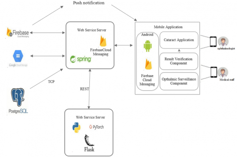
The system architecture (Figure 8) consists of 4 parts:
Figure 8. General system architecture
· Firebase Cloud Messaging (FCM): This is a software as a service that allows us to send push notifications. Once a verification request is created, the Web Service Server tells the FCM the sender and the recipient (The Ophthalmologist) so that they can receive a notification on the smartphone.
· Google Cloud Storage (GCS): This is a cloud service that allows us to store the analyzed fundus images and diagnosis.
· Web Service Server (SWS): There are two servers. A server implements a REST Web Service using the Flask microframework (Version 1.1.1) with the Python programming language (Version 3.6.5). This will have the responsibility of diagnosing the cataract using Neural Networks. The other server implements a REST Web Service using the Spring framework (Version 5.2) with the Java programming language (Version 8). This is responsible for receiving verification and diagnosis requests, storing them in the PostgreSQL database (Version 11.8) and consuming the FCM and GCS services. It also provides methods for user authentication.
· Mobile Application: Mobile application based on the Android operating system (Version 6.0) that implements the functionalities for Cataract Diagnosis, Verification of diagnosis results and Ophthalmic Surveillance as shown in Figure 9. This application will consume the services of the Web Service Server and will be attentive to push notifications from the FCM.
Figure 9. Screen 1 Cataract diagnosis & Screen2: Diagnostic report
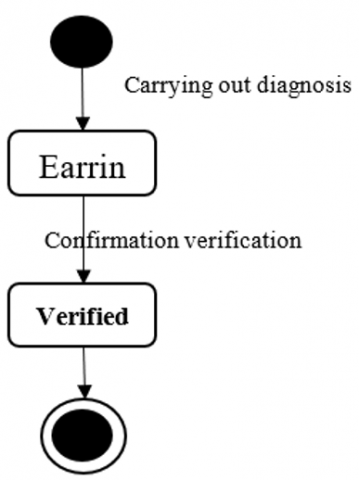
The State Diagram (Figure 10) is also relevant to mention since it is key to the process of verifying the diagnostic result by the Ophthalmologist. When the diagnosis is made, a report is generated with an initial status of “Pending” and when the Ophthalmologist gives his verdict, it moves to a final status of “Verified”.
Figure 10. State diagram
The Deployment Diagram (Figure 11) details how the deployment of each component or part of the defined architecture will be carried out. The Firebase Cloud Messaging service and Google Cloud Storage will be on the Google Cloud Platform. On Heroku, a cloud computing platform as a service, the Web Service and the PostgreSQL database will be deployed. Finally, on a smartphone, with Android operating system, the Cataract Diagnostic System will be installed.
Figure 11. Deployment diagram
4.4 Test plan document
This artefact details the plan for performing software testing. Its scope is to describe the test elements, the approach, deliverables, tasks, risks, etc.
The elements that will provide the basis for correct operation are:
· Requirements Specification Document
· Analysis document
· Design and Architecture Document
· User Manual
· Cataract Diagnostic Module
· Diagnostic Result Verification Module
· Diagnostic Results Consultation Module
A smartphone with an Android operating system with Snapdragon 835 eight-core 2.35GHz hardware and 4GB RAM is required to perform the test. In addition, we identified the risks that may arise during the testing stage, the contingencies that must be carried out if they occur and the impact of each risk.
4.5 Test specification document
This artefact details the software testing specifications for this system. Each test's preconditions, steps and expected results are described for each test.
For use case CU001 - Automatic cataract diagnosis, the following test specifications are defined:
· EP001 – Patient does not exist
· EP002 – Patient already registered
· EP003 – Diagnostic report
For use case CU002 – Diagnostic Result Verification, the following test specifications are defined:
· EP004 – Diagnosis to be verified
· EP005 – Verify cataract diagnosis (Reject)
· EP006 – Verify cataract diagnosis (Confirm)
For use case CU003 – Diagnostic Result Query, the following test specifications are defined:
· EP007 – Patient history
· EP008 – Diagnostic history
· EP009 – Patient has no history
· EP010 – View a diagnostic report
4.6 System use
Cataract Diagnosis: The ophthalmologist or a medical staff opens the Cataract Diagnosis System on their smartphone and displays what the camera is capturing. He places the micro lens on the smartphone camera and fixes it on the patient's eye. The fundus image of the patient's eye to be captured is displayed on the screen. The Ophthalmologist or a medical staff, having captured the image of the fundus of the eye, confirms the application so that the automatic diagnosis begins. Once the diagnosis is completed, the system displays a report with the patient's data, the captured fundus image, the result of the presence and degree of development of the cataract.
Diagnostic verification: The ophthalmologist or a medical staff member with a smartphone and an internet connection sends the diagnostic reports made that day to an ophthalmologist with more experience to verify the result. The most experienced ophthalmologist has been notified of a new verification request. She can view and examine the background image and then give a verdict on the result. Upon giving his ruling, the doctor can provide the corresponding follow-up to the patient to give the respective indications for the treatment.
Validation and Results: This chapter describes the population, the sample, and the unit of analysis selected to carry out this research work, considering a case study and the analysis of the results obtained.
4.6.1 Population and sample
The proposed system uses fundus images to diagnose cataracts (Retina Dataset, 2016). Table 3 presents the population, sample and unit of analysis defined for the validation of this research work.
Table 3. Population, sample and unit of analysis
|
Extent |
Description |
|
Population |
All fundus images of people who have difficulty seeing. |
|
Sample Unit of Analysis |
For this project, only 100 images were extracted from each class. However, given the reduced data set, a data augmentation technique was used to train the model. KERAS performs operations to rotate the image randomly from 0 to 40 degrees and flip the images horizontally and randomly. As a result, 400 images of each class were generated, finally having 1000 images as the training set. |
4.6.2 Model validation
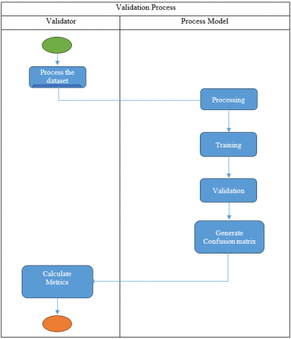
The model validation process has been carried out considering the flow presented in Figure 12.
The model validation process begins when the validator prepares the dataset, dividing it into a group of images for training and another group for validation (80% of the dataset and 20% of the dataset, respectively). The model then preprocesses the image. Once pre-processed, it is introduced to the model for training and validation. When finished with all the images in the dataset, the model generates a confusion matrix containing the number of positive and negative cases predicted. Finally, the validator, using the confusion matrix, calculates the metrics to evaluate the model's performance.
Figure 12. Validation process of the proposed model
4.7 System validation
The system validation process has been carried as the system validation process begins when validator 1 authenticates the system, accessing the system. Then, in the “Diagnosis” section, start registering the patient's personal data. Once registered, proceed with taking the fundus image. The next step is to assign validator 2 to verify the result of the diagnosis and send to diagnose. The system processes the image with the convolutional neural network and determines whether or not it has a cataract. At this point a notification appears on validator 2's phone while validator 1 views the result in a report. Validator 2 attends to the notification where it will display the result. This will be able to decide if the result is correct or not. Finally, an analysis of the results obtained during the validation process is carried out.
4.8 Quality metrics
The quality metrics will be based on the FURSP+, on the functionality attribute and applied to the diagnosis of cataract by the convolutional neural network. These three metrics are defined to evaluate functionality:
· Precision: Percentage of correct precisions.
· Recall: Ability of the classifier to find all positive cases.
· F1-Score: Percentage of positive predictions that were correct.
These metrics are calculated from the data obtained in the Confusion Matrix [12, 14].
· True Positive (TP): Number of positives that were correctly classified as positive by the model [14].
· True Negative (TN): Number of negatives that were correctly classified as negative by the model [14].
· False Positive (FP): Number of positives that were incorrectly classified as negative [14].
· False Negative (FP): Number of negatives that were incorrectly classified as positives [14].
From these values, the metrics are defined with the following formulas [12]:
· Accuracy: $\frac{T P}{T P+F P}$
· Recall: $\frac{T P}{T P+F N}$
· F1-Score: $2 * \frac{\text { Precision } * \text { recall }}{\text { Precision }+ \text { recal }}$
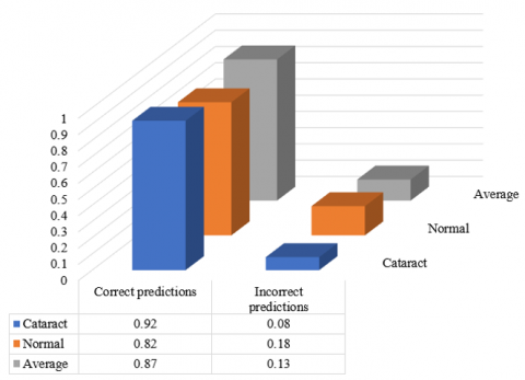
In Figure 13, we find the results of metric 1 (Precision) for the two classifications defined in this work. Applying formula (1) for the “Cataract” classification gives us 92% correct predictions and 8% incorrect predictions. For the “Normal” classification, we have 82% accurate predictions and 18% incorrect predictions. This gives us an average of 87% correct predictions and 13% incorrect predictions.
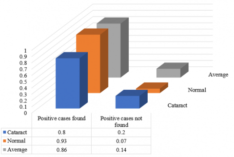
Figure 14 shows the results of metric 2 (Recall) for the two classifications. Applying formula (2) for the “Cataract” classification gives us 80% of positive cases found and 20% of positive cases not found. For the “Normal” classification, we have 93% of positive cases found and 7% of positive cases not found. As an average of positive cases found, we have 86% and 14% of positive cases not found.
In Figure 15 we observe the results of metric 3 (F1-Score) for the two classifications. Applying formula (3), for the "Cataract" classification, gives us 86% of correct positive predictions and 14% of incorrect optimistic predictions. For the "Normal" classification, we have 87% correct positive predictions and 13% incorrect positive predictions. This gives us an average of 87% correct positive predictions and 14% incorrect positive predictions.
Figure 13. Precision metric results
Figure 14. Results of the recall metric
Figure 15. Results of the F1-Score metric
4.9 Discussion
Metrics help us evaluate the performance and ability of the model to predict and identify the defined classifications. In Table 4, we have summarised the results, and we can jointly evaluate the averages. In all cases, we have a percentage greater than 80%. To improve this and equip the model, more images of both classifications are needed. In this way, we avoid overfitting, which only recognises the photos used during the training stage. Furthermore, different degrees of the cataract could be defined if more specimens were available.
Table 4. Summary table of the results
|
Metrics |
Classification |
Average |
|
Precision |
Correct predictions |
0.91 |
|
Incorrect predictions |
0.14 |
|
|
Recall |
Positive cases found |
0.9 |
|
Positive cases not found |
0.15 |
|
|
F1-Score |
Correct positive predictions |
0.9 |
|
Incorrect positive predictions |
0.15 |
The study is limited to the non-experimental, cross-sectional and quantitative methods used in this research. To develop the methodological proposal, the structure of the ResNet-18 and SVM networks was understood, and later improvements were implemented in the inputs and propagation cycles. This involves using the codes of two network structures replicated in other studies. Therefore, the contribution of this research lies in testing each network under different conditions, its subsequent comparative analysis to determine if there are significant differences, and finally, recommending, based on the results, if it is appropriate to develop software for detecting cataracts and glaucoma.
Adhering to the time parameter and confirming what was established by Cifuentes et al. [24], who state that the efficiency of RNCs lies in the ability to extract patterns from images, SVM is the only one that meets both efficiency criteria. Although diagnostic accuracy is an imminent priority in the healthcare field, time is also a crucial factor for the detection of certain diseases/abnormalities. Therefore, the adoption of this artificial intelligence in high-demand ophthalmology centers or in those where the specialist takes more than 62 minutes to diagnose cataracts could be assessed from the operational point of view.
Of the methodological proposals that used the same network structures, the following are contrasted: Li et al. [25] obtained the highest level of prediction using the VGG-19. However, the other algorithms delivered a result above 98% and the others above 99%. The results for diagnosing glaucoma are unsatisfactory since none of the algorithms achieved 91% accuracy. On the other hand, for cataract detection, a network based on the SVM structure did manage to exceed 92% accuracy; in addition, the performance, loss and execution time reinforce the claim that this model is more efficient than the ResNet-18.
Likewise, it is agreed with Perdomo-Charry et al. [26] that convolutional neural network models do not take advantage of all the information provided in ophthalmological data because they resort only to the assessment of the fundus of the eye when there are clinical data and diagnostic reports that can be crucial in the detection of cataract such as visual acuity [27], or glaucoma, such as tearing or the presence of megalocornea. It is suggested that the new methodological proposals acquire an integral character for developing multimodal systems.
The results obtained in the performance and time factors are conclusive in determining that SVM [28-30] is the most appropriate convolutional neural network [29] for detecting cataracts and that none of the proposed networks is appropriate for detecting glaucoma. Likewise, these results showed that during training the SVM network structure processes better the information contained in the ocular photographs. The convolutional layers affect the taking of information from ocular photographs, but the theory that the more training stages, the better predictions are obtained, is discarded.
5.1 Conclusion
One of the most important findings of this research was that the performance of convolutional neural networks is directly related to the indicators of loss and accuracy since, in cases where the networks demonstrated greater performance, they consequently obtained less data loss and greater accuracy in their predictions.
In this study, a System was proposed to diagnose cataracts using Neural Networks. The objective of the present study was to reduce the costs incurred in this evaluation and increase its access in places where there is no presence of ophthalmologists or who do not have sufficient experience, which has been achieved through the development of cataract diagnostic components, ophthalmic surveillance and verification of results. The proposed solution allows the technician to capture the fundus image of the patient through the smartphone camera, obtain an automatic diagnosis and report, assign an ophthalmologist to give the verdict of the result, and review the patient's history. diagnoses made. Additionally, it allows the Ophthalmologist to be notified of any result verification assigned to them and give their verdict.
5.2 Future work
The proposed System could be improved by including the degree of the cataract in the diagnosis to more accurately provide the corresponding treatment to the patient. This is achieved with more specimens of fundus images. Also, improve the ophthalmic surveillance component with functionality so that communication is established between the Ophthalmologist and the technician or patient about the treatment that should be carried out.
[1] Mencucci, R., Stefanini, S., Favuzza, E., Cennamo, M., De Vitto, C., Mossello, E. (2023). Beyond vision: Cataract and health status in old age, a narrative review. Frontiers in Medicine, 10: 1110383. https://doi.org/10.3389/fmed.2023.1110383
[2] Aruljyothi, L., Janakiraman, A., Malligarjun, B., Babu, B.M. (2021). Smartphone applications in ophthalmology: A quantitative analysis. Indian Journal of Ophthalmology, 69(3): 548-553. https://doi.org/10.4103/ijo.IJO_1480_20
[3] Rana, J., Galib, S.M. (2017). Cataract detection using smartphone. In 2017 3rd International Conference on Electrical Information and Communication Technology (EICT), Khulna, Bangladesh, pp. 1-4. https://doi.org/10.1109/EICT.2017.8275136
[4] Nagino, K., Sung, J., Midorikawa-Inomata, A., Eguchi, A., Fujimoto, K., Okumura, Y., Inomata, T. (2023). Clinical utility of smartphone applications in ophthalmology: A systematic review. Ophthalmology Science, 4(1): 100342. https://doi.org/10.1016/j.xops.2023.100342
[5] Wan Zaki, W.M.D., Abdul Mutalib, H., Ramlan, L.A., Hussain, A., Mustapha, A. (2022). Towards a connected mobile cataract screening system: A future approach. Journal of Imaging, 8(2): 41. https://doi.org/10.3390/jimaging8020041
[6] Cheng, J. (2018). Sparse range-constrained learning and its application for medical image grading. IEEE Transactions on Medical Imaging, 37(12): 2729-2738. https://doi.org/10.1109/TMI.2018.2851607
[7] Dixit, A., Pathak, S., Raj, R., Naveen, C., Satpute, V.R. (2018). An efficient fuzzy based edge estimation for iris localization and pupil detection in human eye for automated cataract detection system. In 2018 9th International Conference on Computing, Communication and Networking Technologies (ICCCNT), Bengaluru, India, pp. 1-6. https://doi.org/10.1109/ICCCNT.2018.8493740
[8] Pathak, S., Kumar, B. (2016). A robust automated cataract detection algorithm using diagnostic opinion based parameter thresholding for telemedicine application. Electronics, 5(3): 57. https://doi.org/10.3390/electronics5030057
[9] Hsiao, P.Y., Chou, S.S., Huang, F.C. (2007). Generic 2-D gaussian smoothing filter for noisy image processing. In TENCON 2007-2007 IEEE Region 10 Conference, Taipei, Taiwan, pp. 1-4. https://doi.org/10.1109/TENCON.2007.4428941
[10] Pratap, T., Kokil, P. (2019). Computer-aided diagnosis of cataract using deep transfer learning. Biomedical Signal Processing and Control, 53: 101533. https://doi.org/10.1016/j.bspc.2019.04.010
[11] Li, Z., Wang, L., Wu, X., Jiang, J., Qiang, W., Xie, H., Zhou, H., Wu, S., Shao, Y., Chen, W. (2023). Artificial intelligence in ophthalmology: The path to the real-world clinic. Cell Reports Medicine, 4(7): 101095. https://doi.org/10.1016/j.xcrm.2023.101095
[12] Simanjuntak, R., Fu’adah, Y., Magdalena, R., Saidah, S., Wiratama, A., Ubaidah, I. (2022). Cataract classification based on fundus images using convolutional neural network. JOIV: International Journal on Informatics Visualization, 6(1): 33. https://doi.org/10.30630/joiv.6.1.856
[13] Ganokratanaa, T., Ketcham, M., Pramkeaw, P. (2023). Advancements in cataract detection: The systematic development of LeNet-convolutional neural network models. Journal of Imaging, 9(10): 197. https://doi.org/10.3390/jimaging9100197
[14] Elloumi, Y. (2022). Cataract grading method based on deep convolutional neural networks and stacking ensemble learning. International Journal of Imaging Systems and Technology, 32(3): 798-814. https://doi.org/10.1002/ima.22722
[15] Yadav, S., Yadav, J.K.P.S. (2023). Automatic cataract severity detection and grading using deep learning. Journal of Sensors, 2023: 1-17. https://doi.org/10.1155/2023/2973836
[16] Xu, X., Li, J., Guan, Y., Zhao, L., Zhao, Q., Zhang, L., Li, L. (2021). GLA-Net: A global-local attention network for automatic cataract classification. Journal of Biomedical Informatics, 124: 103939. https://doi.org/10.1016/j.jbi.2021.103939
[17] Zhang, X.Q., Hu, Y., Xiao, Z.J., Fang, J.S., Higashita, R., Liu, J. (2022). Machine learning for cataract classification/grading on ophthalmic imaging modalities: A survey. Machine Intelligence Research, 19(3): 184-208. https://doi.org/10.1007/s11633-022-1329-0
[18] Pratap, T., Kokil, P. (2021). Deep neural network based robust computer-aided cataract diagnosis system using fundus retinal images. Biomedical Signal Processing and Control, 70: 102985. https://doi.org/10.1016/j.bspc.2021.102985
[19] Nizami, A.A., Gurnani, B., Gulani, A.C. (2024). Cataract (Nursing). In: StatPearls [Internet]. Treasure Island (FL): StatPearls Publishing. https://www.ncbi.nlm.nih.gov/books/NBK568765/.
[20] Xiong, Y., He, Z., Niu, K., Zhang, H., Song, H. (2018). Automatic cataract classification based on multi-feature fusion and SVM. In 2018 IEEE 4th International Conference on Computer and Communications (ICCC), Chengdu, China, pp. 1557-1561. https://doi.org/10.1109/CompComm.2018.8780617
[21] Kaur, G., Sharma, N., Chauhan, R., Kukreti, S., Gupta, R. (2023). Eye disease classification using ResNet-18 deep learning architecture. In 2023 2nd International Conference on Futuristic Technologies (INCOFT), Belagavi, Karnataka, India, pp. 1-5. https://doi.org/10.1109/INCOFT60753.2023.10425690
[22] Jiménez, V., Afonso, P., Fernandes, G. (2020). Using agile project management in the design and implementation of activity-based costing systems. Sustainability, 12(24): 10352. https://doi.org/10.3390/su122410352
[23] Lee, W.T., Chen, C.H. (2023). Agile software development and reuse approach with scrum and software product line engineering. Electronics, 12(15): 3291. https://doi.org/10.3390/electronics12153291
[24] Cifuentes, A., Mendoza, E., Lizcano, M., Santrich, A., Moreno-Trillos, S. (2019). Development of a convolutional neural network to recognize patterns in images. Research and Development in ICT, 10(2): 7-17.
[25] Li, S., Zhang, H., Xu, F. (2023). Intelligent detection method for wildlife based on deep learning. Sensors, 23(24): 969.https://doi.org/10.3390/s23249669
[26] Perdomo-Charry, O.J., Pérez-Pérez, A.D., de-la-Pava-Rodríguez, M., Ríos-Calixto, H.A., Arias-Vanegas, V.A., Lara-Ramírez, J.S., González-Osorio, F.A. (2020). SOPHIA: System for ophthalmic image acquisition, transmission, and intelligent analysis. Revista Facultad de Ingeniería, 29(54): e11769. https://doi.org/10.19053/01211129.v29.n54.2020.11769
[27] Zhao, J., Liu, L.P., Cheng, H.H., Li, J.B., Han, X.T., Liu, Y., Wu, M.X. (2020). Accuracy of eight intraocular lens power calculation formulas for segmented multifocal intraocular lens. International Journal of Ophthalmology, 13(9): 1378-1384. https://doi.org/10.18240/ijo.2020.09.07
[28] Iourzikene, Z., Gougam, F., Benazzouz, D. (2024). Performance evaluation of feature extraction and SVM for brain tumor detection using MRI images. Traitement du Signal, 41(4): 1967-1979. https://doi.org/10.18280/ts.410426
[29] Omar, A., Mohamed, F., Mohammed, M., Fouad, B. (2021). Discrete event systems fault’s diagnosis and prognosis using feed-forward neural networks. Journal Européen des Systèmes Automatisés, 54(6): 853-863. https://doi.org/10.18280/jesa.540607
[30] Sajja, V.R., Kalluri, H.K. (2020). Classification of brain tumors using convolutional neural network over various SVM methods. Ingénierie des Systèmes d’Information, 25(4): 489-495. https://doi.org/10.18280/isi.250412