Ankita Patra | Santi Kumari Behera | Nalini Kanta Barpanda | Prabira Kumar Sethy*
© 2022 IIETA. This article is published by IIETA and is licensed under the CC BY 4.0 license (http://creativecommons.org/licenses/by/4.0/).
OPEN ACCESS
Image-based features of breast cancers have an important role in clinical prognostics, such as grading breast invasive carcinoma (BIC). Magnification is useful for investigating poorly defined abnormal tissue present in mammogram images. But, the disadvantage of viewing at a higher magnification is that less of the slide can be viewed. So, the question raised on which scale of magnification is better for grading BIC. In this study, the four scales of magnification such as 4x, 10x, 20x, and 40x are considered to evaluate their contribution towards BIC grading in deep learning, transfer learning, and the traditional machine learning approach. Here, 13 CNN models are considered for the transfer learning approach. In the deep learning approach, the deep feature of 13 CNN models with three classifiers is considered. Further, the handcrafted feature such as LBP, HOG, and GLCM with three classifiers like SVM, KNN, and Naïve Bayes are evaluated for grading of BIC. Finally, the 40x scale of magnification performed better in all classification models.
breast invasive carcinoma, microscopy magnification, breast cancer, deep learning
Breast cancer is one of the leading causes of death among women globally. As per World Health Organization (WHO), breast cancer registers 2.09 million cases with 6,27000 deaths globally in 2019 [1]. Among different types of cancers, it accounts for approximately 13% of breast cancer cases diagnosed among women globally, clearly depicting its fatal nature. Screening mammograms are the preferred technique for detecting abnormalities [2, 3], but at the same time, determining the stages of breast cancer is necessary for its proper diagnosis.
Cancer on breasts is made up of fatty tissue. They contain small chambers called lobules where breast milk is made; the milk travels through tiny ducts to reach the nipples, where lymph nodes are located inside your breasts and under your arms. Cancer that develops inside the milk ducts is called ductal carcinoma (DC). When DC is spreading into surrounding tissue, it is called invasive ductal carcinoma (IDC). A breast biopsy is a test that removes a few cells or a small amount of tissue from the suspicious area. Then, the pathologist analyses the breast biopsy tissue by looking at the cells under a microscope. Breast cancers are classified based on how they behave; this includes non-invasive cancer that has not spread outside the tissue, whereas invasive cancer has spread and invaded healthy tissues. Common types of breast cancer are invasive ductal carcinoma (IDC), and it spread from the area where it started to other areas of the body. Cancers are graded 1 through 3, with 1 being the most similar to normal healthy cells and 3 being the most different from normal cells, and the most aggressive breast cancer. Cancer treatment not only depends on its type but also depends on its stages/ grades. The primary means of breast cancer diagnosis involves manual microscopic examination of stained tissue by a pathologist [4]. Magnification, which is considered to be a relatively high "dose cost" mammographic technique, is a complementary examination performed on women exhibiting breast complaints or abnormalities [5]. For decades, magnification mammographic images of selected breast regions have been considered the most effective diagnostic tool for enhancing the visibility of subtle suspicious breast lesions and microcalcifications, thus providing improved diagnostic sensitivity and specificity [5]. The enhancement of visibility in magnification views is attributed to the increased contrast to noise ratio (CNR) caused by the increased fluence per irradiated area [6]. Again, the slide visibility is different from the scale of magnification. At 40x magnification, the slide visibility is 5mm. At 400x magnification, the slide visibility is 0.45mm, or 450 microns. At 1000x magnification, we will be able to see 0.180mm, or 180 microns. So, the question arises which scale of magnification is better for diagnosis of BIC. This article examines the four scales of microscopy magnified histopathological images in transfer learning, deep learning, and traditional machine learning approaches towards grading breast invasive ductal carcinoma.
The remaining article is organized as follows. First, section 2 describes the background of this research. Then, the material and methodology are presented in section 3. Next, the findings are recorded in section 4 with its proper description. Finally, the article is concluded in section 5.
There has been a lot of research over the last couple of decades into creating a CAD system for early diagnosis of breast cancer. Although, there are still problems and obstacles to overcome in the current process. For instance, the method of staining depends on the cellular component being examined, as different approaches are used to highlight different aspects of the cell's architecture. The H&E staining method in particular has been studied, and a variety of techniques are placed to use. Variability in appearance in H & E stained breast cancer histopathology images is a significant problem for a number of reasons, including tissue and staining preparation, slide digitalization, and the heterogeneity of cancer itself. Malignant and benign cancers are currently classified using one of two methods, each with its own set of advantages and disadvantages that may be found in the literature. Either the morphology of the nuclei and the cellular mitotic structure are specifically segmented to aid in the classification, or the whole image is used in a global manner. A substantial amount of research on breast mass, microcalcification detection, and classification can be found in the literature [7-16]. However, masses are more challenging to detect than microcalcifications because the mass features may be concealed or similar to normal breast parenchyma. Thus, the detection of masses is still an open challenge in breast cancer detection. Apart from mass detection, mammograms' classification and grading of different breast cancers are also important research topics [11]. The new developments must cope and overcome the challenges that existing algorithms exhibit by improving the performance.
The applications of DL have grown tremendously in various fields such as image classification [17], natural language processing [18], gaming [19]; and, in particular, it has become very popular in the medical imaging community for detection and diagnosis of diseases such as skin cancer [20, 21], brain tumor detection [22], and segmentation [23].
Furthermore, it is also important to evaluate the contribution of different scales of micro-magnified mammogram images with varying classification techniques, including machine learning, transfer learning, and deep learning.
Magnification mammography has been required in diagnostic mammography units [24]. The radiology personnel can collect different scales of magnified mammogram images such as 4x, 10x,20x, and 40x. This paper seeks to evaluate the scale of mammogram images over the other for grading of IDC. There is very limited work has been published related to micro magnification for breast cancer diagnosis. Kim et al. [25] compared between zooming method (62.0) of digital mammography vs. digital magnification view (61.8) in full-field digital mammography for the diagnosis of microcalcifications. They found that magnified images were significantly better than ZOOM images in terms of visual imaging quality. Øynes et al. [26] provides a systematic review on magnification and zoom techniques for detecting and diagnosing mammography microcalcifications. Sandor and Nott [27] studied the effect of radiographic magnification on image contrast on blood vessels. Concerning issues associated with magnification, some existing methods have investigated various categorization approaches. Results for models that account for magnification factors are presented by Spanhol et al. [28, 29]. It appears, however, that a single magnification model may not be able to process images at multiple magnifications, and that separate classifiers are needed for each. In addition, relying on a single magnification level to make a conclusion when there is substantial diversity in patient scores is risky. Again, with increasing the scale of magnification, the contrast of blood vessel images rapidly decreases. In this consequence, no doubt the magnification is a good approach towards diagnosing IDC from mammogram images. Still, the question arises on which scale is better, as with increasing the magnification scale, the contrast decreases, and blurs on images are increased. There are many works reported for breast cancer diagnosis based on concatenation of deep feature and handcrafted feature [30], bi-layer fusion [31] and multi-modal fusion [32].
The dataset is collected from Kaggle repository [33]. The dataset consists of histopathological microscopy images of 922 related to 124 patients with IDC introduced. For this work, we have selected 50 specimens from each three grades of IDC. Again, the dataset has four magnification levels of microscopy images. The distribution of microscopy images used is illustrated in Table 1.
Table 1. Details of microscopy images used for Grading of IDC
|
|
4x |
10x |
20x |
40x |
|
Grade 1 |
40 |
40 |
40 |
40 |
|
Grade 2 |
40 |
40 |
40 |
40 |
|
Grade 3 |
40 |
40 |
40 |
40 |
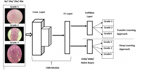
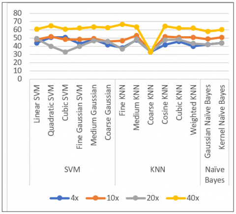
Figure 1. Evaluation of micro magnification effect for grading of breast invasive carcinoma in transfer learning and deep learning approach
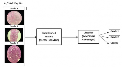
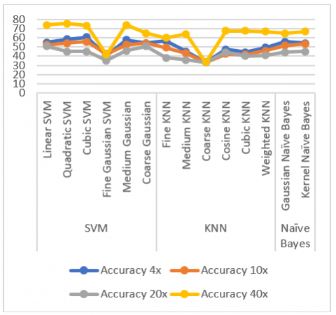
Figure 2. Evaluation of micro magnification effect for grading of breast invasive carcinoma in machine learning approach
The effect of micro magnification for grading IDC is evaluated in transfer learning, deep learning and transfer learning approach. The transfer learning and deep learning approach is illustrated in Figure 1. In both approaches 13 CNN models such as Alexnet, vgg16, vgg19, googlenet, resnet18, resnet50, resnet101, xception, inceptionv3, inceptionresnetv2, densenet201, mobilenetv2 and shufflenet are considered. In transfer learning approach, the last layer is refashioned to classify the three grades if IDC microscopy images. In deep learning approach, the deep features of 13 CNN models are fed to the classifiers to classify the three grades of IDC microscopy images. Again, in machine learning approaches hand-crafted features such as GLCM, HOG and LBP are taken into consideration. The classifiers considered in deep learning and machine learning approaches are SVM, KNN and Naïve bayes with their different paradigms. The machine learning approach is illustrated in Figure 2.
This part of the work presents the experimental results and discussions. This work is executed using, “Acer Predator Helios 300 Core i5 8th Gen - (8 GB/1 TB HDD/128 GB SSD/Windows 10 Home/4 GB Graphics) and equipped with NVIDIA GeForce GTX 1050Ti in MATLAB 2020a”. The effect of micro magnification for grading of IDC using microscopy images is evaluated in transfer learning, deep learning and machine learning approaches in terms of accuracy, TPR and PPV. “The hyper parameters used in deep learning and in transfer learning approaches are: solver type: stochastic gradient descent, initial learning rate is 0.001, learning rate policy: Step (decreases by a factor of 10 every 50/5 epochs), momentum: 0.9, drop out is 0.2, Number of Epochs is 50 and minibatch size:64. The adaptive learning rate is good compared to fixed learning rate. As an adaptive algorithm usually converge much faster than simple back-propagation with a poorly chosen fixed learning rate [34]”.
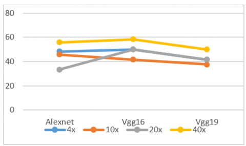
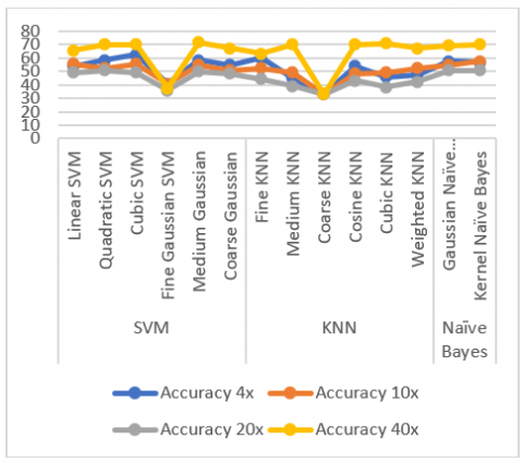
The effect of micro magnification in different approaches are evaluated and recorded. Figure 3 depicts the findings of transfer learning approach. The findings of deep learning approaches, i.e., Alexnet, vgg16 and vgg19 are noted from Figure 4, Figure 5 and Figure 6 respectively. The machine learning approach with GLCM, LBP and HOG features are recorded in Figure 7, Figure 8 and Figure 9 respectively. From all those results, it is implied that, the magnification level 40x is performed well than 4x, 10x and 20x for grading of IDC.
Figure 3. Performance comparison of accuracy in transfer learning approach for grading of IDC
Figure 4. Performance comparison of accuracy in deep learning approach in Alexnet
Figure 5. Performance comparison of accuracy in deep learning approach in VGG16
Figure 6. Performance comparison of accuracy in deep learning approach in VGG19
Figure 7. Performance comparison of accuracy in machine learning approach using GLCM feature
Figure 8. Performance comparison of accuracy in machine learning approach using LBP Feature
Figure 9. Performance comparison of accuracy in machine learning approach using HOG feature
Breast cancer is a fatal and most common cancerous diseases among women. To diagnose these diseases not only detection of abnormal tissue &/ or classification of its types but also very important to grade as per its severity. The radiologists are collected the microscopy images with different magnification levels. For computer aid diagnosis, it is better to know, the beneficence of magnification levels towards grading of IDC. The evaluation of micro magnification effect towards grading of IDC in transfer learning, deep learning and machine learning approaches resulted that, the magnification level 40x is better compared to 4x, 10x and 20x.
A special thanks to Prof. Sanjiv Mittal, Hon’ble Vice-Chancellor of Sambalpuri University for providing fund though SRIC, Sambalpur University and encourage to pursue this research.
[1] Siegel, R.L., Miller, K.D., Jemal, A. (2019). CA: a cancer journal for clinicians. Cancer Statistics, 69(1): 7-34. https://doi.org/10.3322/caac.21551
[2] Pisano, E.D., Gatsonis, C., Hendrick, E., et al. (2005). Diagnostic performance of digital versus film mammography for breast-cancer screening. New England Journal of Medicine, 353(17): 1773-1783. https://doi.org/10.1056/NEJMoa052911
[3] Yang, W.T., Lai, C.J., Whitman, G.J., Murphy, W.A., Dryden, M.J., Kushwaha, A.C., Sahin, A.A., Johnston, D., Dempsey, P.J., Shaw, C.C. (2006). Comparison of full-field digital mammography and screen-film mammography for detection and characterization of simulated small masses. AJR. American Journal of Roentgenology, 187(6): W576. https://doi.org/10.2214/AJR.05.0126. 17114508
[4] Majeed, H., Nguyen, T.H., Kandel, M.E., Kajdacsy-Balla, A., Popescu, G. (2018). Label-free quantitative evaluation of breast tissue using Spatial Light Interference Microscopy (SLIM). Scientific Reports, 8(1): 1-9. https://doi.org/10.1038/s41598-018-25261-7
[5] Koutalonis, M., Delis, H., Pascoal, A., Spyrou, G., Costaridou, L., Panayiotakis, G. (2010). Can electronic zoom replace magnification in mammography? A comparative Monte Carlo study. The British Journal of Radiology, 83(991): 569-577. https://doi.org/10.1038/10.1259/bjr/21753020
[6] Koutalonis, M., Delis, H., Spyrou, G., Costaridou, L., Tzanakos, G., Panayiotakis, G. (2007). Contrast-to-noise ratio in magnification mammography: A Monte Carlo study. Physics in Medicine & Biology, 52(11): 3185. https://doi.org/10.1088/0031-9155/52/11/017
[7] Magna, G., Casti, P., Jayaraman, S.V., Salmeri, M., Mencattini, A., Martinelli, E., Di Natale, C. (2016). Identification of mammography anomalies for breast cancer detection by an ensemble of classification models based on artificial immune system. Knowledge-Based Systems, 101: 60-70. https://doi.org/10.1016/j.knosys.2016.02.019
[8] Wang, H., Zheng, B., Yoon, S.W., Ko, H.S. (2018). A support vector machine-based ensemble algorithm for breast cancer diagnosis. European Journal of Operational Research, 267(2): 687-699. https://doi.org/10.1016/j.ejor.2017.12.001
[9] Sert, E., Ertekin, S., Halici, U. (2017). Ensemble of convolutional neural networks for classification of breast microcalcification from mammograms. In 2017 39th Annual International Conference of the IEEE Engineering in Medicine and Biology Society (EMBC), pp. 689-692. https://doi.org/10.1109/EMBC.2017.8036918
[10] Eltoukhy, M.M., Gardezi, S.J.S., Faye, I. (2014). A method to reduce curvelet coefficients for mammogram classification. In 2014 IEEE Region 10 Symposium, pp. 663-666. https://doi.org/10.1109/TENCONSpring.2014.6863116
[11] Gardezi, S.J.S., Faye, I., Eltoukhy, M.M. (2014). Analysis of mammogram images based on texture features of curvelet Sub-bands. In Fifth International Conference on Graphic and Image Processing (ICGIP 2013), 9069: 421-426. https://doi.org/10.1117/12.2054183
[12] Ramirez-Villegas, J.F., Ramirez-Moreno, D.F. (2012). Wavelet packet energy, Tsallis entropy and statistical parameterization for support vector-based and neural-based classification of mammographic regions. Neurocomputing, 77(1): 82-100. https://doi.org/10.1016/j.neucom.2011.08.015
[13] Wajid, S.K., Hussain, A. (2015). Local energy-based shape histogram feature extraction technique for breast cancer diagnosis. Expert Systems with Applications, 42(20): 6990-6999.
[14] Zhang, X., Homma, N., Goto, S., et al. (2013). A hybrid image filtering method for computer-aided detection of microcalcification clusters in mammograms. Journal of Medical Engineering, 2013: 615254. https://doi.org/10.1155/2013/615254
[15] Abbas, Q., Celebi, M.E., Garcı́a, I.F. (2013). Breast mass segmentation using region-based and edge-based methods in a 4-stage multiscale system. Biomedical Signal Processing and Control, 8(2): 204-214. https://doi.org/10.1016/j.bspc.2012.08.003
[16] Jiang, M., Zhang, S., Li, H., Metaxas, D.N. (2014). Computer-aided diagnosis of mammographic masses using scalable image retrieval. IEEE Transactions on Biomedical Engineering, 62(2): 783-792. https://doi.org/10.1109/TBME.2014.2365494
[17] Krizhevsky, A., Sutskever, I., Hinton, G.E. (2012). Imagenet classification with deep convolutional neural networks. Advances in Neural Information Processing Systems, 25.
[18] Young, T., Hazarika, D., Poria, S., Cambria, E. (2018). Recent trends in deep learning based natural language processing. IEEE Computational Intelligence Magazine, 13(3): 55-75. https://doi.org/10.1109/MCI.2018.2840738
[19] Schuurmans, D., Zinkevich, M.A. (2016). Deep learning games. Advances in Neural Information Processing Systems, 29.
[20] Yang, X., Zeng, Z., Yeo, S.Y., Tan, C., Tey, H.L., Su, Y. (2017). A novel multi-task deep learning model for skin lesion segmentation and classification. arXiv preprint arXiv:1703.01025. https://arxiv.org/abs/1703.01025.
[21] Esteva, A., Kuprel, B., Novoa, R.A., Ko, J., Swetter, S.M., Blau, H.M., Thrun, S. (2017). Dermatologist-level classification of skin cancer with deep neural networks. Nature, 542(7639): 115-118. https://doi.org/10.1038/nature21056
[22] Sethy, P.K., Behera, S.K. (2021). A data constrained approach for brain tumour detection using fused deep features and SVM. Multimedia Tools and Applications, 80(19): 28745-28760. https://doi.org/10.1007/s11042-021-11098-2
[23] Havaei, M., Davy, A., Warde-Farley, D., et al. (2017). Brain tumor segmentation with deep neural networks. Medical Image Analysis, 35: 18-31. https://doi.org/10.1016/j.media.2016.05.004
[24] Perry, N., Broeders, M., de Wolf, C., Törnberg, S., Holland, R., von Karsa, L. (2008). European guidelines for quality assurance in breast cancer screening and diagnosis. Summary document. Oncology in Clinical Practice, 4(2): 74-86.
[25] Kim, M.J., Youk, J.H., Kang, D.R., Choi, S.H., Kwak, J.Y., Son, E.J., Kim, E.K. (2010). Zooming method (× 2.0) of digital mammography vs digital magnification view (× 1.8) in full-field digital mammography for the diagnosis of microcalcifications. The British Journal of Radiology, 83(990): 486-492. https://doi.org/10.1259/bjr/16967819
[26] Øynes, M., Strøm, B., Tveito, B., Hafslund, B. (2020). Digital zoom of the full-field digital mammogram versus magnification mammography: A systematic review. European Radiology, 30(8): 4223-4233. https://doi.org/10.1007/s00330-020-06798-6
[27] Sandor, T., Nott, P. (1980). Effect of radiographic magnification on image contrast of blood vessels. American Journal of Roentgenology, 134(1): 159-192. https://doi.org/10.2214/ajr.134.1.159
[28] Spanhol, F.A., Oliveira, L.S., Petitjean, C., Heutte, L. (2016). A dataset for breast cancer histopathological image classification. IEEE Transactions on Biomedical Engineering, 63(7): 1455-1462. https://doi.org/10.1109/TBME.2015.2496264
[29] Spanhol, F.A., Oliveira, L.S., Petitjean, C., Heutte, L. (2016). Breast cancer histopathological image classification using Convolutional Neural Networks. 2016 International Joint Conference on Neural Networks (IJCNN), pp. 2560-2567. https://doi.org/10.1109/IJCNN.2016.7727519
[30] Sethy, P.K., Behera, S.K. (2022). Automatic classification with concatenation of deep and handcrafted features of histological images for breast carcinoma diagnosis. Multimedia Tools and Applications, Springer. 81: 9631-9643. https://doi.org/10.1007/s11042-021-11756-5
[31] Patra, A., Behera, S.K., Barpanda, N.K., Sethy, P.K. (2022). Two-layer deep feature fusion for detection of breast cancer using thermography images. Onkologia i Radiotherapy, 16(8): 6-8.
[32] Sethy, P.K., Shanthi, S., Anitha, K., Devi, G.A., Biswas, P. (2022). Breast cancer detection using bimodal image fusion: Thermography and mammography. Oncology and Radiotherapy, 16(6): 1-5.
[33] Bolhasani, H., Amjadi, E., Tabatabaeian, M., Jassbi, S.J. (2020). A histopathological image dataset for grading breast invasive ductal carcinomas. Informatics in Medicine Unlocked, 19: 100341. https://doi.org/10.1016/j.imu.2020.100341
[34] Sethy, P.K., Barpanda, N.K., Rath, A.K., Behera, S.K. (2020). Deep feature based rice leaf disease identification using support vector machine. Computers and Electronics in Agriculture, 175: 105527. https://doi.org/10.1016/j.compag.2020.105527