Green Synthesis of Zinc Oxide Nanoparticles Using Conocarpus lancifolius Extract and Their Antibacterial Activity Against Xanthomonas citri

Taqwa Abdel Qader Abdel Dayem*![]() | Enas Hamed AL-Ani
| Enas Hamed AL-Ani![]()
© 2025 The authors. This article is published by IIETA and is licensed under the CC BY 4.0 license (http://creativecommons.org/licenses/by/4.0/).
OPEN ACCESS
The increasing resistance of pathogenic bacteria to conventional antibiotics poses a serious threat to both human health and agriculture, prompting the search for alternative antimicrobial strategies. This study aimed to investigate the antibacterial potential of zinc oxide nanoparticles (ZnO-NPs) synthesized through a green method. Xanthomonas citri, a bacterial pathogen, was isolated from infected plant leaves collected from Baghdad, Babylon, and Kut in Iraq. Zinc oxide nanoparticles were biologically synthesized using Conocarpus lancifolius leaf extract. The biologically synthesized nanoparticles were characterized using Fourier-transform infrared spectroscopy (FTIR), ultraviolet-visible spectroscopy (UV-Vis), and atomic force microscopy (AFM). The antimicrobial activity of the biologically synthesized nanoparticles against Xanthomonas citri was evaluated using the streaking technique. The results of the characterization of the ZnO-NPs revealed an absorption peak at 292 nm and an average particle size of 73.55 nm. Furthermore, the ZnO-NPs demonstrated an 18 mm clear zone against the test bacterial pathogen. At 100 mg/mL, however, the crude C. lancifolius extract had no inhibitory effect, suggesting that the formulation of the nanoparticles greatly increased the antimicrobial activity. These findings highlight the promising role of biologically synthesized ZnO-NPs as an effective alternative approach to combat bacterial resistance, with potential applications in both medical and agricultural fields.
antibacterial, Xanthomonas citri, green synthesis, zinc oxide nanoparticles, Conocarpus lancifolius
Citrus fruits are among the most extensively grown and commercially important fruit varieties in the world. They belong to the Rutaceae family [1]. Citrus species, which originated in Southeast Asia, have effectively spread to other areas because of their adaptability to a variety of soil types and climates as well as their numerous uses in the food industry, medicine, and human nutrition. Beyond their appealing flavor, citrus fruits are recognized for their richness in essential nutrients and biologically active compounds that play a crucial role in promoting health and preventing diseases [2]. Despite their high nutritional value and commercial importance, citrus plants are prone to several diseases caused by pathogens, including bacteria, fungi, viruses, and phytoplasmas. These diseases can lead to considerable reductions in fruit yield and quality, resulting in substantial economic losses for the global citrus industry [3].
One of the most serious diseases affecting citrus is citrus canker, which is triggered by the bacterium Xanthomonas citri subsp. citri. This disease causes premature fruit drop and leaf loss, which significantly reduces crop yield. It appears as elevated lesions on plant surfaces, usually surrounded by a yellow halo [4]. Highly contagious, citrus canker spreads rapidly through wind-blown rain and contaminated tools. Citrus production has suffered greatly as a result of the disease in nations like Brazil and the United States. Strategies to manage and limit its spread include the application of copper-based antibacterial agents, adherence to strict sanitation practices, and, in some regions, the removal of infected trees as a containment measure [5]. Many citrus species are highly vulnerable to citrus canker, a severe plant disease caused by the rod-shaped, Gram-negative bacterium Xanthomonas citri. This microorganism, formally identified as Xanthomonas citri subsp. citri belongs to the family Xanthomonadaceae and is notable for its ability to spread rapidly and thrive in warm, humid climates [6]. Infection symptoms are evident on both upper and lower surfaces of citrus leaves, typically presenting as raised, blister-like lesions encircled by a distinct yellow halo. In advanced cases, significant leaf drop can occur, impairing the plant's photosynthetic efficiency [7]. On stems and branches, similar lesions may develop, often resulting in cracks and eventual dieback of the affected limbs [8]. According to studies, nanoparticles have been shown to control diseases brought on by Xanthomonas species. Biosynthesized zinc oxide nanoparticles from mangosteen peel ethanol extract effectively inhibited Xanthomonas oryzae and enhanced rice growth, highlighting their potential as eco-friendly agricultural nanomaterials [9]. Plant-based ZnO nanoparticles effectively control bacterial leaf blight, showing strong antibacterial activity, mechanistic insights, and good biocompatibility, offering a sustainable nanobiocontrol approach for crops [10].
Plants from the Conocarpus genus, which belongs to the Combretaceae family, are widely recognized for their ability to endure harsh environmental conditions, making them valuable in urban landscaping and ecological rehabilitation projects. Notably, species such as Conocarpus erectus and Conocarpus lancifolius are native to coastal regions in tropical and subtropical zones and exhibit strong tolerance to salinity, drought, and extreme heat [11]. Scientific investigations have revealed that various parts of Conocarpus, including leaves and bark, possess antibacterial, antifungal, and antioxidant properties, suggesting their potential utility in medicinal applications alongside environmental use [12]. In recent years, the eco-friendly synthesis of zinc oxide nanoparticles (ZnO-NPs) using plant materials has garnered significant attention, especially for biomedical purposes [13]. Green ZnONP synthesis is preferred because it is easy, affordable, and safe, allowing for a variety of uses [14]. These nanoparticles have demonstrated promise in various biomedical roles, including drug delivery, anticancer, antidiabetic, and agricultural uses [15]. Zinc oxide nanoparticles exhibit significant promise as antimicrobial agents in light of these advantages.
The present research aimed to synthesize ZnO-NPs using an eco-friendly green synthesis approach with Conocarpus lancifolius leaf extract as the reducing agent and to evaluate their antibacterial activity against Xanthomonas citri, the causal agent of citrus canker.
2.1 Isolation of bacterial strains
Citrus leaves exhibiting symptoms resembling bacterial canker were collected from three different Iraqi provinces: Baghdad, Babylon, and Kut. These leaf samples were selected at random from different orchard trees and rows. After collection, the leaves were placed in plastic bags and transported to the laboratory for further analysis. To get rid of surface impurities, the samples were first rinsed for about ten minutes under running tap water. One lesion and roughly 12 mm of nearby bark were removed and finely chopped with a sterile scalpel. The resulting tissue fragments were macerated in phosphate-buffered saline (PBS, pH 7.2) and left to rest at ambient temperature for 10 to 20 minutes [16]. To culture the target bacteria, the processed samples were inoculated onto nutrient agar and incubated at 37℃ for 48 hours. Subsequently, colonies from these cultures were streaked onto general-purpose media and incubated again for an additional 48 hours at the same temperature. The identification of the bacterial isolates was based on several diagnostic criteria, including microscopic analysis, biochemical tests, and morphological assessment of the colonies, evaluating features such as shape, size, margin, consistency, and color.
2.2 Plant collection and pretreatment
In September 2024, C. lancifolius leaves were collected from local markets in Baghdad, ensuring that environmental factors were considered, such as avoiding plants treated with pesticides. The plant was botanically identified as Conocarpus lancifolius at the Department of Life Sciences, College of Science for Girls, University of Baghdad, based on its morphological characteristics.
2.3 Preparation of the aqueous extract
Leaves from C. lancifolius were carefully detached from the plant, thoroughly rinsed with water, and then chopped into small fragments [17]. To improve the homogeneity of the mixture, 25 grams of the prepared leaves were combined with 100 milliliters of distilled water and sonicated for 15 minutes in an ultrasonic bath. The ultrasonic treatment aided in better extraction through improved dispersion. Afterward, the mixture was filtered using Whatman filter paper to remove solid residues. The resulting aqueous extract was divided into 10 mL portions, which were placed in centrifuge tubes and centrifuged at 400 rpm for 10 minutes to separate the supernatant from the sediment. Following the nanoparticle synthesis process, the filtrate was preserved for further use. Zinc oxide nanoparticles were subsequently biosynthesized using this extract [18].
2.4 Green synthesis of zinc oxide nanoparticles
Zinc oxide nanoparticles were synthesized through an eco-friendly method by mixing 0.5 grams of zinc chloride (ZnCl₂) powder with 10 mL of the aqueous extract derived from Conocarpus lancifolius. The ratio between ZnCl₂ and the plant extract was determined based on the protocol outlined in a recent study [19]. The prepared mixture was maintained at room temperature and placed in a dark environment on a shaker-incubator for 24 hours to prevent any potential photoactivation of ZnCl₂ [20]. During this process, a gradual color change was observed from light yellow to pale white, indicating the formation of ZnO nanoparticles. The pH of the mixture was initially 6.4 and increased to 9.2 after the addition of the plant extract. Following the incubation period, the mixture was centrifuged at 12,000 rpm for 20 minutes. After discarding the supernatant, the remaining pellet was washed with 70% ethanol to eliminate any residual impurities, and centrifugation was repeated. The resulting sediment was then transferred into sterile glass dishes and allowed to air-dry at ambient temperature overnight, resulting in the formation of a dry powder made from zinc oxide nanoparticles. The final yield of ZnO NPs was approximately 0.41 g, corresponding to a synthesis efficiency of 82%.
2.5 Characterization of the plant-based green synthesized zinc oxide nanoparticles
The green synthesized nanoparticles derived from plants were characterized in the laboratory of the Department of Chemistry, University of Baghdad, using the following techniques:
2.5.1 Atomic force microscopy
The surface morphology, granularity, topography, and roughness of the synthesized ZnO-NPs were analyzed using atomic force microscopy (AFM). To prepare the sample for analysis, 100 μl of the nanoparticle suspension was deposited onto a clean glass slide, forming a thin film. This was then left to dry at room temperature prior to examination with the AFM instrument (AA3000, Angstrom Advanced Inc., USA), operated in tapping mode under ambient conditions [21].
2.5.2 Ultraviolet–visible spectroscopy
To conduct the ultraviolet–visible (UV-Vis) spectroscopy, the absorbance spectra of both the ZnO-NP solution and the reference were recorded across a wavelength range of 200 to 1100 nm. The resulting spectra were utilized to determine the maximum absorbance (max). The spectral measurements were repeated to ensure accuracy, enabling the estimation of any potential error in the maximum value [22]. A Shimadzu UV-1800 spectrophotometer (Shimadzu Corporation, Japan) was used with a quartz cuvette (1 cm path length). Measurements were taken at room temperature using a deuterium and tungsten-halogen lamp as light sources.
2.5.3 Fourier transform infrared spectroscopy
Fourier transform infrared spectroscopy (FTIR) was used in the Chemistry Department of Al-Nahrain University's College of Science in Iraq to further characterize the ZnO-NPs. To prepare the sample, 100 μl of the synthesized ZnO-NP solution was subjected to ultrasonic agitation, after which the sample was analyzed at room temperature. The FTIR spectra were recorded within the 400–4000 cm⁻¹ wavenumber range [23]. Spectra were obtained using a Bruker Alpha II FTIR spectrometer equipped with a DTGS detector and a KBr beam splitter. Samples were scanned 32 times at a resolution of 4 cm⁻¹.
2.6 Antimicrobial activity of zinc oxide nanoparticles
Xanthomonas citri was subcultured on Muller–Hinton agar plates using the streaking technique with a sterilized inoculating loop, following growth in nutrient broth. Subsequently, five wells were carefully created in each agar plate. Into each well, 50 μL of varying concentrations of ZnO-NPs was introduced, alongside a control (DMSO). The plates were then sealed to prevent contamination and incubated at 37℃ for 48 hours. Each treatment was performed in triplicate, and data were collected the following day.
2.7 Statistical analysis
Statistical analysis was conducted using the Statistical Package for the Social Sciences (SPSS; IBM, version 24), following a factorial experimental design. The experiment was arranged in a completely randomized block design (CRBD), with three replicates for each concentration. Additionally, GraphPad Prism (version 10) was employed to evaluate the impact of various concentrations of ZnO-NPs and the plant extract on bacterial growth. Statistical significance between treatment means was determined at p = 0.05.
3.1 Plant extract sensitivity to Xanthomonas citri
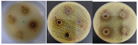
On Muller-Hinton agar, plant extracts at concentrations of S-100, 50, 25, and 12.5 mg/mL were placed in wells, followed by inoculation with an overnight bacterial culture. Upon examining the results the next day, Xanthomonas citri was found to be resistant to the plant extract, as illustrated in Figure 1 and Table 1. These findings align with previous research [24]. This study's objective was to assess the plant extract's antimicrobial activity against Xanthomonas axonopodis pv. Mangifera indica in a laboratory setting. The disc diffusion method was applied to assess the efficacy of the aqueous extract of C. erectus, revealing that the extract was ineffective in inhibiting bacterial growth. The negative control (DMSO) produced no inhibition zone, confirming that the observed activity (or lack thereof) was due to the test sample itself.
A B C
Figure 1. Antibacterial activity of the plant extract against Xanthomonas citri at different concentrations (S-100, 50, 25, 12.5 mg/ml)
3.2 Antibacterial efficacy of biosynthesized zinc oxide nanoparticles
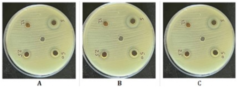
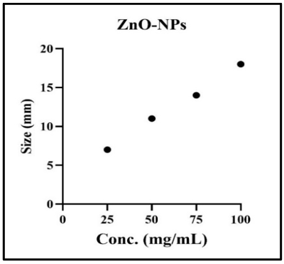
The results of the antibacterial efficacy of biosynthesized ZnO-NPs at varying concentrations (100, 50, 25, and 12.5 mg/mL) are presented in Figures 2 and 3 and Table 1. The results unequivocally show a dose-dependent relationship, with higher ZnO-NP concentrations demonstrating stronger antibacterial activity. Table 1 shows that an inhibition zone of 7 mm was observed at a concentration as low as 12.5 mg/mL, whereas an inhibition zone of 18 mm was produced at a concentration of 100 mg/mL. Although no standard antibiotic (e.g., streptomycin) was included in the present assays, previous reports demonstrated that streptomycin sulfate at 500 ppm produced inhibition zones of approximately 17–19 mm against X. axonopodis pv. citri, suggesting that the ZnO-NPs exhibit comparable antimicrobial potential [25]. In comparison with chemically synthesized ZnO-NPs, the biosynthesized nanoparticles in our study demonstrated comparable or even superior antibacterial efficacy. For instance, study [26] reported that chemically synthesized ZnO-NPs produced inhibition zones ranging from 10 to 16 mm against Xanthomonas campestris at concentrations between 50 and 100 mg/mL. In contrast, our green-synthesized ZnO-NPs achieved an inhibition zone of 18 mm at 100 mg/mL against X. citri, suggesting that the phytochemicals present in the plant extract may enhance the antibacterial activity by improving nanoparticle dispersion, stability, and interaction with bacterial membranes [26].
Figure 2. Antibacterial activity of the biosynthesized zinc oxide nanoparticles against Xanthomonas citri at varying concentrations (S-100, 50, 25, 12.5 mg/mL)
Figure 3. Dose–response curve of biosynthesized zinc oxide nanoparticles against Xanthomonas citri
Table 1. Zones of inhibition induced by Conocarpus extract and zinc oxide nanoparticles against Xanthomonas citri isolates
|
Test Solution |
Concentration (mg/mL) |
Diameter of Inhibition Zone (mm) |
||
|
Isolate 1 |
Isolate 2 |
Isolate 3 |
||
|
ZnO-NPs |
S |
18 |
20 |
18 |
|
50 |
14 |
13 |
14 |
|
|
25 |
11 |
12 |
11 |
|
|
12.5 |
7 |
6 |
7 |
|
|
Plant extract |
S |
Nill |
Nill |
Nill |
|
50 |
Nill |
Nill |
Nill |
|
|
25 |
Nill |
Nill |
Nill |
|
|
12.5 |
Nill |
Nill |
Nill |
|
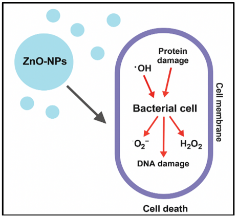
The variation in inhibition zones, as shown in the table, may be attributed to differences in ZnO-NP’s interaction mechanisms with the bacterial cells, as well as the susceptibility of the bacteria used in the study. These observations are consistent with previous research findings [27]. The antibacterial capabilities of ZnO-NPs with varying surface areas against Xanthomonas campestris pv. campestris, the efficiency of ZnO-NPs in managing black mold disease, and their impact on soil bacterial communities were also investigated in that study. The findings revealed that ZnO-NPs with larger surface areas significantly inhibited X. campestris pv. campestris by disrupting bacterial growth and causing cellular damage. The dose-dependent antibacterial activity observed in this study can be explained by the ability of ZnO-NPs to generate reactive oxygen species (ROS), such as hydroxyl radicals, superoxide anions, and hydrogen peroxide, which induce oxidative damage to bacterial cell membranes, proteins, and DNA, ultimately leading to cell death [28, 29]. Additionally, ZnO-NPs interact directly with bacterial membranes, disrupting their integrity and increasing permeability, which causes leakage of cellular contents [30]. These combined mechanisms contribute to the enhanced antimicrobial efficacy observed and are supported by previous literature. The zinc oxide nanoparticles induce bacterial cell death by generating reactive oxygen species (ROS), such as hydroxyl radicals (•OH), superoxide anions (O2⁻), and hydrogen peroxide (H2O2), which cause protein, DNA, and membrane damage. A schematic representation of the proposed antibacterial mechanism is provided in Figure 4.
Figure 4. Illustration of the proposed antibacterial mechanism of zinc oxide nanoparticles
In addition to antimicrobial potency, the long-term stability and practical applicability of ZnO‑NPs are crucial for real-world deployment. ZnO‑NPs are known for their high chemical stability under diverse conditions, including different pH and environmental stress, and can be further enhanced via surface functionalization [31]. Nonetheless, colloidal aggregation and surface oxidation over time may reduce their activity. Therefore, future studies should assess storage stability over extended periods and under variable temperature, humidity, and light exposure. Additionally, techniques such as polymer coating or nanocarrier encapsulation could enhance nanoparticle stability and enable controlled release, making them more suitable for agricultural or biomedical field applications.
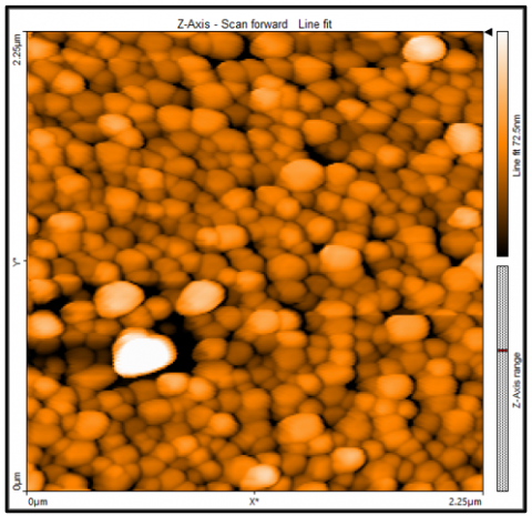
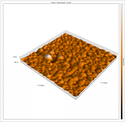
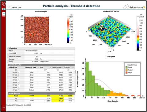
3.3 AFM-based characterization of zinc oxide nanoparticles
The ZnO-NPs' surface roughness was visible to the AFM. As shown in Figure 5, the results demonstrated that the sample's 2D and 3D images had a uniform height distribution with an average of 73.55 nm. The previous study [32] examined the AFM and found that pure ZnO-NPs had an average diameter of 88.17 nm. The results showed that ZnO-NPs are pure and effective, making them suitable for use in a range of industrial and medicinal applications. Smaller particle size enhances antibacterial activity due to increased surface area and better interaction with bacterial cells, which likely contributed to the observed inhibition zones.
(C)
Figure 5. AFM characterization of zinc oxide nanoparticles (ZnO-NPs). (A): The two-dimensional image of ZnO-NPs; (B): The three-dimensional image of ZnO-NPs; (C): AFM analysis report of ZnO-NPs
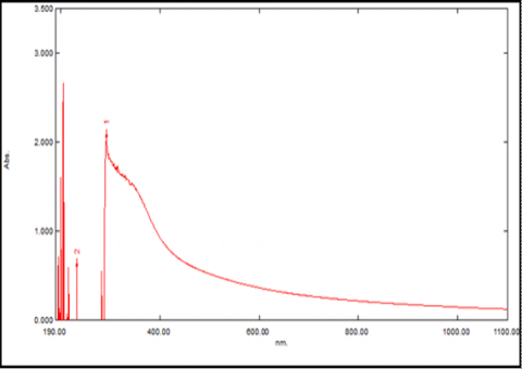
3.4 UV-Visible absorption of zinc oxide nanoparticles
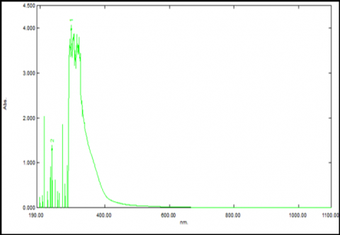
The results presented in Figure 6(A) demonstrate that the UV-visible spectroscopy of the aqueous plant extract exhibited a characteristic absorption peak at 259 nm. The UV-Vis spectrum of the green-synthesized ZnO-NPs using the crude extract, on the other hand, is shown in Figure 6(B). It shows an absorption peak at 292 nm. According to previous findings [33], ZnO-NPs typically exhibit a distinct absorption peak near 275 nm, as determined by UV-visible spectroscopy.
(A)
(B)
Figure 6. UV-visible spectra (A) Aqueous extract and (B) Biosynthesized zinc oxide nanoparticles
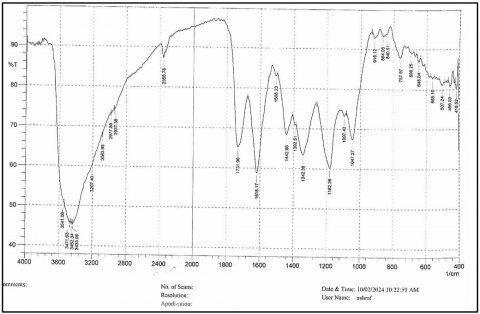
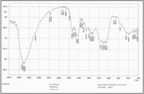
3.5 FTIR spectroscopy-based characterization of zinc oxide nanoparticles
The results of the FTIR spectral analysis of the biosynthesized ZnO-NPs are illustrated in Figure 7(A). The spectra display distinct absorption bands at 3471.63 cm⁻¹ and 3433.06 cm⁻¹, which are attributed to O–H stretching vibrations of alcohol groups and N–H stretching from amine groups. Additionally, a band observed at 1618.17 cm⁻¹ corresponds to N–H bending of amines and C=C stretching vibrations. Another notable peak appears at 1182.28 cm⁻¹, which is associated with C–O stretching. Figure 7(B) presents the FTIR spectrum of the plant extract, showing similar characteristic peaks. These spectral patterns are in agreement with findings reported in previous studies [34].
(A)
(B)
Figure 7. FTIR spectra of (A) Biosynthesized zinc oxide nanoparticles and (B) Plant extract
Using the aqueous extract of C. lancifolius leaves, this study successfully demonstrated the green synthesis of ZnO-NPs and assessed their antibacterial efficacy against Xanthomonas citri. The biosynthesized ZnO-NPs demonstrated significantly enhanced antimicrobial effects, whereas the crude plant extract showed no discernible antibacterial activity. Characterization of the nanoparticles confirmed their successful formation, with AFM revealing an average particle size of 73.55 nm and UV-visible spectroscopy indicating a distinct absorption peak at approximately 292 nm. Fourier transform infrared spectroscopy further verified the presence of functional groups involved in the nanoparticle formation. The ZnO-NPs displayed notable antibacterial activity, achieving a maximum inhibition zone of 18 mm at a concentration of 100 mg/mL. These findings highlight the potential of green-synthesized ZnO-NPs as an effective biocontrol agent against phytopathogenic bacteria, offering a sustainable and eco-friendly alternative to conventional treatments. A key limitation of this study is that the Conocarpus lancifolius leaf extract alone exhibited no antibacterial activity against Xanthomonas citri. This suggests that the antimicrobial effect observed was solely due to the synthesized zinc oxide nanoparticles. Therefore, the independent antibacterial potential of the plant extract remains unverified, limiting its application without nanoparticle formulation. Moreover, the study was limited to specific Iraqi regions, and in vitro results may not accurately reflect real-world field conditions.
The authors express their appreciation to the Department of Biotechnology, University of Al-Nahrain, Iraq, for their valuable assistance.
[1] Saleem, M., Ali, M., Gulshan, A.B. (2024). Nutritional uses of the family Rutaceae. In Phytochemical and Pharmacological Investigation of the Family Rutaceae, Apple Academic Press, pp. 147-160.
[2] Kumari, S., Bhowal, R., Suprasanna, P. (2023). Sustainable approaches for biodiversity and bioprospecting of citrus. Sustainability, 15(9): 7731. https://doi.org/10.3390/su15097731
[3] Satpute, A.D., Fadli, A. (2022). Diseases of citrus and their control. In Citrus Production, pp. 239-266.
[4] Nauman, M., Mushtaq, S., Khan, M.F., Ali, A., Naqvi, S.A.H., Haq, Z., Umar, U.U.D. (2023). Morphological, biochemical, and molecular characterization of Xanthomonas citri subsp. citri, cause of citrus canker disease in Pakistan. Pakistan Journal of Botany, 55(6): 2409-2421. https://doi.org/10.30848/PJB2023-6(14)
[5] Ali, S., Hameed, A., Muhae-Ud-Din, G., Ikhlaq, M., Ashfaq, M., Atiq, M., Wang, Y. (2023). Citrus canker: A persistent threat to the worldwide citrus industry—An analysis. Agronomy, 13(4): 1112. https://doi.org/10.3390/agronomy13041112
[6] Jyothi, V., Shilpa, M.E. (2020). Citrus diseases and their management. In Diseases of Fruits and Vegetable Crops, Apple Academic Press, pp. 41-63.
[7] Osdaghi, E. (2022). Xanthomonas citri pv. citri (Asiatic citrus canker). CABI Compendium. https://doi.org/10.1079/cabicompendium.56921
[8] Zou, X., Du, M., Liu, Y., Wu, L., Xu, L., Long, Q., Chen, S. (2021). CsLOB1 regulates susceptibility to citrus canker through promoting cell proliferation in citrus. The Plant Journal, 106(4): 1039-1057. https://doi.org/10.1111/tpj.15217
[9] Jaithon, T., Atichakaro, T., Phonphoem, W., T-Thienprasert, J., Sreewongchai, T., T-Thienprasert, N.P. (2024). Potential usage of biosynthesized zinc oxide nanoparticles from mangosteen peel ethanol extract to inhibit Xanthomonas oryzae and promote rice growth. Heliyon, 10(1): e24076. https://doi.org/10.1016/j.heliyon.2024.e24076
[10] Chanthapong, P., Maensiri, D., Rangsrisak, P., Jaiyan, T., Rahaeng, K., Oraintara, A., Mahakham, W. (2025). Plant-based ZnO nanoparticles for green nanobiocontrol of a highly virulent bacterial leaf blight pathogen: Mechanistic insights and biocompatibility evaluation. Nanomaterials, 15(13): 1011. https://doi.org/10.3390/nano15131011
[11] Hussein, N.R., El-Dawy, E.G. (2024). Morpho-anatomical attributes of the Egyptian Conocarpus erectus L.(Combretaceae R. Br.) with its phytochemicals and fungal-endophytes. Symbiosis, 92(1): 91-109. https://doi.org/10.1007/s13199-023-00960-6
[12] Ramanjaneyulu, A., Chaitanya, T., Joseph, B. (2023). Conocarpus tree—A boon or bane. Chronicle of Bioresource Management, 7(2): 28-34.
[13] Abdelsattar, A.S., Farouk, W.M., Gouda, S.M., Safwat, A., Hakim, T.A., El-Shibiny, A. (2022). Utilization of Ocimum basilicum extracts for zinc oxide nanoparticles synthesis and their antibacterial activity after a novel combination with phage. Materials Letters, 309: 131344. https://doi.org/10.1016/j.matlet.2021.131344
[14] Pulit-Prociak, J., Banach, M. (2016). Silver nanoparticles–A material of the future…? Open Chemistry, 14(1): 76-91. https://doi.org/10.1515/chem-2016-0005
[15] Irshad, S., Riaz, M., Anjum, A.A., Sana, S., Saleem, R.S.Z., Shaukat, A. (2020). Biosynthesis of ZnO nanoparticles using Ocimum basilicum and determination of its antimicrobial activity. Journal of Animal & Plant Sciences, 30(1): 185-191.
[16] Saleh, A. (2014). Distribution and pathotype identification of Xanthomonas citri subsp. citri recovered from southwestern region of Saudi Arabia. African Journal of Microbiology Research, 8(7): 673-679. https://doi.org/10.5897/AJMR2013.6561
[17] Yaaqoob, L.A., Abed, R.M., Kamona, Z.K., Altaee, M.F. (2022). Evaluation the ability of titanum oxide nanoparticles to increase the production of prodigiosin and phenasine from Serratia marcescens and Pseudomonas aeruginosa respectively. Iraqi Journal of Agricultural Sciences, 53(3): 496-504. https://doi.org/10.36103/ijas.v53i3.1557
[18] Nawar, M.H., Yaaqoob, L.A., Hatem, M.W., Mohammed, A.K. (2024). The effect of nanoparticles preparation from extract of Dodonaea Viscosa L. leaves on the biological performance of the Great Waxworm. AIP Conference Proceedings, 2922(1): 030001. https://doi.org/10.1063/5.0183841
[19] Obeizi, Z., Benbouzid, H., Ouchenane, S., Yılmaz, D., Culha, M., Bououdina, M. (2020). Biosynthesis of Zinc oxide nanoparticles from essential oil of Eucalyptus globulus with antimicrobial and anti-biofilm activities. Materials Today Communications, 25: 101553. https://doi.org/10.1016/j.mtcomm.2020.101553
[20] Lakhan, M.N., Chen, R., Shar, A.H., Chand, K., Shah, A.H., Ahmed, M., Wang, J. (2020). Eco-friendly green synthesis of clove buds extract functionalized silver nanoparticles and evaluation of antibacterial and antidiatom activity. Journal of Microbiological Methods, 173: 105934. https://doi.org/10.1016/j.mimet.2020.105934
[21] Garcia, R. (2020). Nanomechanical mapping of soft materials with the atomic force microscope: Methods, theory and applications. Chemical Society Reviews, 49(16): 5850-5884. https://doi.org/10.1039/D0CS00318B
[22] Wang, Q., Mei, S., Manivel, P., Ma, H., Chen, X. (2022). Zinc oxide nanoparticles synthesized using coffee leaf extract assisted with ultrasound as nanocarriers for mangiferin. Current Research in Food Science, 5: 868-877. https://doi.org/10.1016/j.crfs.2022.05.002
[23] Zaera, F. (2014). New advances in the use of infrared absorption spectroscopy for the characterization of heterogeneous catalytic reactions. Chemical Society Reviews, 43(22): 7624-7663. https://doi.org/10.1039/C3CS60374A
[24] Haq, M.E., Shahbaz, M.U., Kamran, M., Matloob, M.J., Abrar, W., Ali, S., Iqbal, M.A. (2021). Relative potential of different plant extracts and antibiotics against Xanthomonas axonopodis pv. mangiferaeindicae causing bacterial leaf spot of mango in lab conditions. Pakistan Journal of Phytopathology, 33(2): 395-399. https://doi.org/10.33866/phytopathol.033.02.0726
[25] Madavi, P.N., Totawar, M.V., Mane, S.S. (2020). In-vitro efficacy antibiotics amongst the isolates of Xanthomonas axonopodis pv. citri. International Journal of Current Microbiology and Applied Sciences, 9(8): 3494-3505. https://doi.org/10.20546/ijcmas.2020.908.404
[26] Singh, P., Kim, Y.J., Zhang, D., Yang, D.C. (2016). Biological synthesis of nanoparticles from plants and microorganisms. Trends in Biotechnology, 34(7): 588-599. https://doi.org/10.1016/j.tibtech.2016.02.006
[27] Sripo‐ngam, S., Khaengraeng, C., Kasem, S., Chatnaparat, T. (2024). Antibacterial activity of high surface area zinc oxide nanoparticles for controlling black rot disease of Chinese kale. Plant Pathology, 73(7): 1957-1968. https://doi.org/10.1111/ppa.13923
[28] Raghupathi, K.R., Koodali, R.T., Manna, A.C. (2011). Size-dependent bacterial growth inhibition and mechanism of antibacterial activity of zinc oxide nanoparticles. Langmuir, 27(7): 4020-4028. https://doi.org/10.1021/la104825u
[29] Raliya, R., Tarafdar, J.C. (2013). ZnO nanoparticle biosynthesis and its effect on phosphorous-mobilizing enzyme secretion and gum contents in Clusterbean (Cyamopsis tetragonoloba L.). Agricultural Research, 2(1): 48-57. https://doi.org/10.1016/j.agee.2018.01.001
[30] Jones, N., Ray, B., Ranjit, K.T., Manna, A.C. (2008). Antibacterial activity of ZnO nanoparticle suspensions on a broad spectrum of microorganisms. FEMS Microbiology Letters, 279(1): 71-76. https://doi.org/10.1111/j.1574-6968.2007.00945.x
[31] Zhou, X.Q., Hayat, Z., Zhang, D.D., Li, M.Y., Hu, S., Wu, Q., Yuan, Y. (2023). Zinc oxide nanoparticles: Synthesis, characterization, modification, and applications in food and agriculture. Processes, 11(4): 1193. https://doi.org/10.3390/pr1104119
[32] AL-Asady, Z.M., AL-Hamdani, A.H., Hussein, M.A. (2020). Study the optical and morphology properties of zinc oxide nanoparticles. AIP Conference Proceedings, 2213(1): 020061. https://doi.org/10.1063/5.0000259
[33] Jaji, S.B., Ochigbo, S.S., Abubakar, A.Y., Suleiman, M.A.T., Okafor, J.O., Olutoye, A.F. (2024). Terminalia catappa leaf extract as a capping agent for ZnO nanoparticle synthesis via sol-gel method and its microbial effects on selected fungi and bacterial. Journal of Applied Sciences and Environmental Management, 28(4): 1053-1060. https://doi.org/10.4314/jasem.v28i4.6
[34] Shafqat, U., Hussain, S., Shahzad, T., Shahid, M., Mahmood, F. (2023). Elucidating the phytotoxicity thresholds of various biosynthesized nanoparticles on physical and biochemical attributes of cotton. Chemical and Biological Technologies in Agriculture, 10(1): 30. https://doi.org/10.1186/s40538-023-00402-x