Laboratory Evaluation of Artemisia herba-alba Ethanolic Extract and Its Chitosan Nanoparticle Formulation Against Adult Musca domestica L.

Abdullah Fadhil Obaid AL-Zubaidy* | Enas Hamed AL-Ani
© 2026 The authors. This article is published by IIETA and is licensed under the CC BY 4.0 license (http://creativecommons.org/licenses/by/4.0/).
OPEN ACCESS
Adult houseflies, Musca domestica L., are important mechanical vectors of microorganisms. This study evaluated a 70% ethanolic extract prepared from the pre-flowering aerial parts of Artemisia herba-alba and an extract-containing chitosan nanoparticle formulation under laboratory conditions. The formulation was prepared by ionic gelation and characterized by dynamic light scattering, zeta-potential analysis, field-emission scanning electron microscopy (FESEM), and Fourier-transform infrared spectroscopy (FTIR). Across three technical measurements, mean particle size was 238.2 ± 21.6 nm and mean zeta potential was -19.4 ± 3.0 mV; three selected FESEM features averaged 161.89 ± 34.49 nm. At 15,000 ppm and 96 h, mortality reached 23.3% with the crude extract and 83.33% with the nanoparticle formulation. The concentration basis differed, however: crude-extract mass for the free extract and total dried formulation mass for the nanoparticle treatment. In a two-choice assay at 15,000 ppm, negative preference indices indicated greater occupancy of the control arm. Because blank nanoparticles, extract-equivalent doses, and a unified repeated-measures model were unavailable, the observed differences cannot be attributed independently to nanoencapsulation. The formulation represents a laboratory-stage candidate requiring dose-matched controls and separate safety evaluation.
adult mortality, Artemisia herba-alba, chitosan, Musca domestica, nanoparticle formulation, preference index
Houseflies, Musca domestica L., are closely associated with humans, animals, food, and decomposing materials. Their movement between contaminated substrates and clean surfaces enables the mechanical transfer of diverse microorganisms, making adult houseflies relevant to public and animal health [1].
Housefly control has long relied on synthetic insecticides. Repeated exposure can select for resistance, while operational use may also create concerns regarding residues and effects on non-target organisms [2]. These limitations support the laboratory investigation of botanically derived materials, although efficacy and safety must be demonstrated for each preparation rather than assumed.
Artemisia herba-alba Asso is an aromatic medicinal species containing phenolic and other extractable constituents. Studies of its aerial parts have reported phenolic compounds and biological activity in ethanolic fractions [3]. A 70% ethanolic extract of the dried aerial material has also been used in pharmacological studies [4], and an alcoholic extract has shown insecticidal activity against Tribolium castaneum [5]. These reports are more directly relevant to the present ethanolic extract than the essential-oil literature because the two preparations differ in chemical composition and extraction selectivity.
Chitosan can be cross-linked with sodium tripolyphosphate (TPP) by ionic gelation to form particulate carrier systems. Chitosan-based formulations have been studied in insect management [6], and ingested chitosan itself has reduced adult housefly survival under no-choice conditions [7]. Consequently, a chitosan-containing formulation may show biological activity through more than one component. Appropriate blank-particle and dose-matched controls are therefore needed before any difference from a free extract can be attributed specifically to nanoencapsulation.
This study provides an integrated laboratory evaluation of a 70% ethanolic extract prepared from the pre-flowering aerial parts of Artemisia herba-alba and an extract-containing chitosan nanoparticle formulation against adult Musca domestica. Physicochemical characterization, time-dependent mortality screening, and a two-choice behavioral assay were combined to establish a preliminary dataset for the tested formulation. The study was designed to describe the observed responses; it does not isolate the independent contribution of nanoencapsulation.
The experimental work comprised plant collection and extraction, preparation and characterization of an extract-containing chitosan formulation, establishment of a laboratory housefly colony, and mortality and two-choice behavioral assays.
2.1 Plant material and extraction
The aerial parts (leaves and stems) of Artemisia herba-alba were collected from a home garden in Baghdad, Iraq, in October 2025, before flowering. The plant material was botanically authenticated as Artemisia herba-alba Asso (family Asteraceae) by the College of Agriculture, Al-Qasim Green University, Iraq, and an official botanical identification certificate was issued (Certificate No. 866, dated 29 March 2026). A herbarium voucher specimen was not deposited. The collected material was not washed and was air-dried for 14 days before grinding. The dried plant material (50 g) was extracted with 500 mL of 70% aqueous ethanol for 24 h. The use of aqueous ethanol provides a mixed-polarity solvent system suitable for recovering diverse moderately polar plant constituents [3, 4, 8]. The mixture was filtered, and the solvent was removed using a rotary evaporator at 40–45 ℃, followed by oven drying at 50 ℃. The resulting dry extract weighed 4 g, corresponding to an extraction yield of 8% (w/w), calculated as dry-extract mass divided by the initial dry-plant mass × 100. The dried extract was stored at 4 ℃ until use.
2.2 Preparation of the extract-containing chitosan formulation
The formulation was prepared by ionic gelation with reference to established chitosan–TPP preparation approaches [9-13]. Chitosan derived from shrimp shells (Loba Chemie Pvt. Ltd., India; Article No. 02697, CAS 9012-76-4; degree of deacetylation ≥75%; viscosity 200–530 cps for a 1% solution at 20 ℃) was used. The manufacturer's specification sheet does not report molecular weight for this product grade; viscosity was the only rheological parameter provided. Based on the widely used viscosity-based classification for commercial chitosan (low viscosity 20–300 cps ≈ 50,000–190,000 Da; medium viscosity 200–800 cps ≈ 190,000–310,000 Da; high viscosity 800–2,000 cps ≈ 310,000–375,000 Da), the viscosity range of the chitosan used is consistent with a medium molecular weight grade (approximately 190,000–310,000 Da). This estimate is indicative rather than directly measured and represents a limitation for reproducibility. Chitosan (5 g) was dissolved in 1000 mL of 1.5% (v/v) aqueous acetic acid, producing a 0.5% (w/v) chitosan solution, and was stirred at 600 rpm for 24 h at room temperature without heating. Five drops of Tween 80 were added; the corresponding volume was not recorded. The dried ethanolic extract (3.2 g) was then added directly to the chitosan solution.
Separately, TPP (1.25 g) was dissolved in 499 mL of distilled water to prepare an approximately 0.25% (w/v) solution. The entire TPP solution was added dropwise to the chitosan–extract mixture under continuous stirring at 900 rpm, corresponding to an approximate chitosan-solution-to-TPP-solution volume ratio of 2:1. The exact TPP addition rate was not recorded. After TPP addition, the mixture was stirred for 5 h at 900 rpm at room temperature without heating. A 1 M NaOH solution was prepared by dissolving 4 g of NaOH in approximately 50 mL of distilled water and completing the final volume to 100 mL after dissolution. As the final preparation step, pH was adjusted to 5.22 using 215.2 mL of 1 M NaOH. The suspension was centrifuged at 5000 rpm for 15 min; the rotor radius and corresponding relative centrifugal force were unavailable. The collected material was washed three times, each using 75 mL of distilled water and 20 mL of 95% ethanol, followed by centrifugation at 5000 rpm for 15 min. The recovered material was oven-dried at 45 ℃ for 10–12 h to obtain the final powder.
2.3 Physicochemical characterization
Particle size was measured by dynamic light scattering (DLS) using a HORIBA SZ-100 with distilled water as the dispersant. Three technical measurements of the same preparation were available. Zeta potential was measured on the same instrument in three technical replicates. The pH of the DLS dispersion was not recorded; pH 5.22 refers to the formulation before centrifugation and drying. Field-emission scanning electron microscopy (FESEM) was used to examine one dried, agglomerated sample. Three selected features were measured from a high-magnification micrograph and were not treated as a representative particle-size distribution. Fourier-transform infrared spectroscopy (FTIR) was used to describe functional-group bands in the final formulation. DLS and zeta-potential results were interpreted according to the scope and limitations of light-scattering measurements [14, 15].
2.4 Housefly source and rearing
The laboratory colony was established from adult houseflies obtained from a local market in Al-Dora, Baghdad, Iraq. Flies were kept in cages covered with tulle cloth at 25–30 ℃ and 55 ± 5% relative humidity. Adults were supplied with powdered milk and sugar (1:1), and water was provided on cotton. Experimental flies were 3-day-old adults from the second laboratory generation. Males and females were tested together, but the sex ratio was not standardized. Before testing, flies were deprived of food for 6 h while water remained available to prevent dehydration. They were not anaesthetized, cooled, or otherwise treated and were transferred directly from the rearing cages using an aspirator [16-18].
2.5 Adult mortality bioassay
Tests were conducted in nominal 3 L square plastic containers with internal dimensions of 15 × 15 × 13.3 cm. The approximate combined area of the bottom and four walls was 1023 cm². Five to eight spray actuations were applied to the bottom, walls, food, and water-moistened cotton. A dedicated hand sprayer was used for each concentration and control. Spray output was not calibrated; consequently, applied volume and dose per unit area could not be calculated. Treated surfaces were left for 3–5 min until visibly dry before flies were introduced. This procedure involved residual contact and possible oral exposure through treated food and cotton, and these routes were not separated [16, 19, 20]. Each container received 10 adult flies, and three independent containers were used per treatment. Containers were covered with tulle cloth and a perforated lid.
For the crude extract, ppm denotes dry crude-extract mass per final spray volume; a 30,000 ppm (3% w/v) stock was prepared in distilled water. For the nanoparticle formulation, ppm denotes total dried formulation-powder mass per final spray volume; a 15,000 ppm (1.5% w/v) stock was prepared by dispersing 1.5 g of powder in 100 mL of distilled water. This powder comprised chitosan, TPP, and plant material. Test solutions (25 mL) were prepared using C1V1 = C2V2. The crude extract was tested at 1000, 3000, 5000, 10,000, and 15,000 ppm; the nanoparticle formulation was evaluated at the same nominal concentrations in the mortality dataset. These nominal concentrations were not equivalent active-extract doses because encapsulation efficiency and loading capacity were not measured. Distilled water was the negative control and produced 0% mortality at all observations; therefore, Abbott correction was not applied. Mortality was recorded at 24, 48, 72, and 96 h. A fly that did not move after gentle stimulation with a fine brush was recorded as dead. DELTAFLY 25 SC (deltamethrin 2.5% w/v; Saudi Delta) was included as a positive control at 1.25 mL in 250 mL of water (125 mg/L) and was applied by the same spraying procedure. It caused 100% mortality at 24 h, so later readings were not taken. Blank chitosan–TPP particles and formulation-component controls were not prepared.
2.6 Two-choice behavioral assay
Behavior was evaluated at 15,000 ppm in a custom static-air two-choice apparatus with a total length of 65 cm, two arms of approximately 27 cm each, a central chamber of approximately 11 cm, and an internal diameter of approximately 7 cm. Two grams of a milk:sugar mixture (1:1, w/w) were placed at the terminal end of each arm. The treatment food received two spray actuations of crude extract or nanoparticle formulation; the control food received two actuations of distilled water. Spray output was not calibrated. Food was dried for 10 min before testing. The exact distance from the release point to each food source was not measured. The design followed general principles for two-choice orientation assays [20-22].
Ten 3-day-old, mixed-sex adults from the second generation were placed in the central chamber after a 6 h food-deprivation period with water available. Following 30 s of acclimation, both arms remained open for 60 min. The same flies were counted in the treatment arm, control arm, and central chamber at 30 and 60 min. Counts at 30 and 60 min were treated as repeated measures on the same cohort and are presented descriptively. Arm orientation was alternated among replicates. Tests were conducted in darkness, in still air, at 25–30 ℃ and 55 ± 5% relative humidity. A dedicated apparatus was used for each replicate; three replicates were conducted per formulation. No separate blank side-preference assay was performed. Behavioral response was summarized by the preference index (PI) = (T − C)/(T + C), where T and C are the numbers in the treatment and control arms, respectively. Central non-responders were excluded from the denominator. Negative PI values indicate preference for the control arm.
2.7 Statistical analysis
Mortality percentages were analyzed in GenStat using separate conventional two-way Analysis of Variance (ANOVA) models for the crude extract and nanoparticle formulation, with concentration, time, and their interaction as fixed effects and no blocking. The replicate container (10 flies) was the statistical unit (n = 3 per concentration–time combination), and untransformed replicate-level mortality percentages were analyzed. The original procedure was a two-way ANOVA with replication; time was not treated as a repeated measure. Means were compared using the least significant difference (LSD) at the 5% level [23]. Because the same containers were observed repeatedly over time and mortality arose from binary outcomes, the independence and distributional assumptions of conventional ANOVA were not fully met; the models did not explicitly account for within-container temporal correlation or a binomial error structure. Results are therefore interpreted as exploratory within-formulation analyses and are not used to establish definitive temporal effects. Confirmatory inference would require a binomial repeated-measures generalized linear mixed model with replicate container included as a random effect. No unified formulation × concentration × time model was fitted, so cross-formulation differences are descriptive and do not establish statistical superiority. Behavioral counts and replicate-level PI values were summarized descriptively without inferential lettering. Pooled concentration–mortality data for the nanoparticle formulation at 96 h (n = 30 per concentration) were additionally analyzed by probit regression via maximum-likelihood estimation (Python 3, SciPy v1.17) to estimate LC50, LC90, and the slope of the concentration–response line. Model fit was assessed using a Pearson chi-square goodness-of-fit test; because this test indicated a significant lack of fit, Wald-based confidence intervals for LC50 and LC90 are not reported, as they would not be statistically meaningful for a poorly fitting model. For the crude extract, mortality did not exceed 23.3% at the highest tested concentration, precluding Probit estimation within the tested range.
3.1 Physicochemical characterization
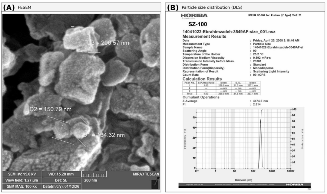
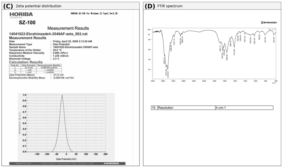
The FESEM, DLS, zeta-potential, and FTIR outputs are presented in Figures 1(A)–(D), respectively. Across three technical DLS measurements (Table 1), mean particle size was 238.2 ± 21.6 nm (range 216.2–259.3 nm), and each measurement showed a single predominant intensity-based peak. The representative run shown in Figure 1(B) had a mean of 239.0 nm, mode of 237.1 nm, and standard deviation of 21.5 nm. The available instrument outputs did not provide a consistent, valid polydispersity index (PDI); therefore, PDI is not reported.
Table 1. Physicochemical measurements of the extract-containing chitosan formulation
|
Parameter |
Value |
|
DLS mean size (nm) |
238.2 ± 21.6 (n = 3 technical) |
|
DLS range (nm) |
216.2–259.3 |
|
Representative DLS mode (nm) |
237.1 |
|
Zeta potential (mV) |
-19.4 ± 3.0 (n = 3 technical) |
|
Zeta potential range (mV) |
-21.5 to -16.0 |
|
Representative mobility (cm²/V·s) |
-0.000166 |
|
Selected FESEM features (nm) |
161.89 ± 34.49 (n = 3 selected) |
|
Selected FESEM range (nm) |
134.32–200.57 |
Figure 1. Characterization of the extract-containing chitosan formulation: (A) field-emission scanning electron microscopy (FESEM) image with three selected measurements, (B) representative dynamic light scattering (DLS) size distribution, (C) representative zeta-potential distribution, and (D) Fourier-transform infrared spectroscopy (FTIR) spectrum
The DLS results demonstrate a predominant nanoscale hydrodynamic population in the tested dispersion, consistent with the formation of a chitosan–TPP particulate system. They do not, by themselves, demonstrate how much extract was incorporated. The agreement between the representative DLS mean and mode describes that run only and should not be interpreted as a validated PDI measurement.
FESEM examination of one dried sample showed extensive agglomeration and irregular larger structures with smaller features on their surfaces. Three selected features measured 134.32, 150.79, and 200.57 nm, giving 161.89 ± 34.49 nm. This small selected set is presented as illustrative morphology rather than as a representative size distribution. The lower FESEM values relative to DLS may reflect the difference between selected dried-state features and hydrodynamic diameter in dispersion [14, 15].
The three technical zeta-potential measurements were -16.0, -21.5, and -20.8 mV, yielding -19.4 ± 3.0 mV. These values describe a moderately negative surface potential under the measurement conditions but do not establish storage stability. The available FTIR spectrum showed bands in regions associated with O–H/N–H, aliphatic C–H, amide, and C–O–C vibrations. Because spectra of blank chitosan–TPP particles and free extract were not obtained under matched conditions, FTIR was used descriptively and not as proof of encapsulation.
3.2 Adult mortality
For the crude extract, the original exploratory two-way ANOVA returned a concentration term (F = 13.34, p < 0.001), whereas time (F = 0.32, p = 0.808) and the concentration × time interaction (F = 0.08, p = 1.000) were not significant. No mortality occurred at 1000 or 5000 ppm. The largest response was 23.3% at 15,000 ppm after 96 h (Table 2). The modest crude-extract response is within the broader variability reported for plant-derived treatments against adult houseflies [17, 20, 24]. Related botanical-loaded chitosan nanoparticle systems have demonstrated repellent or insecticidal activity against other insect species [25, 26].
Table 2. Mortality (%) of adult Musca domestica exposed to the crude ethanolic extract
|
Concentration |
24 h |
48 h |
72 h |
96 h |
Mean |
|
Water control |
0.0 |
0.0 |
0.0 |
0.0 |
0.0 |
|
1000 ppm |
0.0 |
0.0 |
0.0 |
0.0 |
0.0 |
|
3000 ppm |
3.3 |
3.3 |
3.3 |
6.7 |
4.2 |
|
5000 ppm |
0.0 |
0.0 |
0.0 |
0.0 |
0.0 |
|
10,000 ppm |
3.3 |
3.3 |
3.3 |
6.7 |
4.2 |
|
15,000 ppm |
16.7 |
20.0 |
20.0 |
23.3 |
20.0 |
|
Time mean |
4.7 |
5.3 |
5.3 |
7.3 |
— |
For the nanoparticle formulation, the original exploratory two-way ANOVA returned terms for concentration (F = 64.54, p < 0.001), time (F = 49.79, p < 0.001), and their interaction (F = 4.99, p < 0.001). Mortality reached 83.33% at 15,000 ppm after 96 h (Table 3). Within this formulation, the concentration × time LSD0.05 was 12.284 percentage points. These results describe the total dried formulation and cannot be converted to an extract-equivalent dose because loading was not measured.
Table 3. Mortality (%) of adult Musca domestica exposed to the extract-containing chitosan nanoparticle formulation
|
Concentration |
24 h |
48 h |
72 h |
96 h |
Mean |
|
Water control |
0.00 |
0.00 |
0.00 |
0.00 |
0.00 |
|
1000 ppm |
6.67 |
10.00 |
18.33 |
30.00 |
16.25 |
|
3000 ppm |
6.67 |
6.67 |
13.33 |
20.00 |
11.67 |
|
5000 ppm |
16.67 |
10.00 |
20.00 |
53.33 |
25.00 |
|
10,000 ppm |
23.33 |
30.00 |
20.00 |
46.67 |
30.00 |
|
15,000 ppm |
30.00 |
40.00 |
70.00 |
83.33 |
55.83 |
Probit regression of the pooled nanoparticle-formulation mortality data at 96 h yielded LC50 = 5687.03 ppm, LC90 = 83149.82 ppm, and a slope of 1.100 ± 0.266 (Table 4). The Pearson chi-square goodness-of-fit test indicated significant lack of fit (χ² = 12.01, df = 3, p = 0.0073); Wald-based confidence intervals for LC50 and LC90 are therefore not reported, as they would not be statistically meaningful for a poorly fitting model. LC90 lies outside the tested concentration range and should be interpreted as an extrapolation with low confidence. For the crude extract, mortality did not exceed 23.3% at the highest tested concentration, precluding Probit estimation within the tested range.
Table 4. Probit analysis of the extract-containing chitosan nanoparticle formulation against adult Musca domestica after 96 h
|
Parameter |
Estimate |
|
LC50 (ppm) |
5687.03 |
|
LC90 (ppm) |
83149.82* |
|
Slope ± SE |
1.100 ± 0.266 |
|
Pearson χ² (df = 3) |
12.01 (p = 0.0073) |
At the common nominal concentration of 15,000 ppm and 96 h, the crude extract produced 23.3% mortality and the nanoparticle formulation produced 83.33%. This is a descriptive difference between non-equivalent concentration bases: crude-extract mass versus total formulation-powder mass. Moreover, chitosan itself can influence fly survival when ingested [7]. In the absence of blank chitosan–TPP particles, formulation-matched free extract, measured loading, and a unified statistical model, the higher observed mortality cannot be assigned specifically to nanoencapsulation. The deltamethrin positive control caused 100% mortality at 24 h.
3.3 Two-choice behavioral response
Raw arm counts and replicate-level PI summaries are shown in Table 5. Negative PI values occurred for both preparations at 30 and 60 min, indicating greater occupancy of the control arm among responding flies. The most negative descriptive mean was -0.67 ± 0.06 for the nanoparticle formulation at 60 min. Responder proportions ranged from 76.7% to 90.0%. These observations are presented without letters or inferential comparisons because the same flies were read at both times and no validated factorial behavioral model was fitted.
Table 5. Raw two-choice counts and preference index (PI) at 15,000 ppm
|
Preparation |
Time |
T |
C |
Central |
Responders (%) |
PI |
|
Crude extract |
30 min |
7 |
16 |
7 |
76.7 |
-0.38 ± 0.09 |
|
Crude extract |
60 min |
8 |
19 |
3 |
90.0 |
-0.39 ± 0.11 |
|
Nanoparticle formulation |
30 min |
5 |
18 |
7 |
76.7 |
-0.57 ± 0.05 |
|
Nanoparticle formulation |
60 min |
4 |
20 |
6 |
80.0 |
-0.67 ± 0.06 |
The control-arm preference is compatible with avoidance of the treated food, but the test does not separate olfactory, contact, or gustatory contributions. Because no independent blank side-preference assay or calibrated spray dose was available, the behavioral response should be regarded as preliminary and apparatus-specific [20-22].
3.4 Study limitations and scope
The present findings should be interpreted within the scope of the experimental design. The formulation was characterized using DLS, zeta-potential measurements, FESEM, and FTIR; however, blank chitosan–TPP nanoparticles, a formulation-matched free-extract treatment, encapsulation efficiency, loading capacity, validated PDI measurements, comparative FTIR spectra, release kinetics, and storage-stability data were not available. In addition, the nanoparticle-formulation concentrations were based on total dried formulation mass rather than verified extract-equivalent doses. Consequently, the results characterize the tested extract-containing chitosan formulation and its observed bioactivity but do not establish the independent effect or mechanism of nanoencapsulation.
The spray output was not calibrated, and residual-contact exposure could not be separated from possible oral exposure. Mortality was recorded repeatedly from the same replicate containers, while the original analyses used separate conventional ANOVA models for the two preparations. These models did not explicitly account for within-container temporal correlation or the binomial structure of mortality; therefore, their concentration, time, and interaction results are interpreted only as exploratory within-formulation findings. Confirmatory inference would require a binomial repeated-measures generalized linear mixed model with replicate containers included as a random effect. The behavioral assay also lacked a separate blank side-preference test. Therefore, cross-formulation and behavioral differences should be interpreted cautiously. The study represents a preliminary laboratory evaluation that provides a basis for future experiments using formulation-matched controls, calibrated dosing, equivalent active-extract concentrations, an appropriate repeated-measures model, expanded dose-response testing, and separate environmental and non-target safety assessments.
Under the present laboratory conditions, the 70% ethanolic extract produced a maximum adult mortality of 23.3% at 15,000 ppm after 96 h. Separately, the extract-containing chitosan nanoparticle formulation produced a maximum mortality of 83.33% at the same nominal concentration and exposure period. Because the nanoparticle concentration was based on total formulation mass rather than extract-equivalent dose, and encapsulation efficiency was not determined, these values should not be interpreted as a direct quantitative comparison between the two preparations. Negative two-choice preference indices indicated greater occupancy of the water-control arm. These results establish the observed bioactivity of the tested preparations, but they do not demonstrate that nanoencapsulation independently caused the difference because the concentration bases were not extract-equivalent and formulation-matched controls were absent.
Probit analysis of the nanoparticle formulation at 96 h yielded LC50 = 5687.03 ppm and LC90 = 83149.82 ppm (slope = 1.100 ± 0.266); however, the model showed significant lack of fit (Pearson χ² = 12.01, df = 3, p = 0.0073), so these estimates, and particularly the extrapolated LC90, should be interpreted with caution. For the crude extract, mortality did not exceed 23.3% at the highest tested concentration, precluding reliable LC50/LC90 estimation within the tested range. The extract-containing chitosan nanoparticle formulation represents a laboratory-stage candidate. Its environmental fate, degradation, residue behavior, mammalian safety, and effects on non-target organisms require separate evaluation before any practical application can be considered.
The authors thank the College of Biotechnology, Al-Nahrain University, for supporting the experimental work and the College of Agriculture, Al-Qasim Green University, for botanical identification of the plant material.
[1] Nayduch, D., Neupane, S., Pickens, V., Purvis, T., Olds, C. (2023). House flies are underappreciated yet important reservoirs and vectors of microbial threats to animal and human health. Microorganisms, 11(3): 583. https://doi.org/10.3390/microorganisms11030583
[2] Norris, R.H., Baker, O.S., Burgess, E.R., et al. (2023). Selection for, and characterization of, fluralaner resistance in the house fly, Musca domestica. Pesticide Biochemistry and Physiology, 191: 105355. https://doi.org/10.1016/j.pestbp.2023.105355
[3] Mohammed, M.J., Anand, U., Altemimi, A.B., Tripathi, V., Guo, Y., Pratap-Singh, A. (2021). Phenolic composition, antioxidant capacity and antibacterial activity of white wormwood (Artemisia herba-alba). Plants, 10(1): 164. https://doi.org/10.3390/plants10010164
[4] Abdel Jaleel, G.A.R., Abdallah, H.M.I., Gomaa, N.E.L.S. (2016). Pharmacological effects of ethanol extract of Egyptian Artemisia herba-alba in rats and mice. Asian Pacific Journal of Tropical Biomedicine, 6(1): 44-49. https://doi.org/10.1016/j.apjtb.2015.10.005
[5] Jaber, N.K., Khalaf, A.A., Shafeeq, M.A.A. (2025). Comparison of the toxicity of the pesticide imidacloprid and the bioactivity of Artemisia herba-alba alcohol extract against Tribolium castaneum. Journal of Applied and Natural Science, 17(2): 537-544. https://doi.org/10.31018/jans.v17i2.6456
[6] Abenaim, L., Conti, B. (2023). Chitosan as a control tool for insect pest management: A review. Insects, 14(12): 949. https://doi.org/10.3390/insects14120949
[7] Stoffolano, J., Wong, R., Lo, T., Ford, B., Geden, C.J. (2020). Effect of chitosan on adult longevity when fed, in no-choice experiments, to Musca domestica L., Tabanus nigrovittatus Macquart, and Phormia regina (Meigen) adults and its consumption in adult Musca domestica L. Pest Management Science, 76(12): 4293-4300. https://doi.org/10.1002/ps.5996
[8] Abubakar, A.R., Haque, M. (2020). Preparation of medicinal plants: Basic extraction and fractionation procedures for experimental purposes. Journal of Pharmacy and Bioallied Sciences, 12(1): 1-10. https://doi.org/10.4103/jpbs.JPBS_175_19
[9] Calvo, P., Remunan-Lopez, C., Vila-Jato, J.L., Alonso, M.J. (1998). Novel hydrophilic chitosan-polyethylene oxide nanoparticles as protein carriers. Journal of Applied Polymer Science, 63(1): 125-132. https://doi.org/10.1002/(SICI)1097-4628(19970103)63:1<125::AID-APP13>3.0.CO;2-4
[10] Hoang, N.H., Le Thanh, T., Sangpueak, R., et al. (2022). Chitosan nanoparticles-based ionic gelation method: A promising candidate for plant disease management. Polymers, 14(4): 662. https://doi.org/10.3390/polym14040662
[11] Kain, D., Kumar, S. (2020). Synthesis and characterization of chitosan nanoparticles of Achillea millefolium L. and their activities. F1000Research, 9: 1297. https://doi.org/10.12688/f1000research.26446.1
[12] Sharma, A., Sood, K., Kaur, J., Khatri, M. (2019). Agrochemical loaded biocompatible chitosan nanoparticles for insect pest management. Biocatalysis and Agricultural Biotechnology, 18: 101079. https://doi.org/10.1016/j.bcab.2019.101079
[13] Salama, R., Osman, H., Ibrahim, H.M. (2022). Preparation of biocompatible chitosan nanoparticles loaded with Aloe vera extract for use as a novel drug delivery mechanism to improve the antibacterial characteristics of cellulose-based fabrics. Egyptian Journal of Chemistry, 65(3): 589-604. https://doi.org/10.21608/ejchem.2021.96434.4512
[14] Bhattacharjee, S. (2016). DLS and zeta potential – What they are and what they are not? Journal of Controlled Release, 235: 337-351. https://doi.org/10.1016/j.jconrel.2016.06.017
[15] Carvalho, P.M., Felicio, M.R., Santos, N.C., Goncalves, S., Domingues, M.M. (2018). Application of light scattering techniques to nanoparticle characterization and development. Frontiers in Chemistry, 6: 237. https://doi.org/10.3389/fchem.2018.00237
[16] Insecticide Resistance Action Committee (IRAC). (2011). IRAC susceptibility test method 026: Musca domestica adults (feeding), version 1. https://irac-online.org/methods/musca-domestica-adults/.
[17] Pavela, R. (2007). Insecticidal properties of several essential oils on the house fly (Musca domestica L.). Phytotherapy Research, 22(2): 274-278. https://doi.org/10.1002/ptr.2300
[18] Ali, R.M.M., Bashir, M.H., Shan, M.I., et al. (2023). Comparative efficacy of botanical extracts along with their biosynthesized zinc oxide nanoparticles against house fly Musca domestica (Diptera: Muscidae). University of Sindh Journal of Animal Sciences, 7(4): 61-68. https://doi.org/10.57038/usjas.v7i04.6648
[19] Burgess, E.R., King, B.H., Geden, C.J. (2020). Oral and topical insecticide response bioassays and associated statistical analyses used commonly in veterinary and medical entomology. Journal of Insect Science, 20(6): 1-9. https://doi.org/10.1093/jisesa/ieaa041
[20] Tian, Y.X., Hogsette, J.A., Norris, E.J., Hu, X.P. (2024). Topical toxicity and repellency profiles of 17 essential oil components against insecticide-resistant and susceptible strains of adult Musca domestica (Diptera: Muscidae). Insects, 15(6): 384. https://doi.org/10.3390/insects15060384
[21] Roberts, J.M., Clunie, B.J., Leather, S.R., Harris, W.E., Pope, T.W. (2023). Scents and sensibility: Best practice in insect olfactometer bioassays. Entomologia Experimentalis et Applicata, 171(11): 808-820. https://doi.org/10.1111/eea.13351
[22] Carr, A.L., Denning, S.S., Figurskey, A.C., Hung, K.Y., Reiskind, M.H., Watson, D.W. (2025). Response of adult Cochliomyia macellaria, Musca domestica, and Sarcophaga bullata (Diptera: Calliphoridae, Muscidae, Sarcophagidae) to odors produced by commercial fly baits in a two-choice olfactometer bioassay. Journal of Insect Science, 25(2): 11. https://doi.org/10.1093/jisesa/ieaf020
[23] Payne, R. (2020). A Guide to ANOVA and Design in Genstat (21st Edition). VSN International. https://genstat.kb.vsni.co.uk/wp-content/uploads/sites/2/AnovaGuide.pdf.
[24] Nisar, M.S., Ismail, M.A., Ramzan, H., et al. (2021). The impact of different plant extracts on biological parameters of housefly [Musca domestica (Diptera: Muscidae)]: Implications for management. Saudi Journal of Biological Sciences, 28(7): 3880-3885. https://doi.org/10.1016/j.sjbs.2021.03.070
[25] Sanei-Dehkordi, A., Moemenbellah-Fard, M.D., Sereshti, H., Shahriari-Namadi, M., Zarenezhad, E., Osanloo, M. (2021). Chitosan nanoparticles containing Elettaria cardamomum and Cinnamomum zeylanicum essential oils; repellent and larvicidal effects against a malaria mosquito vector, and cytotoxic effects on a human skin normal cell line. Chemical Papers, 75: 6545-6556. https://doi.org/10.1007/s11696-021-01829-y
[26] Ziaee, M., Sheikhzadeh Takabi, A., Ebadollahi, A. (2023). Fabrication of Carum copticum essential oil-loaded chitosan nanoparticles and evaluation of its insecticidal activity for controlling Rhyzopertha dominica and Tribolium confusum. Frontiers in Plant Science, 14: 1187616. https://doi.org/10.3389/fpls.2023.1187616