Afrah M. Al Hussainey* | Randa Kamel Hussain | Aseel Mustafa Abdul Majeed
© 2026 The authors. This article is published by IIETA and is licensed under the CC BY 4.0 license (http://creativecommons.org/licenses/by/4.0/).
OPEN ACCESS
The metallic materials used in medical implants have witnessed remarkable advances due to ongoing developments in materials science and biomedical engineering. Selecting the appropriate material has become crucial to implant success, as it determines the implant's ability to perform its vital functions within the human body. Scientific research has focused on developing metals that possess a balanced combination of mechanical, chemical, and biological properties. This ensures their ability to withstand various mechanical loads, resist corrosion in the biological environment, and achieve biocompatibility with body tissues without triggering immune or inflammatory reactions. Metals are among the most widely used materials in medical implants due to their high mechanical strength and durability, making them suitable for applications that require resistance to prolonged stress. This field has led to the development of many types of metal alloys, most notably stainless steel, cobalt-chromium-molybdenum (CoCrMo) alloys, and titanium (Ti) alloys. However, advances in materials engineering have led to new trends aimed at improving the performance of metal implants through the use of composites or functionally graded materials (FGMs). These materials enable a gradation of mechanical and chemical properties within the material itself. This gradation helps minimize differences between the implant and surrounding biological tissues, thereby reducing stress concentrations and improving the implant's long-term stability. Therefore, continuous development in the design of metal alloys is essential for improving the effectiveness and lifespan of medical implants and enabling further advancements in modern medical and surgical fields.
functionally graded materials, hydroxyapatite, surface modification, osseointegration, cobalt-chromium alloys, titanium alloys, corrosion resistance, biocompatibility
Biomedical implants are among the most important technologies in modern medicine, as they are implanted inside the human body to replace lost tissues or damaged organs and restore their normal functions. Such implants are successful mainly because of the properties of the materials used in their manufacture, which should have a balanced combination of mechanical strength, durability, and biocompatibility. In addition to the mechanical requirements, these materials should have a high ability to interact with living tissues without causing undesirable immune or inflammatory responses, and also chemical stability and high resistance to corrosion in the complex biological environment of the body, thus ensuring patient safety and extending the operational lifespan of the implant [1]. The development of bio-metallic materials applied in medical implants has been rather slow over the last few decades. Medical applications initially used conventional metal alloys such as stainless steel, cobalt-chromium (CoCr) alloys, and titanium (Ti) alloys for their high mechanical strength, good corrosion resistance, and ability to withstand a variety of physiological loads. However, despite their clinical success, these materials suffered from significant limitations, including a higher modulus of elasticity than natural bone, the potential for stress shielding, the release of metal ions, and the formation of wear-and-tear products [2]. To address the limited biocompatibility of metals, subsequent research has focused on modifying metal surfaces with bioactive coatings, particularly hydroxyapatite (HA), owing to their chemical similarity to the mineral component of natural bone and their ability to promote osseointegration. However, these coatings have encountered problems, including poor adhesion between the coating and the metal surface, mismatches in thermal expansion coefficients, and the potential for detachment or cracking during service [3]. Later, metal–ceramic composite materials such as Ti/HA and CoCr/HA systems were developed to integrate the mechanical strength of metals with the biocompatibility of ceramics. These materials improved the biocompatibility of the bone around the implant, but the clear separation between the components sometimes led to stress concentrations and problems at the metal–ceramic contact area [4]. To overcome these challenges, functionally graded materials (FGMs) have been developed as advanced-generation biomaterials. FGMs are characterized by a continuous gradient in chemical composition, microstructure, and mechanical properties throughout the material volume, with no sharp interfaces between components [5]. The gradient allows the combination of a strong load-bearing metallic core with an active biomaterial surface that enhances biocompatibility and osseointegration. It also reduces stress concentrations and improves load transfer between the implant and bone, thereby enhancing the long-term stability and functional performance of medical implants [6].
The development from traditional metal alloys to bio-coatings, then to composite materials, and finally to FGMs reflects the ongoing effort for the optimal balance between mechanical performance and biological compatibility. Therefore, the importance of studying the development of biomaterials used in medical implants, their properties, challenges, and prospects lies in increasing the efficiency of medical implants, extending their operational lifespan, and supporting continuous development in modern medical and surgical applications.
2.1 Stainless steel
The term “stainless steel” is a generic term for a group of iron-based alloys that contain significant amounts of chromium and nickel. Stainless steel is also used in medical practice, including fracture plates and certain components of joint replacements, notably femoral and tibial knee implants [7].
Stainless steels are primarily divided into four main classes: martensitic (body-centered cubic crystal structure, the hardest), ferritic stainless steel (body-centered cubic crystal structure), austenite (face-centered cubic crystal structure), and duplex stainless steel (an austenitic phase (face-centered cubic) plus a ferrite phase (body-centered cubic)). The three main classes of stainless-steel underscore their importance in medical equipment and their widespread use in implant applications. The advantages of stainless steel include its availability, low cost, high-quality production, good biocompatibility, durability, and excellent corrosion resistance [8].
However, the main limitation of stainless steel is wear corrosion, caused by damage to the protective oxide film on the surface. When proper surface treatment is not applied, stainless steel can trigger unwanted biological reactions in the human body, such as inflammation or allergic responses, thereby compromising its use in medical implants and devices. It is therefore important to analyze the surface modification of stainless steel to achieve improved surface engineering, including chemical stability, mechanical properties, corrosion resistance, biocompatibility, and longevity [8].
2.2 Cobalt–chromium–molybdenum alloys
The wide range of clinical uses for cobalt-chromium-molybdenum (CoCrMo) alloys in artificial hard tissue replacement includes both femoral heads for hip joints and artificial knee joints, as the materials exhibit high strength, corrosion resistance, and outstanding wear behavior, in addition to their biocompatibility. CoCrMo alloys represent the best combination of three crucial implant material properties, as they can meet strength requirements and resist both wear and corrosion while handling load-bearing applications. CoCrMo alloys exhibit higher mechanical strength than stainless steels. These materials have a fatigue strength exceeding 500 MPa and are therefore suitable for load-bearing applications involving constant, cyclic, or heavy loading [9].
Cobalt-based alloys are characterized by the potential presence of two primary crystalline phases: the face-centered cubic structure and the hexagonal close-packed structure. The stability and distribution of these phases are influenced by the alloy's chemical composition as well as heat treatment and manufacturing conditions. Differences in crystal structure lead to variations in plastic deformation mechanisms-specifically dislocation slip and twinning and the phase transformation between face-centered cubic and hexagonal close-packed can also contribute to modifying the alloy's mechanical behavior [10]. It has been found that when Co-based alloys have a chromium content of 26–30 wt%, this ratio increases biocompatibility and corrosion resistance due to the formation of a passive oxide layer of Cr₂O₃. Molybdenum (Mo), at a concentration of 5–10 wt%, contributes to improved corrosion resistance by enhancing the stability of the passive layer; it also influences the alloy's microstructure and phase composition [11].
The fact that CoCrMo alloys have enhanced corrosion resistance does not, by itself, imply improved fatigue performance, since fatigue can occur only in regions of pitting corrosion. The CoCr alloys have much greater corrosion resistance than stainless steels due to their high Cr content and the formation of a Cr₂O₃ passive layer. This passive coating, however, is subject to pitting corrosion in a Cl-rich environment [12]. It is vital to note that the corrosion behavior of Co-based alloys can be enhanced through heat treatment and surface treatments.
2.3 Titanium alloys
Ti alloys are widely used in human body applications to replace rigid tissues because they exhibit exceptional mechanical properties, excellent corrosion resistance, low density, and high biocompatibility [13]. Professional orthopedists use Ti-based implants comprising Commercial Pure (CP) Ti for both dental and spinal use, as well as the Ti-6Al-4V (ASTM F136) alloy for artificial knee, hip, and shoulder joints and bone fixators [14]. Ti-based alloys have a higher specific strength than any other metallic implants [15]. Heat treatment affects the hardness of Ti by altering its microstructure and phases [16].
Ti-6Al-4V exhibits high strength comparable to many stainless steels and CoCr-based alloys, depending on processing route and alloy condition. Its ultimate tensile strength exceeds 930 MPa, while its yield strength ranges between 795 and 860 MPa [17]. Young's modulus and ultimate tensile strength vary with alloy composition and production method.
Ti alloys may be susceptible to fatigue degradation, particularly under fretting or aggressive physiological conditions. Improvements in surface α-phase layers lead to decreased fatigue strength and reduced ductility. Due to the formation of a passive TiO₂ film, Ti exhibits outstanding corrosion resistance; however, this resistance depends strongly on both its chemical composition and structural characteristics [18].
Medical experts consider Ti one of the most biocompatible metals. Formation of a TiO₂ passive oxide film confers high biocompatibility and low cytotoxicity. The primary alloying elements in Ti-based metal systems are V and Al, as they adversely affect the biological properties of CP Ti. Research has shown that vanadium can adversely affect surrounding tissues, including inflammation and toxicity, thereby compromising the biocompatibility of Ti-based alloys. Scientists can modify the elastic moduli of Ti-based alloys by adjusting their chemical and phase compositions. Standard Ti-6Al-4V has an elastic modulus of 110 GPa due to its superior strength relative to bone tissue, yet it has lower stiffness than steel and CoCr-based metals [19]. Substitution of high-modulus metals with those with lower modulus values results in a β-phase microstructure and decreases the modulus. Among all other alloy types, the Ti-35Nb β phase exhibited the lowest elastic modulus of 42 GPa [20].
Table 1. Mechanical properties of biomaterials used in joint replacements [21]
|
Materials |
Yield Strength (MPa) |
Ultimate Tensile Strength (MPa) |
|
Cobalt-chromium (CoCr) alloy |
450–520 |
780–1080 |
|
Austenitic stainless steel |
220 |
500–540 |
|
Ti-6Al-4V |
951 |
1020 |
|
NiTi SMA |
195–690 |
895 |
|
Mg-Ca |
148 |
240 |
Table 1 compares the mechanical properties of the most important metallic materials used in joint and bone prostheses, including CoCr alloys, austenitic stainless steel, Ti-6Al-4V alloys, nickel-titanium alloys (NiTi SMA), and magnesium-calcium alloys (Mg-Ca). Yield strength and ultimate tensile strength are among the most important indicators of a material's ability to withstand mechanical loads in the living body. Ti-6Al-4V alloys exhibited the highest yield strength of approximately 951 MPa and the highest tensile strength of 1020 MPa, reflecting their high capacity to withstand repeated mechanical stresses and their resistance to permanent deformation. This explains why they are so widely used in bone grafting and dental implants. CoCr alloys are also known for their high mechanical strength, with a yield strength of 450–520 MPa and a tensile strength of 780–1080 MPa, making them suitable for applications requiring high wear resistance, such as artificial joint surfaces. The austenitic stainless steel exhibited relatively low yield strength (220 MPa) and tensile strength (500–540 MPa). It is widely used for its low cost, ease of fabrication, and clinically acceptable properties. The NiTi shape memory alloys exhibited a broad range of yield strengths (195–690 MPa), with the highest tensile strength of 895 MPa. This is due to phase transitions that confer shape memory and improved elasticity, giving them an excellent ability to absorb dynamic loads [21].
Table 2 compares the most significant materials in terms of advantages, disadvantages, and medical uses. Stainless steel is one of the most commonly used materials because of its low cost and commercial availability. It may also form a layer of metal oxides on its surface that sometimes acts as a lubricant, reducing friction. However, it has a high coefficient of friction and poor wear resistance. Its high elasticity modulus may also lead to stress shielding, resulting in loosening of the bone surrounding the implant. It is therefore commonly used in artificial hip joints, fracture fixation plates, and surgical instruments. Compared with stainless steel and Ti alloys, cobalt alloys offer high corrosion and abrasion resistance, high tensile strength, and a low coefficient of friction. Nevertheless, they have a high modulus of elasticity, which can lead to more serious consequences, including stress blockage. In addition, particles formed during microscopic abrasion can cause tissue inflammation or sensitization. They are also denser than other materials. Artificial hip joints, dental implants, and artificial knee joints are common uses of these alloys. Ti alloys, on the other hand, have a high strength-to-weight ratio. Compared with steel and cobalt alloys, they also have excellent biocompatibility and good osseointegration.
Their lower modulus of elasticity also reduces stress shielding. However, their wear and abrasion resistance is only moderate compared with cobalt alloys. Therefore, they are widely used in artificial hip and knee joint components and many orthopedic implants. In general, the table shows that the choice of biomaterial depends on balancing mechanical strength, biocompatibility, corrosion resistance, and weight to ensure the material's properties suit the intended medical application.
Table 2. Comparison of orthopedic metallic biomaterials in terms of their advantages, disadvantages, and applications [22]
|
Metals and Alloys |
Advantages |
Disadvantages |
Applications |
|
Stainless Steel (316L) |
Low cost, high strength, easy fabrication, good corrosion resistance. |
Stress shielding, lower corrosion resistance than Ti alloys, possible ion release. |
Plates, screws, intramedullary nails, temporary fixation devices. |
|
Cobalt-Chromium (CoCr) Alloys |
Excellent wear resistance, high hardness, superior fatigue strength. |
High elastic modulus, difficult machining, potential ion release. |
Hip femoral heads, knee prostheses, bearing surfaces. |
|
Titanium Alloys (Ti-6Al-4V) |
Excellent biocompatibility, corrosion resistance, low density, enhanced osseointegration. |
Lower wear resistance and higher cost. |
Dental implants, orthopedic implants, spinal fixation systems, prosthetic stems. |
FGMs are advanced materials characterized by a gradual variation in their chemical composition and microstructure across their dimensions, resulting in corresponding changes in their physical and mechanical properties. Unlike conventional composite materials, which have sharp boundaries between their components, FGMs provide a smooth transition in properties, reducing stress concentrations and minimizing the likelihood of mechanical failure at material interfaces. These materials have attracted considerable interest across various branches of engineering, including aerospace, materials, industrial, and biomedical applications.
FGMs can exhibit spatially varying properties that meet the functional performance needs of each component. They are motivated by observations in natural biological systems, where graded structure is found in a variety of tissues, such as bone and teeth. Their main features are a hierarchical structure that ensures efficient distribution of mechanical loads and preserves the structure's strength and stability [23].
In natural materials, biological gradients serve several important mechanical functions, including load support in bones and plant stems and shock and abrasion resistance in the teeth of some marine animals and fish scales. These gradients also increase interface stability and resistance to damage, and they provide functional properties such as light collection and transmission, response to diverse environmental factors, and regulation of fluid convection in biological structures.
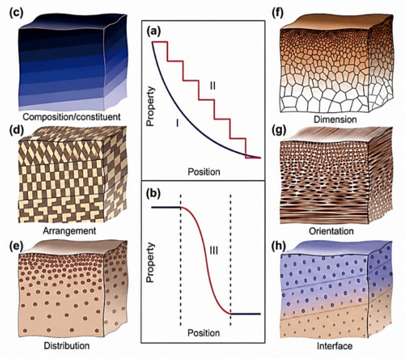
The biological systems of gradient materials have multi-level structural diversity and organization. These properties are associated with the spatial distribution of components in the material, variations in density and microstructure, and a gradation of scales and structures between the micro- and nanoscales. Structural-unit direction and successive-layer patterns are also important in forming these gradient structures, as are partial gradual contacts between different components and the combination of several structural gradients within a single material, as shown in Figure 1. In biomedical applications, FGMs offer an effective solution to problems associated with traditional metal implants, especially the mechanical incompatibility between the implant and the surrounding bone tissue, which can lead to stress shielding. The gradation in mechanical properties of FGMs reduces the modulus mismatch between the implant and bone, thereby improving load distribution and increasing the bone response around the implant [24].
Figure 1. Local property profiles and basic forms of gradients in biological materials. a) Local properties change either gradually (I) or in a stepwise manner (II) through the entire material volume. b) Local properties vary continuously across the interface between dissimilar components. c–h) Gradients in biological materials are fundamentally associated with changes in chemical composition and constituents, and c) structural characteristics, including the arrangement (d), distribution (e), dimensions (f), and orientations (g) of building units; h) Gradient interfaces in biological materials [24]
Figure 1 provides a conceptual illustration of structural gradient patterns and mechanical properties in biomaterials. It demonstrates how the local properties of biomaterials vary with changes in chemical composition and microstructure, either within the material or across interfaces between components. The concept of gradients in biomaterials is an important design paradigm employed by nature to create materials with improved mechanical performance, such as the combination of stiffness, toughness, and fracture resistance. The first part of the figure shows how the mechanical or physical properties of a material vary with position within a structure. In the first case (I), the property changes continuously and gradually throughout the material's volume, varying its value from one region to another without any sharp boundaries. This gradual change allows for a balanced distribution of the stresses, limiting the build-up of localized stresses that can lead to cracking or structural failure. The second case (II) describes a change between successive steps or levels. The material consists of several layers with slightly different properties, so that the change is gradual rather than a discrete transition. The third case (III) depicts the property dependence at the interface of two different materials. The properties are very similar in each material but gradually change across the transition zone between them. Such gradual change is essential in biological systems, as it minimizes mechanical incompatibility between different materials and prevents stress concentration at the interface. Many biological structures exhibit such phenomena, for example, the junction between bone and cartilage or between different layers of teeth. The second part of the figure depicts the basic structural forms of gradients in biological materials, which may manifest at several levels of organization. The first of these is the gradient in chemical composition or in the ratios of components (Composition/Constituent). In this case, the proportions of organic and mineral or other components gradually change throughout the material. This, in turn, leads to changes in mechanical and physical properties such as hardness, modulus of elasticity, and abrasion resistance. Additionally, the building blocks of the material may be patterned in a graded manner, with the same components present but different spatial patterns. This structural arrangement directly affects stress transmission and crack propagation in the material, thereby improving fracture resistance. There may also be gradation in the distribution of components, where particle, pore, or fiber density varies through the material. This leads to a gradual change in mechanical and physical properties such as density, stiffness, and permeability. Another type of gradation is variation in the size of structural units, where the cells, sheets, grains, or fibers may vary in size from one region to another. This difference in microscopic size affects the material's overall mechanical behavior because the microstructure is critical to its strength and durability. A gradient in the orientation of structural units, such as fibers or crystalline sheets, may be observed, with the angles of these structures varying gradually throughout the material. This change increases the material’s ability to withstand multi-directional loads and decreases the likelihood of crack propagation. The microscopic images in the figure show real examples of these gradients in natural biomaterials, with changes in the layered structure, the orientation of building blocks, and their distribution and dimensions at the micro- and nano-scale. These observations support the idea that structural gradients constitute an effective natural design approach for biomaterials to achieve an optimal balance between rigidity, durability, and energy absorption capacity. Hence, knowledge of these transition zones in biological materials provides an important basis for biomaterials engineering and the development of FGMs that mimic these natural strategies, aiming to create synthetic materials with improved mechanical and functional properties, especially in medical applications such as bone and dental implants.
To achieve a better balance between mechanical properties and biocompatibility of metal implants, recent research has focused on combining biomaterials with metals, and HA is one of the most prominent biomaterials due to its chemical similarity to the mineral phase of natural bone. HA is a calcium phosphate (CaP) compound with the chemical formula Ca10(PO4)6(OH)2. It is the most abundant component of the mineral phase of human bone [25]. This structural and chemical similarity confers a high degree of biocompatibility and enables it to interact favorably with bone tissue without eliciting significant immune or inflammatory responses. HA possesses unique biological properties, making it a bioactive material that can form direct chemical bonds with surrounding bone tissue, promoting osseointegration between the implant and bone. Also, its surface provides an optimal environment for bone cell adhesion and proliferation, thereby accelerating new bone formation around the implant. It is therefore widely used in dental and orthopedic applications both as a ceramic material and as a coating on metallic implants.
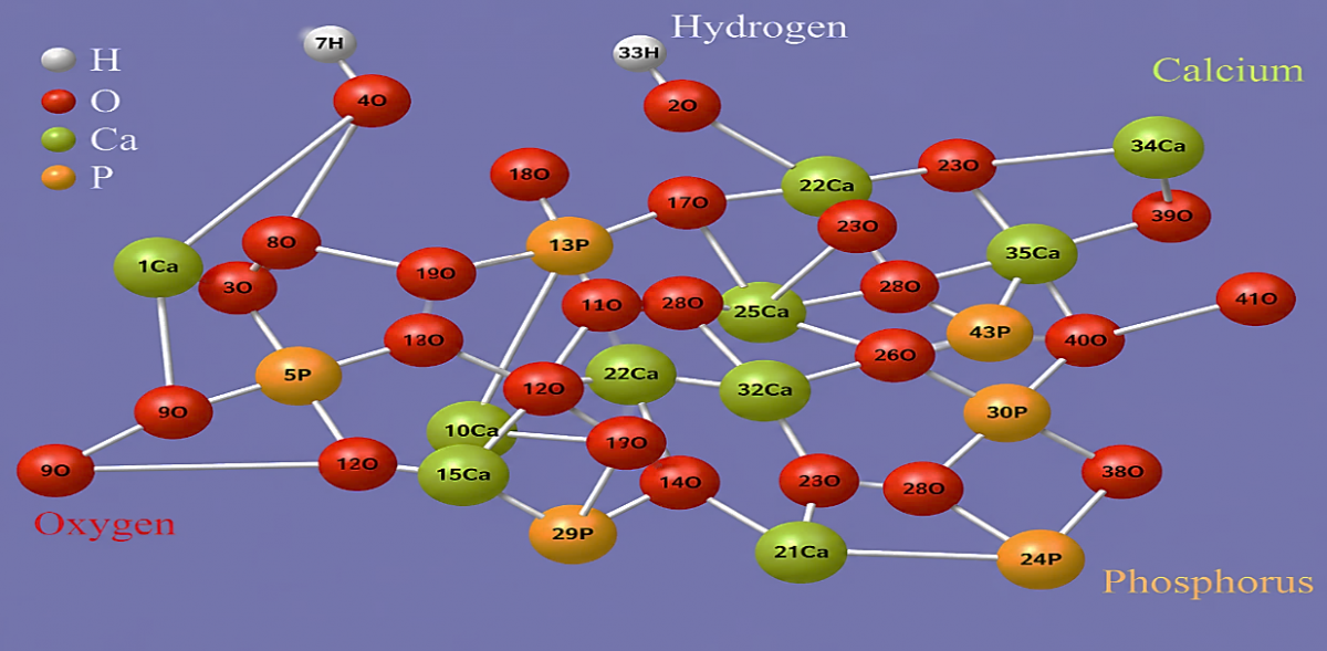
HA has a hexagonal crystal structure with space group P63/m; the angles α and β are 90°, and γ is approximately 120°. The lattice constants are about a = 9.4 A° and c = 6.88 A°. In this system, the unit cell contains six groups of phosphate ions (PO₄³⁻) and two hydroxyl ions (OH⁻) in the center of the cell. The spatial distribution of calcium ions (Ca2+), phosphate groups, and hydroxyl ions in the network is shown in Figure 2. Pure HA has two distinct sites for calcium ions: Site 1, Ca(I), at the center of the hexagonal arrangement of ions. The second site, Ca(II), is at the corners of the hexagonal lattice and has different crystallographic positions. Surrounding the OH⁻ ion channel are six calcium atoms, which reflects the regular geometric character of the material’s hexagonal structure [3].
Figure 2. The crystalline structure of hydroxyapatite (HA) [3]
The interface between the metallic and ceramic phases is a critical region in composites and FGMs used for bone replacement applications, as it plays a key role in ensuring the uniform transfer of mechanical loads between the various components and preventing stress concentrations. The efficiency of this interface depends on the strength of the interfacial bonding and the degree of physical and chemical compatibility between the material's constituent phases. However, differences in the elastic modulus and coefficient of thermal expansion between the metal and the ceramic can generate residual stresses during manufacturing or service, leading to interfacial defects such as microporosity, cracking, and interfacial delamination.
The mechanical performance of composites and FGMs depends not only on the individual properties of their constituents but is also significantly influenced by the distribution of phases and the strength of the interfacial bonding between them. In metal–ceramic systems, the metallic phase contributes to toughness and fracture resistance, while the ceramic phase enhances hardness and resistance to bio-corrosion. A graded distribution of constituents minimizes abrupt changes in mechanical properties between the surface and the core, thereby limiting stress concentrations and improving load transfer across the structure. Consequently, evaluating the mechanical performance of FGMs must encompass analyses of failure mechanisms, fatigue behavior, and crack propagation as well as corrosion and wear resistance rather than being limited solely to hardness or compressive strength.
3.1 Fabrication routes for functionally graded materials
The manufacturing process is a significant parameter affecting the microstructure, mechanical, and biological properties of composite and graded materials used in medical implants. The manufacturing process determines the material's shape and controls the phase distribution, structural gradation, porosity, and quality of the metal–ceramic interface. Powder metallurgy (PM) is one of the most widely used methods for producing graded materials, in which several layers with different compositions can be built up by controlling the proportions of powders before pressing and sintering. This technique is characterized by low cost and the ability to control compositional gradation, but it may suffer from residual porosity and incomplete density in some areas [26].
Spark plasma sintering (SPS) is an advanced technique that allows for the production of fine-grained structures while reducing sintering time. It also limits grain growth and preserves the stability of heat-sensitive phases such as HA, but the limited sample volume restricts its use compared to conventional manufacturing methods [27].
In surface applications, plasma spraying is used to produce gradient coatings on metal implant surfaces. This technique allows for a continuous gradient between the metal and the bio-phase; however, residual thermal stresses may lead to cracking or stratification in some cases.
In contrast, cold spraying produces high-density coatings without exposing the material to high temperatures, thus reducing powder oxidation and preserving the chemical composition of sensitive materials. It also provides excellent adhesion to the metal surface and minimizes thermal cracking associated with plasma spraying [28].
In recent years, additive manufacturing (AM) has become one of the most promising trends in medical implants. These technologies allow for the production of porous, variable-particle structures with complex geometries, customized for each patient. They also offer precise control over porosity, chemical composition, and material distribution. However, high costs and the need for precise control of the microstructure remain major challenges to widespread industrial application [29].
Table 3 compares the main fabrication techniques for FGMs, outlining the types of grading achievable, key operating parameters, and potential drawbacks of each technique. PM and SPS have demonstrated efficiency in producing homogeneous structural grades and improving mechanical properties, while AM techniques such as selective laser melting (SLM) provide high control over structure, porosity, and complex implant geometry. The table also shows that defect types, including residual porosity, thermal stresses, and microcracks, vary by fabrication method. In general, these technologies are promising tools for developing bone implants that combine high biocompatibility with improved mechanical performance, thereby promoting bone integration and increasing the clinical lifespan of implants.
Table 3. Comparison of fabrication routes for functionally graded materials (FGMs) [26-31]
|
Fabrication Method |
Type of Gradient Produced |
Key Processing Parameters |
Typical Defects/Limitations |
Scalability |
|
Powder Metallurgy (PM) |
Compositional gradient, layered structures |
Powder particle size, compaction pressure, sintering temperature, and time |
Residual porosity, incomplete densification, interfacial voids |
High |
|
Spark Plasma Sintering (SPS) |
Fine-grained compositional gradients |
Pulsed current, heating rate, pressure, dwell time |
Localized overheating, size limitations |
Moderate |
|
Plasma Spraying |
Surface and coating gradients |
Plasma power, spray distance, particle velocity, feed rate |
Delamination, microcracks, residual stresses |
High |
|
Cold Spraying |
Surface compositional gradients |
Gas pressure, particle velocity, spray angle |
Incomplete bonding, non-uniform deposition |
High |
|
Additive Manufacturing (AM) |
Composition, porosity, and structural gradients |
Laser/electron beam power, layer thickness, scan speed, powder feed rate |
Lack of fusion, anisotropy, residual stresses |
Moderate to high |
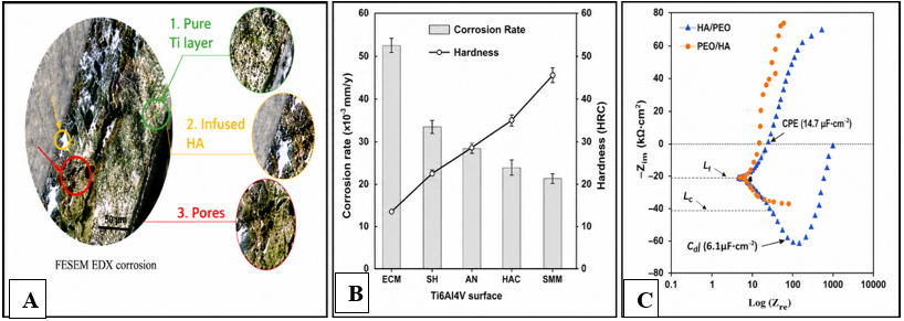
Marsh et al. [32] demonstrated that long-term orthopedic implant failure results from the interaction of multiple mechanical and biological factors, including fatigue stress, abrasive wear, and electrochemical corrosion. These factors, together with the release of particles and corrosion products, stimulate an inflammatory response and bone resorption around the graft. Therefore, improving implant surface properties is crucial for promoting osseointegration, increasing graft stability, and reducing the likelihood of long-term clinical failure. Ul Haq et al. [33] conducted a study to develop a graded titanium-hydroxyapatite (Ti-HA) bio-composite using PM techniques. The aim was to reduce mechanical incompatibility between the implant and bone and improve long-term biocompatibility. The graded structure was designed so that Ti in the inner region provides load-bearing capacity, while HA in the surface regions enhances biocompatibility and osseointegration, thereby improving implant performance. Structural analysis revealed a stable interface between the metallic and ceramic phases, with a graded distribution of components throughout the structure, supporting the composite’s structural integrity. Electrochemical tests also demonstrated a lower corrosion rate than pure porous Ti, attributable to enhanced oxide layer stability and reduced physiological permeability, as shown in Figure 3, indicating improved durability.
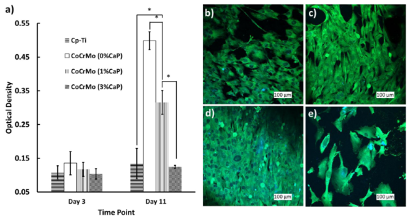
A study [34] investigated the effect of adding CaP to CoCrMo and pure titanium (Cp-Ti) alloy surfaces on osteoblast biocompatibility. 3-(4,5-dimethylthiazol-2-yl)-2,5-diphenyltetrazolium bromide (MTT) assay results showed increased cell viability and metabolic activity over time, with no cytotoxic effects. ALP and confocal microscopy analyses also showed improved cell adhesion, proliferation, and osteogenic differentiation on the CaP-containing surfaces. In addition, the surface treatments reduced bacterial adhesion while maintaining high osteoblast biocompatibility, confirming CaP surface modification as an effective strategy for promoting osseointegration and reducing the risk of infection associated with medical implants, as shown in Figure 4.
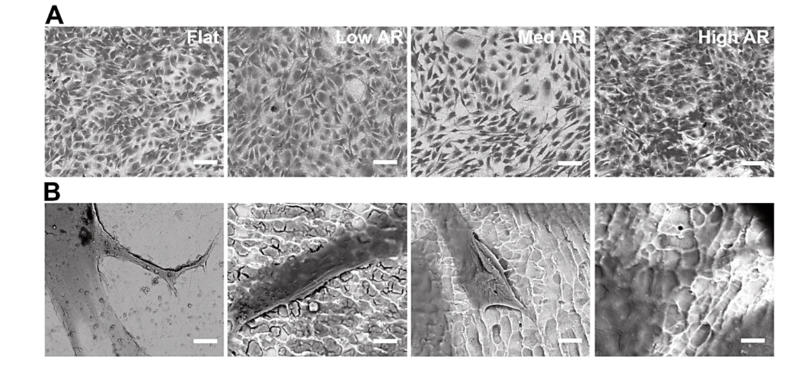
This study investigated the effect of the surface nanostructure of CoCrMo alloys on the behavior of human mesenchymal stem cells (hMSCs). The results showed that increasing the aspect ratio of the nanostructures improved cell proliferation and elongation and enhanced their adhesion to the surface. The electron microscopy images showed more pronounced filopodia on the nanostructured surfaces, indicating more focal adhesion sites and better cell-surface interaction. The findings indicate that modifications in nanoscale surface topography could promote cell adhesion and proliferation, resulting in increased osseointegration and performance of medical implants [34] (Figure 5).
The study showed that CaP-reinforced CoCrMo alloys had significantly better osseointegration around implants than unmodified CoCrMo alloys and Cp-Ti. Histological results showed enhanced new bone formation and improved bone–implant bonding over time, especially at higher CaP concentrations. This effect is attributed to CaP's ability to promote osteoblast adhesion, activate cellular signaling pathways associated with osteoproliferative and differentiating processes, stimulate the polarization of immune cells toward the M2 regenerative mode, and increase the secretion of growth factors that promote healing and vascularization. These results suggest that CaP surface modification of CoCrMo is an effective strategy to improve biocompatibility and accelerate osseointegration of medical implants [35], as shown in Figure 6.
Figure 5. Effect of surface topography of cobalt-chromium-molybdenum (CoCrMo) alloy on cell adhesion and spreading [34]
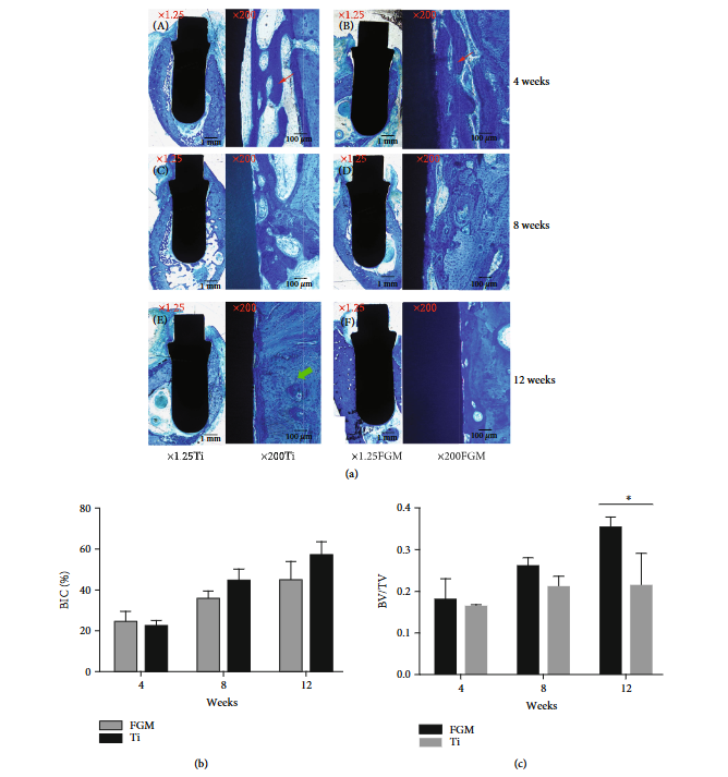
The study by Wang et al. [25] aimed to develop titanium-hydroxyapatite (Ti-HA FGM) graded dental implants to reduce implant-bone mechanical incompatibility and improve osseointegration. Bioassays and histological tests showed that the graded implants achieved a significant increase in peri-implant bone formation and a higher bone-to-volume ratio (BV/TV) compared to conventional Ti implants. The results also demonstrated improved biocompatibility and long-term stability due to the combination of Ti's mechanical strength and HA’s bioactivity, as shown in Figure 7.
Figure 7. Histological analysis of Ti-HA FGM dental implants in vivo. (a) (A, B) Images illustrate that the calcified tissues around the implant were immature (white arrow) and surrounded by vascular spaces; (C, D) images obtained at week 8 show greater contact between the implant and the denser osseous structure; and (E, F) at week 12 in the Ti group and week 8 in the Ti-HA FGM group, mature osseous tissue can be seen gradually surrounding the implant surface (green arrow). (b) From weeks 4–12, bone–implant contact (BIC) (%) increased faster in the Ti-HA FGM group than in the Ti group. (c) The Ti-HA FGM dental implant exhibited higher bone-to-volume ratio (BV/TV) values [25]
Figure 7 illustrates the osseointegration evaluation of Ti-HA-graded implants compared with conventional Ti implants at 4, 8, and 12-weeks post-implantation. The results showed a gradual increase in bone formation and improved BIC and BV/TV values over time, with higher values in the graded implants, indicating enhanced osseointegration and improved implant retention compared to conventional Ti [36].
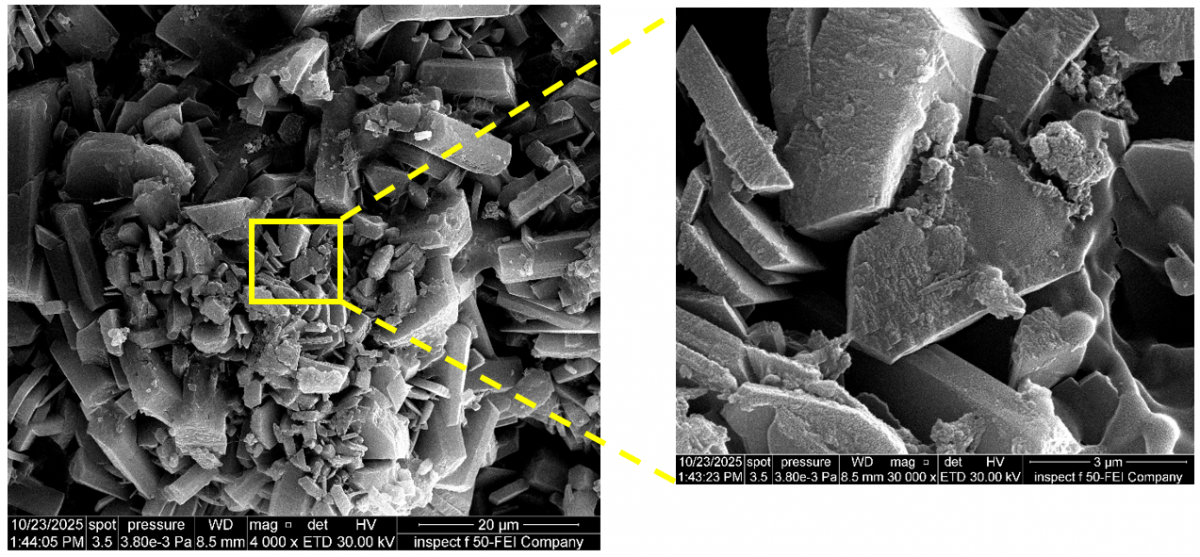
Al Hussainey et al. [37] developed a graded biomaterial composed of an F75 (Co–Cr–Mo) alloy reinforced with HA and 4% Ti using PM, as shown in Figure 8. The graded structure reduced mechanical incompatibility between the implant and bone by lowering the modulus of elasticity to 34.37 ± 0.49 GPa, which is close to that of cortical bone. The material also exhibited improved hardness (375.63 ± 17.8 HV) and a significant reduction in wear rate (0.413 ± 0.015 mpy) due to the formation of a stable TiO₂ layer. Furthermore, bioactivity tests demonstrated high bioactivity capacity and antibacterial activity exceeding 95%, indicating that this graded system offers an effective balance between mechanical strength, wear resistance, and biocompatibility for bone–implant applications [37].
Table 4 compares the aforementioned studies on the development of functionally graded and surface-modified metallic biomaterials for bone grafting applications. These studies showed that incorporating HA into Ti or CoCr alloys, together with surface enhancement through functionally graded design and surface nano-modification, improved biocompatibility, osseointegration, and osteoblast activity. Marsh et al. [32] also demonstrated this. In addition, resistance to wear and fatigue, as well as reduced long-term implant failure, are key factors in improving the clinical performance of implants. Taken together, the results indicate that improving the structural composition and surface properties of metal implants contributes to enhanced osseointegration, improved biostability, and extended operational lifespan of bone implants.
Figure 8. Surface morphology of apatite formation on the functionally graded materials (FGMs) at different magnifications [37]
Table 4. Comparative summary of recent studies on functionally graded and surface-modified metallic biomaterials
|
Study |
Material System |
Method |
Main Findings |
Biomedical Significance |
|
[32] |
Ti, CoCrMo, and stainless-steel orthopedic implants |
Metal evaluation |
Identified wear, corrosion, and fatigue failure |
Supports the development of surface modifications |
|
[33] |
Ti-HA bio-composite |
Powder metallurgy (PM) |
Improved corrosion resistance and bioactivity |
Better biocompatibility and mechanical balance |
|
[34] |
Nanostructured CoCrMo |
Nano-topographical modification |
Enhanced cell adhesion and proliferation |
Improved implant integration |
|
[35] |
CoCrMo + CaP |
CaP incorporation |
Higher osteoblast viability and lower bacterial adhesion |
Enhanced biocompatibility |
|
[36] |
Ti-HA FGM |
Functionally graded design |
Higher BIC and BV/TV; enhanced bone formation |
Improved osseointegration |
|
[37] |
F75 CoCrMo/HA/Ti FGM |
Functionally graded design |
Reduced elastic modulus, increased hardness, and antibacterial activity |
Reduced stress shielding |
Note: Titanium-hydroxyapatite (Ti-HA), cobalt–chromium–molybdenum (CoCrMo), calcium phosphate (CaP), functionally graded material (FGM), bone–implant contact (BIC), bone-to-volume ratio (BV/TV).
This review shows that bio-metallic materials used in medical implants have evolved from traditional single-phase metals to advanced systems with bio-coatings and FGMs. A review of the evolution of orthopedic implant materials reveals that stainless steel, cobalt, and titanium alloys, despite their varying degrees of mechanical suitability and corrosion resistance, do not individually satisfy the comprehensive mechanical and biological requirements of an implant. While the use of HA has demonstrated a clear ability to enhance bioactivity and osseointegration, its direct integration with metals can pose challenges regarding bonding and mechanical stability.
In this context, FGMs emerge as a more integrated solution, enabling a gradual transition in the implant's composition and properties from the mechanically supportive metal to the bioactive HA. This minimizes the abrupt discontinuity at the interface and enhances the implant's compatibility with the bone. However, achieving a controlled and stable gradient while simultaneously minimizing porosity and defects and managing reactions during sintering remains the critical factor for the successful fabrication and clinical application of these materials.
The authors extend their sincere thanks and appreciation to Mustansiriyah University for its continuous academic support and encouragement of scientific research.
[1] Ladani, L., Palmieri, M. (2024). Review of the use of metals in biomedical applications: Biocompatibility, additive manufacturing technologies, and standards and regulations. Metals, 14(9): 1039. https://doi.org/10.3390/met14091039
[2] Jaafar, A., Hecker, C., Árki, P., Joseph, Y. (2020). Sol-gel derived hydroxyapatite coatings for titanium implants: A review. Bioengineering, 7(4): 127. https://doi.org/10.3390/bioengineering7040127
[3] Kareem, R.O., Bulut, N., Kaygili, O. (2024). Hydroxyapatite biomaterials: A comprehensive review of their properties, structures, medical applications, and fabrication methods. Journal of Chemical Reviews, 6(1): 1-26. https://doi.org/10.48309/jcr.2024.415051.1253
[4] Suansuwan, N., Swain, M.V. (2002). Determination of elastic properties of metal alloys and dental porcelains. Journal of Oral Rehabilitation, 28(2): 133-139. https://doi.org/10.1046/j.1365-2842.2001.00642.x
[5] Naebe, M., Shirvanimoghaddam, K. (2016). Functionally graded materials: A review of fabrication and properties. Applied Materials Today, 5: 223-245. https://doi.org/10.1016/j.apmt.2016.10.001
[6] Shi, H.Y., Zhou, P., Li, J., Liu, C.Z., Wang, L.Q. (2021). Functional gradient metallic biomaterials: Techniques, current scenery, and future prospects in the biomedical field. Frontiers in Bioengineering and Biotechnology, 8: 616845. https://doi.org/10.3389/fbioe.2020.616845
[7] Teoh, S.H. (2000). Fatigue of biomaterials: A review. International Journal of Fatigue, 22(10): 825-837. https://doi.org/10.1016/S0142-1123(00)00052-9
[8] Findik, F. (2020). Recent developments of metallic implants for biomedical applications. Periodicals of Engineering and Natural Sciences, 8(1): 33-57. https://doi.org/10.21533/pen.v8.i1.1029
[9] Gurappa, I. (2002). Characterization of different materials for corrosion resistance under simulated body fluid conditions. Materials Characterization, 49(1): 73-79. https://doi.org/10.1016/S1044-5803(02)00320-0
[10] Hermawan, H. (2012). Introduction to metallic biomaterials. In Biodegradable Metals, pp. 1-11. https://doi.org/10.1007/978-3-642-31170-3_1
[11] Niinomi, M. (2003). Recent research and development in titanium alloys for biomedical applications and healthcare goods. Science and Technology of Advanced Materials, 4(5): 445-454. https://doi.org/10.1016/j.stam.2003.09.002
[12] Antunes, R.A., de Oliveira, M.C.L. (2012). Corrosion fatigue of biomedical metallic alloys: Mechanisms and mitigation. Acta Biomaterialia, 8(3): 937-962. https://doi.org/10.1016/j.actbio.2011.09.012
[13] Arifin A, Sulong AB, Muhamad N, Syarif J, Ramli MI. Material processing of hydroxyapatite and titanium alloy (HA/Ti) composite as implant materials using powder metallurgy: A review. Materials & Design. 2014;55:165-75.https://doi.org/10.1016/j.matdes.2013.09.045.
[14] Zhou, Y.L., Niinomi, M., Akahori, T. (2004). Effects of Ta content on Young's modulus and tensile properties of binary Ti-Ta alloys for biomedical applications. Materials Science and Engineering: A, 371(1-2): 283-290. https://doi.org/10.1016/j.msea.2003.12.011
[15] Wang, K. (1996). The use of titanium for medical applications in the USA. Materials Science and Engineering: A, 213(1-2): 134-137. https://doi.org/10.1016/0921-5093(96)10243-4
[16] Geetha, M., Singh, A.K., Muraleedharan, K., Gogia, A.K., Asokamani, R. (2001). Effect of thermomechanical processing on microstructure of a Ti-13Nb-13Zr alloy. Journal of Alloys and Compounds, 329(1-2): 264-271. https://doi.org/10.1016/S0925-8388(01)01604-8
[17] Gorsse, S., Miracle, D.B. (2003). Mechanical properties of Ti-6Al-4V/TiB composites with randomly oriented and aligned TiB reinforcements. Acta Materialia, 51(9): 2427-2442. https://doi.org/10.1016/S1359-6454(02)00510-4
[18] Hansen, D.C. (2008). Metal corrosion in the human body: The ultimate bio-corrosion scenario. The Electrochemical Society Interface, 17(2): 31-34. https://doi.org/10.1149/2.F04082IF
[19] Hao, Y.L., Yang, R., Niinomi, M., et al. (2002). Young's modulus and mechanical properties of Ti-29Nb-13Ta-4.6Zr in relation to α″ martensite. Metallurgical and Materials Transactions A, 33: 3137-3144. https://doi.org/10.1007/s11661-002-0299-7
[20] Ankem, S., Greene, C.A. (1999). Recent developments in microstructure/property relationships of beta titanium alloys. Materials Science and Engineering: A, 263(2): 127-131. https://doi.org/10.1016/S0921-5093(98)01170-8
[21] Davis, R., Singh, A., Jackson, M.J., et al. (2022). A comprehensive review on metallic implant biomaterials and their subtractive manufacturing. The International Journal of Advanced Manufacturing Technology, 120: 1473-1530. https://doi.org/10.1007/s00170-022-08770-8
[22] Solanke, S., Gaval, V. (2021). Most commonly used metallic biomaterials for plasma sprayed hydroxyapatite coatings. IOP Conference Series: Materials Science and Engineering, 1168: 012013. https://doi.org/10.1088/1757-899X/1168/1/012013
[23] Habeeb, A.M., Salih, N.A. (2024). Fabrication and mechanical characterization of functionally graded NiTi/HA alloys. Revue des Composites et des Matériaux Avancés-Journal of Composite and Advanced Materials, 34(1): 77-86. https://doi.org/10.18280/rcma.340110
[24] Li, Y., Feng, Z.Y., Hao, L., et al. (2020). A review on functionally graded materials and structures via additive manufacturing: From multi-scale design to versatile functional properties. Advanced Materials Technologies, 5(6): 1900981. https://doi.org/10.1002/admt.201900981
[25] Wang, X.H., Wan, C.P., Feng, X.X., Zhao, F.Y., Wang, H.M. (2021). In vivo and in vitro analyses of titanium-hydroxyapatite functionally graded material for dental implants. BioMed Research International, 2021(1): 8859945. https://doi.org/10.1155/2021/8859945
[26] Hamweendo, A., Malama, T., Botef, I. (2016). Titanium-nickel alloys for bone tissue engineering application via cold spray. In International Conference on Competitive Manufacturing (COMA’16), Stellenbosch, South Africa, pp. 273-279. https://doi.org/10.13140/RG.2.1.4046.9527
[27] Sun, L.M., Berndt, C.C., Gross, K.A., Kucuk, A. (2002). Material fundamentals and clinical performance of plasma-sprayed hydroxyapatite coatings: A review. Journal of Biomedical Materials Research, 58(5): 570-592. https://doi.org/10.1002/jbm.1056
[28] Sun, W., Chu, X., Lan, H.M., et al. (2022). Current implementation status of cold spray technology: A short review. Journal of Thermal Spray Technology, 31: 848-865. https://doi.org/10.1007/s11666-022-01382-4
[29] Sidambe, A.T. (2014). Biocompatibility of advanced manufactured titanium implants—A review. Materials, 7(12): 8168-8188. https://doi.org/10.3390/ma7128168
[30] Svetlizky, D., Das, M., Zheng, B.L., et al. (2021). Directed energy deposition (DED) additive manufacturing: Physical characteristics, defects, challenges and applications. Materials Today, 49: 271-295. https://doi.org/10.1016/j.mattod.2021.03.020
[31] Gao, B.W., Zhao, H.J., Peng, L.Q., Sun, Z.X. (2022). A review of research progress in selective laser melting (SLM). Micromachines, 14(1): 57. https://doi.org/10.3390/mi14010057
[32] Marsh, A.C., Pajares Chamorro, N., Chatzistavrou, X. (2019). Long-term performance and failure of orthopedic devices. In Bone Repair Biomaterials, Elsevier, pp. 379-410. https://doi.org/10.1016/B978-0-08-102451-5.00015-9
[33] Ul Haq, E., Ahmed, F., Rehman, F.U., et al. (2023). Synthesis and characterization of a titanium-based functionally graded material-structured biocomposite using powder metallurgy. ACS Omega, 8(32): 28976-28983. https://doi.org/10.1021/acsomega.3c01471
[34] Man, K., Mazumder, S., Dahotre, N.B., Yang, Y. (2023). Surface nanostructures enhanced biocompatibility and osteoinductivity of laser-additively manufactured CoCrMo alloys. ACS Omega, 8(50): 47658-47666. https://doi.org/10.1021/acsomega.3c04305
[35] Bandyopadhyay, A., Shivaram, A., Isik, M., Avila, J.D., Dernell, W.S., Bose, S. (2019). Additively manufactured calcium phosphate reinforced CoCrMo alloy: Bio-tribological and biocompatibility evaluation for load-bearing implants. Additive Manufacturing, 28: 312-324. https://doi.org/10.1016/j.addma.2019.04.020
[36] Sadeq, J.A., Hussain, R.K., Majeed, A.M.A. (2023). Numerical analysis of PMMA/HAP/MgO composite for dental applications. AIP Conference Proceedings, 2834: 090013. https://doi.org/10.1063/5.0161578
[37] Al Hussainey, A.M., Mustafa, A., Hussain, R.K. (2026). Synthesis and characterization of Ti-Enhanced F75/HA functionally graded materials fabricated by powder metallurgy. Karbala International Journal of Modern Science, 12(2): 191-203. https://doi.org/10.33640/2405-609X.3453