Antibacterial Efficacy of Crude Postbiotics from Enterobacter sp., Bacillus thuringiensis, and Lactobacillus plantarum against Marine Fish Bacterial Pathogens

Chun Yao Ang | Mohd Fadhli Zil Ikram Sidiqin Md Saffie | Leong Seng Lim | Annita Seok Kian Yong | Sujjat Al Azad | Motohiko Sano | Nor Azman Kasan | Francis Yong | Ilham Misbakudin Al Zamzami | Mohammad Tamrin Mohamad Lal*
© 2026 The authors. This article is published by IIETA and is licensed under the CC BY 4.0 license (http://creativecommons.org/licenses/by/4.0/).
OPEN ACCESS
This study evaluates crude cell-free postbiotic extracts derived from three shrimp gut–associated probiotic isolates, Enterobacter sp. P17, Bacillus thuringiensis, P18, and Lactobacillus plantarum P20 as a sustainable disease-control strategy in marine fish aquaculture. Crude extracts of the organic, interphase, and aqueous layers obtained after ethyl-acetate partitioning of sterile cell-free supernatants (CFS) were screened against four marine fish pathogens (Vibrio alginolyticus ATCC 17749, V. anguillarum ATCC 19264, V. parahaemolyticus SS07, and Photobacterium damselae SS16). All three isolates produced extracts active against the four pathogens and were therefore advanced to the full characterization workflow. Across the three isolates, the minimum inhibitory concentration (MIC) ranged from 10% to 25% and the minimum bactericidal concentration (MBC) from 10% to 30%. The aqueous-layer extracts of all three isolates exhibited the lowest MIC (10%) against every tested pathogen, and the aqueous extract of B. thuringiensis P18 was the most potent overall, producing the lowest MBC (10%) against three of the four pathogens. At 1× and 2× MIC, the selected extracts suppressed the exponential growth phase of V. parahaemolyticus SS07 over a 12-h period. Antibacterial activity was retained after heating from 40 ℃ to 121 ℃, indicating high thermal stability. These in vitro findings support the potential of crude postbiotic extracts as an antimicrobial strategy in aquaculture, but in vivo fish-challenge trials, toxicity assessment, water/feed-matrix stability tests, and dose optimization are still required before farm-level application.
crude postbiotic extracts, cell-free metabolites, antibiotic resistance, aquaculture disease management, sustainable aquaculture, circular microbiology
Marine fish aquaculture plays a significant role in Malaysia’s economy. In 2024, the wholesale value of marine finfish production in Malaysia was approximately RM2.02 billion, with seabass alone contributing over RM1.04 billion [1]. Despite its economic importance, marine fish farming is highly vulnerable to various diseases. Several disease control approaches have been implemented, including improved disease and effluent management practices as well as chemical and antimicrobial treatments [2]. The excessive use of antibiotics is of particular concern, as it can promote the emergence of antibiotic-resistant bacteria that pose serious risks to human health [3]. Although probiotics are commonly applied as biological control agents in aquaculture, they may harbor mobile genetic elements carrying antibiotic resistance genes, which can potentially be transferred to environmental microorganisms [4]. Consequently, alternative disease management strategies, such as postbiotics, are being increasingly explored.
Various alternative disease control strategies have been investigated in aquaculture. In line with sustainability goals, there is a growing trend toward utilizing waste-derived materials in aquaculture applications [5, 6]. Nevertheless, information regarding the use of waste materials specifically for disease management remains limited. Among these materials, crude cell-free metabolites produced by beneficial bacteria have received comparatively little attention in aquaculture. These preparations, collectively termed postbiotics and also referred to in the literature as metabiotics, biogenics, bacterial metabolites, or processed cell-free supernatants (CFS), consist of soluble compounds secreted by live cells or released after controlled cell lysis, which can exert beneficial physiological effects on the host [7, 8]. In the present study, the term crude postbiotic extract refers specifically to the concentrated organic, interphase, and aqueous fractions recovered from ethyl-acetate partitioning of sterile-filtered CFS, rather than to undefined microbial waste. Postbiotics offer several advantages, including well-defined chemical compositions, established safe dosage ranges, and extended shelf life. Unlike probiotics, postbiotics do not pose a risk of antibiotic resistance gene transfer to environmental microbes, do not require the maintenance of bacterial viability during production and storage, exhibit stability under varying environmental conditions, and provide greater safety with the potential for controlled and standardized application methods [7, 9-11].
Beyond the immediate question of in vitro antibacterial activity, the use of crude postbiotic extracts is directly relevant to three pillars of sustainable marine aquaculture. First, antibacterial preparations that act through non-antibiotic mechanisms, such as short-chain organic acids, bacteriocin-like peptides, and small secondary metabolites, can displace a portion of the prophylactic and metaphylactic antibiotic use that currently drives the selection of antibiotic-resistant Vibrio and Photobacterium strains in coastal farms [3, 4]. Second, because the active material is cell-free, it does not introduce viable bacteria, plasmids, or transferable resistance genes into the rearing water, reducing the environmental load and the risk of resistance-gene spread to wild microbial communities [4, 7]. Third, the producer cultures used to generate postbiotic extracts can be grown on low-value substrates, including processing side-streams and aquaculture-system effluents [12-15], so the workflow described here is compatible with a circular-microbiology framing in which microbial by-products that would otherwise be discarded are valorized into functional antimicrobials. The present study addresses the first step in that workflow, demonstrating that crude postbiotic extracts from three shrimp gut-associated probiotic isolates can inhibit four key marine fish pathogens under controlled in vitro conditions.
2.1 Bacterial culture
Three probiotic isolates previously recovered from the gastrointestinal tract of pond-cultured Litopenaeus vannamei in Tuaran, Sabah, Malaysia, and characterized in our earlier work [16], namely Enterobacter sp. P17, Bacillus thuringiensis P18, and Lactobacillus plantarum P20 were used throughout the present study. These three isolates were the sole probiotic candidates examined; no additional isolates were screened or excluded. All subsequent assays (antibacterial screening, minimum inhibitory concentration (MIC), minimum bactericidal concentration (MBC), growth curve, and thermal-stability test) were performed on crude postbiotic extracts derived from these three strains. Bacterial cultivation was conducted with minor modifications of previously described methods [17]. Briefly, bacterial glycerol stocks were inoculated into de Man, Rogosa and Sharpe (MRS) broth (Merck, Darmstadt, Germany) and incubated for 96 h at room temperature (28 ± 2 ℃) using an orbital shaker operating at 130 rpm to obtain the working cultures.
Cell free supernatant (CFS) was obtained by centrifuging the producer cultures at 5,000 rpm for 15 min and filtering the supernatant through a 0.45 µm syringe filter. The sterile CFS was then partitioned against an equal volume of ethyl acetate (1:1, v/v) as described by Burianek and Yousef [18] and Islam et al. [19], with solvent substitution. The mixture was vigorously shaken in a separatory funnel and allowed to stand until three distinct phases formed: (i) an upper organic phase containing predominantly hydrophobic metabolites partitioned into the ethyl-acetate layer, (ii) an interphase layer in which amphiphilic peptide- or lipopeptide-like molecules tend to accumulate, and (iii) a lower aqueous phase retaining water-soluble metabolites such as organic acids, low-molecular-weight peptides, and polar secondary metabolites. The organic and interphase fractions were collected separately and concentrated to dryness under reduced pressure on a rotary evaporator at 40 ℃ until no visible solvent remained; residual ethyl acetate was further removed by overnight evaporation under a gentle nitrogen stream in a fume hood, after which each dried residue was resuspended in sterile Tris buffer (pH 7.0) at a CFS-equivalent volume ratio of 5:1 (CFS: buffer). The aqueous fraction was sterile-filtered (0.22 µm) and stored without further concentration. In this study, all three fractions are collectively referred to as crude postbiotic extracts because no purification step beyond solvent partitioning was performed; each fraction was evaluated independently against the four target pathogens so that the contribution of hydrophobic (organic), amphiphilic (interphase), and water-soluble (aqueous) metabolites to the overall antibacterial activity could be compared. All preparations were stored at −20 ℃ until use.
Crude postbiotic extracts obtained from the organic and interphase layers were resuspended in Tris buffer adjusted to pH 7 at a ratio of 5:1 (CFS: buffer). All crude postbiotic preparations were stored at −20 ℃ until further analysis.
2.2 Antibacterial screening and spectrum assays
The antibacterial effect of postbiotic extracts was determined through an antibacterial screening assay against four aquaculture bacterial pathogens, Vibrio parahaemolyticus SS07 [20], V. alginolyticus ATCC 17749, V. anguillarum ATCC 19264, and Photobacterium damselae SS16 [20]. Both the screening test for antibacterial effect and the antibacterial spectrum determination test were done using the agar spot assay method [21]. Briefly, bacterial culture of the pathogen was prepared with inoculation of bacterial glycerol stock in 2% NaCl supplemented Tryptone Soy Broth (TSB), followed by incubation at room temperature for 24 h in an orbital shaker at 130 rpm. About 100 µL of bacterial pathogen culture was inoculated in molten 0.5% Tryptone Soy Agar (TSA), swirled gently to mix, and poured onto 2% NaCl TSA plate. The molten 0.5% TSA was allowed to evenly coat the surface of the 2% NaCl TSA plate. After the molten TSA solidified, 10 µL of the postbiotic extract was pipetted onto the agar plate. The agar plate was set aside for the postbiotic extract to dry. Once the postbiotic extract had dried out, the agar plate was sealed with parafilm and incubated at room temperature for 16 h. The positive and negative controls were oxytetracycline and tris buffer at pH 7, respectively. Each postbiotic extract from organic, interphase, and aqueous layers was tested for each bacterial pathogen. After incubation, a transparent inhibition zone was observed and recorded as a positive result. Postbiotics with the highest spectrum were selected for further characterization.
2.3 Minimum inhibitory concentration and minimum bactericidal concentration
The MIC of the selected crude postbiotic extract against the tested pathogens was determined by adapting previously described methods by researchers [22-24]. As no standardized method is currently available for quantifying crude postbiotic concentrations, the undiluted extract was defined as 100% concentration. The 100% is the extract volume that equivalent to the original CFS. Pathogenic bacteria were cultured in TSB supplemented with 2% NaCl and incubated at room temperature for 24 h. Serial two-step dilutions of the crude postbiotic extract were prepared in 2%-NaCl TSB to obtain a broader final-concentration series of 50%, 40%, 30%, 25%, 20%, 15%, and 10% (v/v) for MIC determination. The dilution series was narrowed to 5%-interval steps between 10% and 30% after initial trials showed that MIC values fell within this range.
The pathogenic bacterial suspension was adjusted to an optical density of 1.0 at 600 nm (≈ 1 × 10⁸ CFU mL⁻¹) and diluted 1:100 in fresh 2%-NaCl TSB to a final working inoculum of ≈ 1 × 10⁶ CFU mL⁻¹, which was added 1:1 (v/v) to each crude postbiotic dilution in sterile glass tubes. Cultures were incubated at room temperature for 24 h. Bacterial growth was scored both visually (turbidity) and spectrophotometrically by measuring OD₆₀₀ before and after incubation; the MIC was defined as the lowest crude postbiotic concentration at which (i) no turbidity was visible and (ii) the post-incubation OD₆₀₀ increase relative to the t = 0 reading was ≤0.05 absorbance units (≥90% growth inhibition compared with the growth control). Each assay was carried out in three independent biological replicates, each with two technical replicates. Controls included: (i) growth control pathogen in TSB without postbiotic; (ii) sterility control TSB without pathogen; (iii) extract control postbiotic dilution series without pathogen, to verify extract sterility and to allow background-OD subtraction; and (iv) solvent control Tris buffer (pH 7) at the highest equivalent concentration carried over from the ethyl-acetate extraction step. Data are reported as mean ± SD of the three biological replicates.
The MBC of the selected crude postbiotic extract was also evaluated with modifications based on the methods reported by Parvekar et al. [23] and Fasolato et al. [24]. Following MIC determination, an aliquot from MIC tubes showing no visible bacterial growth was streaked onto TSA supplemented with 2% NaCl and incubated at room temperature for 24 h. MBC was defined as the lowest crude postbiotic concentration at which no bacterial colonies were observed on the TSA plates.
2.4 Growth curve assays
The bacterial growth curve assay was performed based on the method described by Huang et al. [25], with minor modifications. Vibrio parahaemolyticus was cultivated in TSB supplemented with 2% NaCl and incubated at room temperature for 24 h. The resulting bacterial suspension was standardized to an optical density of 1.0 at 600 nm. The adjusted culture was then inoculated into fresh TSB containing 2% NaCl and supplemented with crude postbiotics at concentrations equivalent to 1× and 2× the previously determined MIC. The cultures were incubated at room temperature for 12 h, during which optical density readings were recorded at 2 h intervals. All experiments were carried out in triplicate.
2.5 Temperature stability test
The influence of temperature and buffer type on the antimicrobial activity of the selected crude postbiotics was evaluated by subjecting the extracts to different thermal conditions and by suspending them in various buffer systems. To assess thermal stability, the crude postbiotics were heated using a block heater (Cole-Palmer, Stone, UK) at 40 ℃, 60 ℃, 80 ℃, and 100 ℃ for 20 min [26], as well as at 121 ℃ for 15 min [27]. Following thermal treatment, the remaining antibacterial activity was assessed against Vibrio parahaemolyticus SS07 using the soft overlay agar method. Untreated crude postbiotic served as the positive control. All assays were conducted in triplicate.
3.1 Screening of antibacterial activity of crude postbiotics
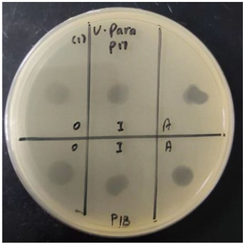
The antibacterial screening was carried out on crude organic, interphase, and aqueous-layer extracts obtained from the three probiotic isolates examined in this study, against the four marine fish pathogens listed in Section 2.2. All nine extracts (three isolates × three solvent fractions) produced a transparent inhibition zone against every tested pathogen on the soft-overlay agar; quantitative inhibition-zone diameters (mean ± SD, n = 3) are reported in Table 1, and representative plate images are shown in Figure 1. Because all three isolates are Enterobacter sp. P17, B. thuringiensis P18, and L. plantarum P20 exhibited broad-spectrum activity against the four target pathogens; the corresponding crude postbiotic extracts were carried forward into the MIC, MBC, growth-curve, and thermal-stability assays.
Table 1. Antibacterial activity of crude postbiotics against aquaculture bacterial pathogens
|
Pathogens |
Crude Postbiotics* |
||||||||
|
P17-O |
P17-I |
P17-A |
P18-O |
P18-I |
P18-A |
P20-O |
P20-I |
P20-A |
|
|
V. alginolyticus ATCC 17749 |
+ |
+ |
+ |
+ |
+ |
+ |
+ |
+ |
+ |
|
V. anguillarum ATCC 19264 |
+ |
+ |
+ |
+ |
+ |
+ |
+ |
+ |
+ |
|
V. parahaemolyticus SS07 |
+ |
+ |
+ |
+ |
+ |
+ |
+ |
+ |
+ |
|
P. damselae SS16 |
+ |
+ |
+ |
+ |
+ |
+ |
+ |
+ |
+ |
Figure 1. Antibacterial activity of crude postbiotics (P17 and P18) against aquaculture bacterial pathogens (V. parahaemolyticus)
3.2 Determination of minimum inhibitory concentration and MBC
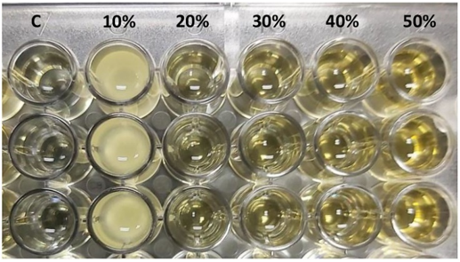
The selected crude postbiotics derived from isolates P17, P18, and P20 exhibited MIC values ranging from 10% to 25%. The MIC determination results are summarized in Table 2. Inhibition of bacterial growth was observed at crude postbiotic concentrations between 10% and 25%, as indicated by the absence of turbidity following inoculation with aquaculture bacterial pathogens (Figure 2).
Table 2. Results from the determination of MIC of crude postbiotics from selected isolates of Enterobacter sp. P17, B. thuringiensis P18, and L. plantarum P20 against aquaculture bacterial pathogens
|
Pathogens |
Probiotic Extracts* |
||||||||
|
P17 (%) |
P18 (%) |
P20 (%) |
|||||||
|
O |
I |
A |
O |
I |
A |
O |
I |
A |
|
|
V. alginolyticus ATCC 17749 |
15 |
20 |
10 |
15 |
15 |
10 |
15 |
20 |
10 |
|
V. anguillarum ATCC 19264 |
15 |
25 |
10 |
10 |
15 |
10 |
15 |
20 |
10 |
|
V. parahaemolyticus SS07 |
15 |
20 |
10 |
15 |
15 |
10 |
15 |
20 |
10 |
|
P. damselae SS16 |
20 |
20 |
10 |
15 |
20 |
10 |
15 |
25 |
10 |
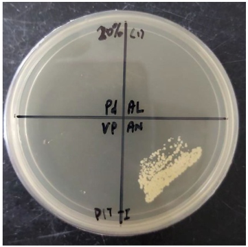
The MBC of crude postbiotics from isolates P17, P18, and P20 ranged from 10% to 30%, with detailed results presented in Table 3. Bactericidal activity was confirmed at these concentrations by the absence of colony formation on TSA supplemented with 2% NaCl (Figure 3), following subculturing from MIC tubes onto TSA.
Table 3. MBC (% v/v, mode of three independent biological replicates; range in parentheses where replicates differed) of the same crude postbiotic extracts, determined by sub-culturing aliquots from MIC tubes onto 2%-NaCl TSA
|
Pathogens |
Probiotic Extracts* |
||||||||
|
P17 (%) |
P18 (%) |
P20 (%) |
|||||||
|
O |
I |
A |
O |
I |
A |
O |
I |
A |
|
|
V. alginolyticus ATCC 17749 |
15 |
20 |
20 |
15 |
20 |
10 |
15 |
20 |
20 |
|
V. anguillarum ATCC 19264 |
15 |
25 |
10 |
15 |
15 |
10 |
15 |
20 |
10 |
|
V. parahaemolyticus SS07 |
15 |
20 |
10 |
15 |
15 |
10 |
15 |
20 |
20 |
|
P. damselae SS16 |
20 |
20 |
20 |
20 |
20 |
20 |
20 |
30 |
20 |
3.3 Growth curve assay
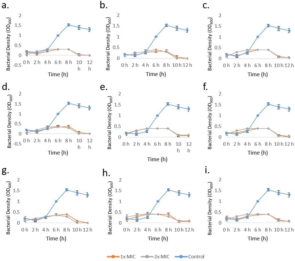
The growth curve assay further demonstrated that the exponential growth phase of V. parahaemolyticus SS07 was effectively inhibited by postbiotics derived from isolates P17, P18, and P20. As illustrated in Figure 4, the growth profiles of V. parahaemolyticus SS07 cultured in TSB supplemented with 1× and 2× MIC of crude postbiotics were monitored over a 12 h period at 2 h intervals. Compared to the untreated control, cultures exposed to crude postbiotics exhibited marked suppression of exponential bacterial growth.
3.4 Temperature stability tests
The effects of temperature and buffer type on the activity of crude postbiotics are summarized in Table 4. Antibacterial activity of the crude postbiotics against V. parahaemolyticus SS07 was retained following thermal treatment across a temperature range of 40 ℃ to 121 ℃, indicating high thermal stability. These results demonstrate that crude postbiotics derived from isolates P17, P18, and P20 remain stable under elevated temperature conditions.
Table 4. The effects of different temperatures on the antibacterial activities of the crude postbiotics against V. parahaemolyticus SS07
|
Temperature (℃) |
P17 (%) |
P18 (%) |
P20 (%) |
||||||
|
O |
I |
A |
O |
I |
A |
O |
I |
A |
|
|
40 |
+ |
+ |
+ |
+ |
+ |
+ |
+ |
+ |
+ |
|
60 |
+ |
+ |
+ |
+ |
+ |
+ |
+ |
+ |
+ |
|
80 |
+ |
+ |
+ |
+ |
+ |
+ |
+ |
+ |
+ |
|
100 |
+ |
+ |
+ |
+ |
+ |
+ |
+ |
+ |
+ |
|
121 |
+ |
+ |
+ |
+ |
+ |
+ |
+ |
+ |
+ |
This study demonstrated that postbiotics derived from probiotic bacteria exhibit antibacterial activity against a range of aquaculture bacterial pathogens. Crude postbiotics produced by isolates P17 (Enterobacter sp.), P18 (Bacillus thuringiensis), and P20 (Lactobacillus plantarum) showed inhibitory effects against all tested pathogens. These findings indicate that the postbiotics evaluated in this study are not species-specific. The antibacterial activities observed from these three isolates were not limited to Vibrio spp. or Photobacterium damselae, suggesting a broader antimicrobial spectrum.
Previous studies have reported that Enterobacter spp. possess antibacterial activity against several major aquaculture pathogens. For instance, supernatants from Enterobacter amnigenus and Enterobacter sp. PIC15 was shown to inhibit Flavobacterium psychrophilum [28-30]. Similarly, unfractionated CFS and ≤3 kDa fractions derived from Enterobacter sp. C6-6 exhibited inhibitory effects against F. psychrophilum [31]. In addition, a partially purified N-acyl-homoserine lactone lactonase enzyme obtained from the cell-free lysate of Enterobacter sp. was reported to suppress biofilm formation by Aeromonas hydrophila [32]. Antibacterial activity of B. thuringiensis has also been documented, where the supernatant of B. thuringiensis G5-8-3T02 inhibited Vibrio mimicus XQ [33]. Likewise, the supernatant of B. thuringiensis KL1 demonstrated inhibitory activity against other important aquaculture pathogens, including Aeromonas caviae ATCC 15468 [34]. The antimicrobial properties of L. plantarum have been widely investigated, with reports showing inhibition against V. parahaemolyticus, V. vulnificus, V. alginolyticus, V. mimicus, V. harveyi, and V. anguillarum [35-38]. However, to date, there are no published studies reporting antibacterial activity of Enterobacter sp. against Vibrio spp. and P. damselae. Therefore, the present study provides the first evidence of antibacterial activity of crude postbiotics derived from Enterobacter sp. against these aquaculture pathogens.
Among the three isolates examined, the inhibitory potential of L. plantarum against major aquaculture bacterial pathogens has been the most extensively studied. Neutralized and filtered supernatants of L. plantarum were reported to inhibit Pseudomonas fluorescens [39]. Moreover, CFS from L. plantarum exhibited antimicrobial activity against Edwardsiella tarda, Streptococcus iniae, Aeromonas veronii, Pse. fluorescens, and A. hydrophila [40-44]. Further investigation by Tremonte et al. [44] revealed that most L. plantarum strains produced organic acids as the primary inhibitory compounds, with only strain W_TA8 retaining antimicrobial activity against Pse. fluorescens after CFS neutralization to pH 7. Additional studies showed that CFS adjusted to pH 6 [45] or pH 5.5 [46] was still effective in inhibiting Streptococcus agalactiae and Pse. fluorescens, respectively. Enzyme-treated and pH-adjusted CFS of L. plantarum was also reported to inhibit Lactococcus garvieae [47]. Giri et al. [40] further demonstrated that intracellular products of L. plantarum VSG3, obtained through cell lysis and filtration, exhibited inhibitory activity against A. hydrophila. In addition, crude ethyl acetate extracts of L. plantarum CY 1-1 showed biofilm inhibition against Aeromonas sobria [48]. Purified bacteriocins produced by L. plantarum have also been reported to inhibit Pse. fluorescens [49-51], A. sobria, and A. hydrophila [48, 52].
The observed MIC and MBC values demonstrate that the selected crude postbiotics are effective against the tested bacterial pathogens. Specifically, crude postbiotics from isolates P17, P18, and P20 required concentrations of 10–25% to inhibit bacterial growth and 10–30% to achieve complete bactericidal effects. Determination of MIC and MBC is essential for subsequent application of postbiotics as disease control agents in aquaculture systems. Given that industrial-scale extraction and purification of postbiotic metabolites remain technically challenging [53, 54], establishing in vitro MIC and MBC values for crude extracts provides a first reference range for the relative potency of different producer–fraction combinations; these crude-extract percentages should not, however, be used directly as application doses in vivo until the active components have been identified and quantified. This approach is consistent with recommendations by Sahoo et al. [55], who emphasized the importance of dose determination for bacteriocin-based disease management in aquaculture.
Comparison of MIC and MBC values across the three solvent fractions (Tables 2 and 3) provides preliminary indications about the chemical nature of the active metabolites in each crude postbiotic preparation. For all three producer isolates, the aqueous-layer extracts consistently produced the lowest MIC (10% v/v) and, in most cases, the lowest MBC against the four target pathogens, whereas the organic- and interphase-layer extracts required higher crude-extract concentrations (15–25% v/v) to achieve equivalent inhibition. The disproportionately high activity of the aqueous fraction is consistent with the well-documented role of water-soluble metabolites, particularly short-chain organic acids (lactic, acetic, and formic acid) and small polar peptides as the principal antibacterial effectors of L. plantarum CFS [39, 44-46], and with the observation that pH-neutralized CFS often loses the bulk of its inhibitory activity [44]. For Bacillus thuringiensis P18, the additional activity recovered in the organic and interphase fractions is compatible with the presence of hydrophobic or amphiphilic secondary metabolites such as cyclic lipopeptides (e.g., iturin- and surfactin-family compounds [10]) and proteinaceous bacteriocin-like substances [33, 34], which partition preferentially into ethyl acetate or accumulate at the water/solvent interphase. For Enterobacter sp. P17, the observation that all three fractions retain measurable activity is consistent with the broader metabolite spectrum reported for this genus, including siderophore-like polar molecules and quorum-sensing-disrupting AHL-lactonases recovered from cell-free lysates [30, 32]. We emphasize that these assignments remain inferential; targeted chemical fractionation (HPLC-MS, NMR), heat- and protease-sensitivity profiling, and pH-neutralization assays of each layer are needed to confirm the identity of the active components in each fraction, and are listed in the Conclusion as priority follow-up work.
The growth curve assay is conducted to understand how the crude postbiotics affect the pathogens’ growth throughout the culture period. Although the growth of the bacterial pathogens treated with 1× and 2× MIC of postbiotics is almost the same, the presence of viable and non-viable cells might be different. Since 2× MIC has a higher concentration than the MBC of crude postbiotics, it is bactericidal to bacterial pathogens, and it is expected that no viable cells will be available. However, when it comes to an aquaculture environment, it is almost impossible to have an environment free of disease-causing agents. Assefa and Abunna [56] agreed that due to the aquaculture environment, the disease is difficult to detect and diagnose, and it allows the disease to transmit quickly. But the disease-causative agents can be brought to a controlled level to prevent any occurrence of disease outbreaks. Therefore, Assefa and Abunna [56] suggested that disease prevention strategies should be focused on instead. Based on the results of this study, postbiotics are a potential disease control strategy in aquaculture and have multiple benefits in comparison with other disease control strategies [7, 9-11]. However, the combination of different disease control strategies with postbiotics may give maximum disease protection to aquaculture animals [56].
Comparable findings have been reported for bacteriocins produced by lactic acid bacteria. Lim [26] reported that bacteriocins from Pediococcus acidilactici MCL11, Leuconostoc mesenteroides MCL12, and Enterococcus faecium MCL13 remained stable up to 80 ℃ after 10 min of heating, whereas bacteriocins from Lactobacillus sakei MCL14 and L. acidophilus MCL15 were stable up to 100 ℃ and 120 ℃, respectively. However, partial inactivation of bacteriocins from P. acidilactici MCL11, Leu. mesenteroides MCL12, and Ent. faecium MCL13 was observed at 100 ℃ [26].
Similarly, bacteriocin-like substances (BLS) produced by Ent. faecium Arla-18, ASR-6, JFR-1, OKA-14, and Yog-3S, Streptococcus thermophilus ASR-1 and TSB-8, as well as Lactobacillus casei JFR-5, exhibited thermal stability at 100 ℃ for up to 90 min [28]. Nevertheless, the activity of BLS from S. thermophilus ASR-1 declined following prolonged exposure at 100 ℃, and heating at 121 ℃ for 15 min resulted in complete inactivation of BLS from both S. thermophilus ASR-1 and L. casei JFR-5 [28]. Thermal stability up to 100 ℃ for 120 min has also been reported for bacteriocins produced by Lactococcus lactis subsp. lactis [57]. In contrast, bacteriocin-like inhibitory substances (BLIS) from Ent. faecium B3-8 showed a marked reduction in activity after 30 min at 90 ℃ and 15 min at 121 ℃, while BLIS from Ent. thailandicus B3-22 rapidly decreased after 15 min exposure at 121 ℃ [58]. Bacteriocins produced by Lactobacillus murinus AU06 were stable between 30 ℃ and 80 ℃, although a gradual decline in activity was observed with increasing temperature and exposure time starting from 40 ℃ [59]. In addition, bacteriocin from Weissella confusa A3 remained heat stable at 100 ℃ for 20 min [29].
Despite the extensive literature on the thermal stability of bacteriocins, studies addressing the temperature stability of postbiotic components, particularly organic acids, remain limited. Therefore, to the best of our knowledge, this study provides novel evidence regarding the thermal stability of bacteria-derived organic acids across a wide range of temperatures, supporting their potential application as robust antimicrobial agents in marine fish aquaculture systems.
This study demonstrates, under in vitro conditions, that crude cell-free postbiotic extracts produced by three probiotic isolates of Enterobacter sp. P17, Bacillus thuringiensis P18, and Lactobacillus plantarum P20 inhibit the growth of four economically relevant marine fish pathogens (Vibrio alginolyticus ATCC 17749, V. anguillarum ATCC 19264, V. parahaemolyticus SS07, and Photobacterium damselae SS16) at MIC values of 10–25% and MBC values of 10–30%, with the aqueous-layer extract of B. thuringiensis P18 being the most potent. The extracts also suppressed the exponential growth phase of V. parahaemolyticus SS07 over 12 h and retained measurable, although not unchanged, antibacterial activity after heating up to 121 ℃. These findings support the potential of crude postbiotic extracts as a complementary, antibiotic-sparing tool for marine fish disease management. However, the present evidence is strictly in vitro and does not yet establish farm-level efficacy. Before translational use, the following studies are required: (i) optimization of postbiotic production (fermentation conditions, biomass yield), recovery (ethyl-acetate alternatives and green solvents), and downstream purification of the bioactive metabolites; (ii) dose–response optimization in aquaculture-relevant matrices such as seawater, pelleted feed, and biofilm-coated tank surfaces; (iii) stability testing of the active fractions under storage, salinity, pH, and UV conditions encountered in farms; (iv) toxicity and biosafety evaluation in target species and non-target organisms, including hemolysis, organ histopathology, and impact on the host microbiome; and (v) in vivo challenge trials in seabass and other commercially important marine finfish. Pending these validations, the present results position crude postbiotic extracts as a promising contribution to sustainable, circular-microbiology-based disease management in marine aquaculture.
This research was supported by the Higher Institution Centre of Excellence (HICoE) Research Grant Scheme (Approval Letter No. JPT(BKPI)1000/016/018/35(2), Grant Code HIC2403) and Fundamental Research Grant Scheme (FRGS/1/2019/WAB01/UMS/02/1) provided by the Ministry of Higher Education, Malaysia.
[1] Annual Fisheries Statistics, Fisheries Department of Malaysia. (2024). https://www.dof.gov.my/en/resources/i-extension-en/annual-statistics/.
[2] Lakshmi, B., Viswanath, B., Sai Gopal, D.V.R. (2013). Probiotics as antiviral agents in shrimp aquaculture. Journal of Pathogens, 2013(1): 424123. https://doi.org/10.1155/2013/424123
[3] Ben, Y., Fu, C., Hu, M., Liu, L., Wong, M.H., Zheng, C. (2019). Human health risk assessment of antibiotic resistance associated with antibiotic residues in the environment: A review. Environmental Research, 169: 483-493. https://doi.org/10.1016/j.envres.2018.11.040
[4] Montassier, E., Valdés-Mas, R., Batard, E., et al. (2021). Probiotics impact the antibiotic resistance gene reservoir along the human GI tract in a person-specific and antibiotic-dependent manner. Nature microbiology, 6(8): 1043-1054. https://doi.org/10.1038/s41564-021-00920-0
[5] Azad, S. (2020). Characterization of finfish hatchery waste for value added product. Advances in Bioscience and Biotechnology. https://doi.org/10.4236/abb.2020.113006
[6] Lim, L.S., Tan, K.S., Fu, M.Y., Au, H.L., Ebi, I., Lal, M.T.M., Lam, S.S. (2021). Valorization of Bokashi leachate as feed additive in tilapia farming. Environmental Research, 198: 110472. https://doi.org/10.1016/j.envres.2020.110472
[7] Aguilar-Toalá, J.E., Garcia-Varela, R., Garcia, H.S., et al. (2018). Postbiotics: An evolving term within the functional foods field. Trends in Food Science & Technology, 75: 105-114. https://doi.org/10.1016/j.tifs.2018.03.009
[8] Aguilar-Toalá, J.E., Arioli, S., Behare, P., et al. (2021). Postbiotics—When simplification fails to clarify. Nature Reviews Gastroenterology & Hepatology, 18(11): 825-826. https://doi.org/10.1038/s41575-021-00521-6
[9] Phister, T.G., O'Sullivan, D.J., McKay, L.L. (2004). Identification of bacilysin, chlorotetaine, and iturin a produced by Bacillus sp. strain CS93 isolated from pozol, a Mexican fermented maize dough. Applied and Environmental Microbiology, 70(1): 631-634. https://doi.org/10.1128/AEM.70.1.631-634.2004
[10] Shenderov, B.A. (2013). Metabiotics: Novel idea or natural development of probiotic conception. Microbial Ecology in Health and Disease, 24(1): 20399. https://doi.org/10.3402/mehd.v24i0.20399
[11] Shigwedha, N., Sichel, L., Jia, L., Zhang, L. (2014). Probiotical cell fragments (PCFs) as “novel nutraceutical ingredients”. Journal of Biosciences and Medicines, 2(3): 43-55. https://doi.org/10.4236/jbm.2014.23007
[12] Kareem, K.Y., Loh, T.C., Foo, H.L., Akit, H., Samsudin, A.A. (2016). Effects of dietary postbiotic and inulin on growth performance, IGF1 and GHR mRNA expression, faecal microbiota and volatile fatty acids in broilers. BMC Veterinary Research, 12(1): 163. https://doi.org/10.1186/s12917-016-0790-9
[13] Kareem, K.Y., Loh, T.C., Foo, H.L., Asmara, S.A., Akit, H. (2017). Influence of postbiotic RG14 and inulin combination on cecal microbiota, organic acid concentration, and cytokine expression in broiler chickens. Poultry Science, 96(4): 966-975. https://doi.org/10.3382/ps/pew362
[14] Loh, T.C., Choe, D.W., Foo, H.L., Sazili, A.Q., Bejo, M.H. (2014). Effects of feeding different postbiotic metabolite combinations produced by Lactobacillus plantarum strains on egg quality and production performance, faecal parameters and plasma cholesterol in laying hens. BMC Veterinary Research, 10(1): 149. https://doi.org/10.1186/1746-6148-10-149
[15] Izuddin, W.I., Loh, T.C., Foo, H.L., Samsudin, A.A., Humam, A.M. (2019). Postbiotic L. plantarum RG14 improves ruminal epithelium growth, immune status and upregulates the intestinal barrier function in post-weaning lambs. Scientific Reports, 9(1): 9938. https://doi.org/10.1038/s41598-019-46076-0
[16] Ang, C.Y., Lal, T.M. (2019). Isolation and characterization of probiotic bacteria from the gastrointestinal tract of pond-cultured Litopenaeus vannamei in Tuaran, Sabah. International Journal of Aquatic Science, 10(1): 60-73. https://cabidigitallibrary.org/doi/full/10.5555/20203148241.
[17] Musikasang, H., Sohsomboon, N., Tani, A., Maneerat, S. (2012). Bacteriocin-producing lactic acid bacteria as a probiotic potential from Thai indigenous chickens. Czech Journal of Animal Science, 57(3): 137-149. https://cjas.agriculturejournals.cz/pdfs/cjs/2012/03/05.pdf.
[18] Burianek, L.L., Yousef, A.E. (2000). Solvent extraction of bacteriocins from liquid cultures. Letters in Applied Microbiology, 31(3): 193-197. https://doi.org/10.1046/j.1365-2672.2000.00802.x
[19] Islam, M.R., Jeong, Y.T., Lee, Y.S., Song, C.H. (2012). Isolation and identification of antifungal compounds from Bacillus subtilis C9 inhibiting the growth of plant pathogenic fungi. Mycobiology, 40(1): 59-65. https://doi.org/10.5941/MYCO.2012.40.1.059
[20] Lal, M.B.M., Rukimin, R. Comparative study on length-weight relationship and Vibrio composition between normal and stunted growth in pond-cultured whiteleg shrimp, Litopenaeus vannamei. Egyptian Journal of Aquatic Biology and Fisheries, 26(4): 1435 -1457.
[21] Hockett, K.L., Baltrus, D.A. (2017). Use of the soft-agar overlay technique to screen for bacterially produced inhibitory compounds. Journal of Visualized Experiments: JoVE, (119): 55064. https://doi.org/10.3791/55064
[22] Mogana, R., Adhikari, A., Tzar, M.N., Ramliza, R., Wiart, C.J.B.C.M. (2020). Antibacterial activities of the extracts, fractions and isolated compounds from Canarium patentinervium Miq. against bacterial clinical isolates. BMC Complementary Medicine and Therapies, 20(1): 55. https://doi.org/10.1186/s12906-020-2837-5
[23] Parvekar, P., Palaskar, J., Metgud, S., Maria, R., Dutta, S. (2020). The minimum inhibitory concentration (MIC) and minimum bactericidal concentration (MBC) of silver nanoparticles against Staphylococcus aureus. Biomaterial Investigations in Dentistry, 7(1): 105-109. https://doi.org/10.1080/26415275.2020.1796674
[24] Fasolato, L., Cardazzo, B., Balzan, S., Carraro, L., Taticchi, A., Montemurro, F., Novelli, E. (2015). Minimum bactericidal concentration of phenols extracted from oil vegetation water on spoilers, starters and food-borne bacteria. Italian Journal of Food Safety, 4(2): 4519. https://doi.org/10.4081/ijfs.2015.4519
[25] Huang, W., Wang, J.Q., Song, H.Y., Zhang, Q., Liu, G.F. (2017). Chemical analysis and in vitro antimicrobial effects and mechanism of action of Trachyspermum copticum essential oil against Escherichia coli. Asian Pacific Journal of Tropical Medicine, 10(7): 663-669. https://doi.org/10.1016/j.apjtm.2017.07.006
[26] Lim, E.S. (2016). Inhibitory effect of bacteriocin-producing lactic acid bacteria against histamine-forming bacteria isolated from Myeolchi-jeot. Fisheries and Aquatic Sciences, 19(1): 42. https://doi.org/10.1186/s41240-016-0040-x
[27] Thuy Pham, T., Hong Nhi Ho, T., Nguyen, V.D. (2014). Screening for bacteriocin-like antimicrobial activity against shrimp pathogenic Vibrios and molecular identification of marine bacteria from otter clam Lutraria philippinarum. The Thai Journal of Veterinary Medicine, 44(3): 345-353. https://doi.org/10.56808/2985-1130.2581
[28] Yang, E., Fan, L., Jiang, Y., Doucette, C., Fillmore, S. (2012). Antimicrobial activity of bacteriocin-producing lactic acid bacteria isolated from cheeses and yogurts. Amb Express, 2(1): 48. https://doi.org/10.1186/2191-0855-2-48
[29] Goh, H.F., Philip, K. (2015). Purification and characterization of bacteriocin produced by Weissella confusa A3 of dairy origin. PloS One, 10(10): e0140434. https://doi.org/10.1371/journal.pone.0140434
[30] Burbank, D.R., LaPatra, S.E., Fornshell, G., Cain, K.D. (2012). Isolation of bacterial probiotic candidates from the gastrointestinal tract of rainbow trout, Oncorhynchus mykiss (W albaum), and screening for inhibitory activity against Flavobacterium psychrophilum. Journal of Fish Diseases, 35(11): 809-816. https://doi.org/10.1111/j.1365-2761.2012.01432.x
[31] Schubiger, C.B., Orfe, L.H., Sudheesh, P.S., Cain, K.D., Shah, D.H., Call, D.R. (2015). Entericidin is required for a probiotic treatment (Enterobacter sp. strain C6-6) to protect trout from cold-water disease challenge. Applied and Environmental Microbiology, 81(2): 658-665. https://doi.org/10.1128/AEM.02965-14
[32] Shastry, R.P., Rekha, P.D., Rai, V.R. (2019). Biofilm inhibitory activity of metallo-protein AHL-lactonase from cell-free lysate of endophytic Enterobacter species isolated from Coscinium fenestratum Gaertn. Biocatalysis and Agricultural Biotechnology, 18. https://doi.org/10.1016/j.bcab.2019.01.047
[33] Ariole, C.N., Agulanna, E.U. (2020). Bioprospecting of an indigeneous Bacillus thuringiensis strain G5-8-3T02 for shrimp culture system. South Asian Journal of Research in Microbiology, 6(1): 33-43. https://doi.org/10.9734/SAJRM/2020/v6i130142
[34] Roy, S., Yasmin, S., Ghosh, S., Bhattacharya, S., Banerjee, D. (2016). Anti-infective metabolites of a newly isolated Bacillus thuringiensis KL1 associated with kalmegh (Andrographis paniculata Nees.), a traditional medicinal herb. Microbiology Insights, 9: MBI-S33394. https://doi.org/10.4137/MBI.S33394
[35] Koga, T., Mizobel, T., Takumi, K. (1998). Antibacterial activity of Lactobacillus species against Vibrio species. Microbiological Research, 153(3): 271-275. https://doi.org/10.1016/S0944-5013(98)80011-6
[36] Kongnum, K., Hongpattarakere, T. (2012). Effect of Lactobacillus plantarum isolated from digestive tract of wild shrimp on growth and survival of white shrimp (Litopenaeus vannamei) challenged with Vibrio harveyi. Fish & Shellfish Immunology, 32(1): 170-177. https://doi.org/10.1016/j.fsi.2011.11.008
[37] Nguyen Thi Truc, L., Trinh Ngoc, A., Tran Thi Hong, T., et al. (2019). Selection of lactic acid bacteria (LAB) antagonizing Vibrio parahaemolyticus: The pathogen of acute hepatopancreatic necrosis disease (AHPND) in whiteleg shrimp (Penaeus vannamei). Biology, 8(4): 91. https://doi.org/10.3390/biology8040091
[38] Yang, Z.Q., Jin, C.J., Gao, L., et al. (2013). Alleviating effects of Lactobacillus strains on pathogenic Vibrio parahaemolyticus-induced intestinal fluid accumulation in the mouse model. FEMS Microbiology Letters, 339(1): 30-38. https://doi.org/10.1111/1574-6968.12050
[39] Mahrous, H., Hussein, H., El-Bagoury, A.M. (2015). Bio-control of Pseudomonas fluorescens in Domiati Cheese. Advances in Dairy Research, 3(2): 136. https://doi.org/10.4172/2329-888X.1000136
[40] Giri, S.S., Sukumaran, V., Sen, S.S., Vinumonia, J., Banu, B.N., Jena, P.K. (2011). Antagonistic activity of cellular components of potential probiotic bacteria, isolated from the gut of Labeo rohita, against Aeromonas hydrophila. Probiotics and Antimicrobial Proteins, 3(3): 214-222. https://doi.org/10.1007/s12602-011-9078-3
[41] Kang, C.H., Shin, Y., Kim, Y., So, J.S. (2016). Isolation of Lactobacillus strains from shellfish for their potential use as probiotics. Biotechnology and Bioprocess Engineering, 21(1): 46-52. https://doi.org/10.1007/s12257-015-0518-x
[42] Kumar, H., Rangrez, A.Y., Dayananda, K.M., Atre, A.N., Patole, M.S., Shouche, Y.S. (2011). Lactobacillus plantarum (VR1) isolated from an Ayurvedic medicine (Kutajarista) ameliorates in vitro cellular damage caused by Aeromonas veronii. BMC Microbiology, 11(1): 152. https://doi.org/10.1186/1471-2180-11-152
[43] Pimentel, S.S., Katagiri, T. (2008). Differences of probiotic effects on Edwardsiella tarda challenged Nile tilapia (Oreochromis niloticus) fed with four Lactobacillus species. Aquaculture Science, 56(3): 401-408. https://doi.org/10.11233/aquaculturesci.56.401
[44] Tremonte, P., Pannella, G., Succi, M., Tipaldi, L., Sturchio, M., Coppola, R., Sorrentino, E. (2017). Antimicrobial activity of Lactobacillus plantarum strains isolated from different environments: A preliminary study. International Food Research Journal, 24(2): 852-859. http://www.ifrj.upm.edu.my/24 (02) 2017/(54).pdf.
[45] Todorov, S.D., Prévost, H., Lebois, M., Dousset, X., LeBlanc, J.G., Franco, B.D. (2011). Bacteriocinogenic Lactobacillus plantarum ST16Pa isolated from papaya (Carica papaya)—From isolation to application: Characterization of a bacteriocin. Food Research International, 44(5): 1351-1363. https://doi.org/10.1016/j.foodres.2011.01.027
[46] Du, H., Yang, J., Lu, X., Lu, Z., Bie, X., Zhao, H., Lu, F. (2018). Purification, characterization, and mode of action of plantaricin GZ1-27, a novel bacteriocin against Bacillus cereus. Journal of Agricultural and Food Chemistry, 66(18): 4716-4724. https://doi.org/10.1021/acs.jafc.8b01124
[47] Pérez-Sánchez, T., Mora-Sánchez, B., Vargas, A., Balcázar, J.L. (2020). Changes in intestinal microbiota and disease resistance following dietary postbiotic supplementation in rainbow trout (Oncorhynchus mykiss). Microbial Pathogenesis, 142: 104060. https://doi.org/10.1016/j.micpath.2020.104060
[48] Lv, X., Lin, Y., Jie, Y., Sun, M., Zhang, B., Bai, F., Li, J. (2018). Purification, characterization, and action mechanism of plantaricin DL3, a novel bacteriocin against Pseudomonas aeruginosa produced by Lactobacillus plantarum DL3 from Chinese Suan-Tsai. European Food Research and Technology, 244(2): 323-331. https://doi.org/10.1007/s00217-017-2958-3
[49] Liu, H., Zhang, L., Yi, H., Han, X., Chi, C. (2016). Identification and characterization of plantaricin Q7, a novel plantaricin produced by Lactobacillus plantarum Q7. LWT-Food Science and Technology, 71: 386-390. https://doi.org/10.1016/j.lwt.2016.04.009
[50] Zhao, S., Han, J., Bie, X., Lu, Z., Zhang, C., Lv, F. (2016). Purification and characterization of plantaricin JLA-9: A novel bacteriocin against Bacillus spp. produced by Lactobacillus plantarum JLA-9 from Suan-Tsai, a traditional Chinese fermented cabbage. Journal of Agricultural and Food Chemistry, 64(13): 2754-2764. https://doi.org/10.1021/acs.jafc.5b05717
[51] Zhou, F., Zhao, H., Bai, F., Piotr, D., Liu, Y., Zhang, B. (2014). Purification and characterisation of the bacteriocin produced by Lactobacillus plantarum, isolated from Chinese pickle. Czech Journal of Food Sciences, 32(5): 430. https://doi.org/10.17221/270/2013-CJFS
[52] Hu, M., Zhao, H., Zhang, C., Yu, J., Lu, Z. (2013). Purification and characterization of plantaricin 163, a novel bacteriocin produced by Lactobacillus plantarum 163 isolated from traditional Chinese fermented vegetables. Journal of Agricultural and Food Chemistry, 61(47): 11676-11682. https://doi.org/10.1021/jf403370y
[53] Bakkal, S., Riley, M.A., Robinson, S.M. (2012). Bacteriocins of aquatic microorganisms and their potential applications in the seafood industry. https://doi.org/10.5772/28302
[54] Rather, I.A., Galope, R., Bajpai, V.K., Lim, J., Paek, W.K., Park, Y.H. (2017). Diversity of marine bacteria and their bacteriocins: Applications in aquaculture. Reviews in Fisheries Science & Aquaculture, 25(4): 257-269. https://doi.org/10.1080/23308249.2017.1282417
[55] Sahoo, T.K., Jena, P.K., Patel, A.K., Seshadri, S. (2016). Bacteriocins and their applications for the treatment of bacterial diseases in aquaculture: A review. Aquaculture Research, 47(4): 1013-1027. https://doi.org/10.1111/are.12556
[56] Assefa, A., Abunna, F. (2018). Maintenance of fish health in aquaculture: Review of epidemiological approaches for prevention and control of infectious disease of fish. Veterinary Medicine International, 2018(1): 5432497. https://doi.org/10.1155/2018/5432497
[57] Biscola, V., Todorov, S.D., Capuano, V.S.C., Abriouel, H., Gálvez, A., Franco, B.D.G.D.M. (2013). Isolation and characterization of a nisin-like bacteriocin produced by a Lactococcus lactis strain isolated from charqui, a Brazilian fermented, salted and dried meat product. Meat Science, 93(3): 607-613. https://doi.org/10.1016/j.meatsci.2012.11.021
[58] Lin, Y.H., Chen, Y.S., Wu, H.C., et al. (2013). Screening and characterization of LAB‐produced bacteriocin‐like substances from the intestine of grey mullet (Mugil cephalus L.) as potential biocontrol agents in aquaculture. Journal of Applied Microbiology, 114(2): 299-307. https://doi.org/10.1111/jam.12041
[59] Elayaraja, S., Annamalai, N., Mayavu, P., Balasubramanian, T. (2014). Production, purification and characterization of bacteriocin from Lactobacillus murinus AU06 and its broad antibacterial spectrum. Asian Pacific Journal of Tropical Biomedicine, 4: S305-S311. https://doi.org/10.12980/APJTB.4.2014C537