Anti-Inflammatory Activities of Chitosan-TPP Nanoformulated Wrightia pubescens R.Br Leaf Extract in Carrageenan-Induced Paw Edema of Wistar Rats

Ida Ayu Raka Astiti Asih*![]() | Stepanus Ngongo
| Stepanus Ngongo![]() | Ida Bagus Putra Manuaba
| Ida Bagus Putra Manuaba![]()
© 2025 The authors. This article is published by IIETA and is licensed under the CC BY 4.0 license (http://creativecommons.org/licenses/by/4.0/).
OPEN ACCESS
The use of herbal medicines like riksusu leaves (Wrightia pubescens R.Br) as anti-inflammatory agents is limited by the low bioavailability of their active compounds. This herb was traditionally used in East Nusa Tenggara (Indonesia) to treat bruises and purify blood. Nanotechnology, particularly chitosan-NaTPP nanoparticles, offers a promising solution due to their biocompatibility and controlled-release properties. This study evaluated the anti-inflammatory activity of riksusu leaf extract nanoparticles in an in vivo model. The extract was obtained through maceration and analyzed using phytochemical screening and LC-MS/MS. Nanoparticles were synthesized using ionic gelation (extract:chitosan:NaTPP = 1:0.2:1) and characterized using a Particle Size Analyzer and TEM. The extract yield was 12.7%, with 6.84 ± 0.69% moisture content. Phytochemical tests confirmed the presence of saponins, flavonoids, alkaloids, and tannins. LC-MS/MS identified isoindoline, morin, kaempferol, luteolin, and eugenin as active compounds. In vivo testing on carrageenan-induced Wistar rats showed that the nanoparticle group (250 mg/kg BW) achieved an average inhibition of 80.13%, compared to 62.30% (p < 0.05) for the non-nanoparticle extract group. These results demonstrate that nanoparticle formulation significantly enhances the anti-inflammatory potential of riksusu leaf extract.
anti-inflammatory, chitosan, nanoparticles, NaTPP, Wrightia pubescens R.Br
Living organisms respond to external disturbances through inflammation, a natural immune response characterized by redness, heat, swelling, and pain [1, 2]. While acute inflammation plays a protective role, chronic inflammation can lead to long-term tissue damage and disease [3, 4]. Non-steroidal anti-inflammatory drugs (NSAIDs) inhibit cyclooxygenase (COX) enzymes involved in prostaglandin synthesis, but long-term use may cause gastric, renal, and cardiovascular side effects due to non-selective COX-1 inhibition [5, 6].
Plant-based medicines are traditionally used to treat various inflammatory conditions due to their content of flavonoids, alkaloids, tannins, and saponins. Flavonoids act as anti-inflammatory agents by inhibiting COX and lipoxygenase enzymes [7]. Wrightia pubescens R.Br. (locally known as riksusu) is traditionally used in East Nusa Tenggara (Indonesia) for treating bruises and purifying blood. Its anti-inflammatory activity has been attributed to oleanolic acid, which inhibits cell adhesion molecule (CAM) expression [8]. A previous study showed that a 70% ethanol extract of riksusu leaves exhibited 54.03% inhibition in anti-inflammatory assays.
To enhance its efficacy, a nanoparticle formulation is proposed. Nanoparticles offer advantages such as increased solubility, stability, absorption, and targeted delivery of phytochemicals, while reducing the required dosage and side effects [9, 10]. Chitosan is a biocompatible and mucoadhesive polymer commonly used in nanoparticle systems. When crosslinked with sodium tripolyphosphate (NaTPP) via ionic gelation, it forms stable nanoparticles that enable controlled release [11]. Similar studies using curcumin or ginger extract nanoparticles have shown improved anti-inflammatory activity compared to conventional extracts [12, 13].
Despite promising traditional use and preliminary bioactivity data, there remains a significant gap in research regarding the development of advanced delivery systems for Wrightia pubescens extracts. Conventional formulations of herbal medicines are often limited by low aqueous solubility, poor gastrointestinal absorption, and degradation of active compounds, which collectively reduce therapeutic efficacy. Nanoparticle-based delivery systems, particularly those using chitosan, offer an innovative approach by enhancing mucosal permeability, protecting bioactive components, and providing sustained release properties that are advantageous in managing chronic inflammatory conditions. Chitosan-based nanoparticles (ChNPs), in particular, serve as efficient nanocarriers capable of encapsulating therapeutic agents and transporting them to target tissues, while ensuring gradual and controlled drug release. Additionally, these systems can potentially reduce toxic side effects by allowing lower therapeutic dosages and improving the selectivity of bioactive compound delivery, thus enhancing the overall safety profile [14].
This study aims to develop and evaluate a chitosan-based nanoparticle system loaded with Wrightia pubescens leaf extract as a novel anti-inflammatory therapy. By integrating traditional herbal knowledge with modern nanotechnology, this research contributes to advancing evidence-based phytopharmaceutical development and valorising local medicinal plants through a scientifically grounded and therapeutically efficient platform.
2.1 Research material
Riksusu leaves (Wrightia pubescens R.Br.) taken from Bondo Ukka Village, South Wewewa District, Southwest Sumba Regency, NTT, ethanol (C2H5OH), ferric chloride (FeCl3), magnesium powder (Mg), hydrochloric acid (HCl), Mayer's reagent, anhydrous acetic acid (CH4CO)2O, sulfuric acid (H2SO4), distilled water, chitosan, 1% glacial acetic acid, sodium tripolyphosphate solution (NaTPP), carrageenan, 1% NaCMC, and male Wistar.
2.2 Sample preparation
A 2000-gram dry sample powder was macerated with ethanol for twice 24-hour periods. The combined filtrates were then evaporated using a rotary vacuum evaporator. The resulting thick extract was weighed, placed in a sterile container, and subsequently subjected to phytochemical tests, LC-MS/MS analysis of its compounds, preparation of nanoparticles, and testing of both the extract and nanoparticles for activity.
2.3 Preparation of nanoparticle of Riksusu leaves
One gram of thick extract from Riksusu leaves was weighed and dissolved in 35 mL of ethanol and 15 mL of distilled water. Simultaneously, 0.2 grams of chitosan were dissolved in 100 mL of 1% glacial acetic acid. The ratios of extract, chitosan, and NaTPP in the solution were maintained at 1:0.2:1. The mixture was stirred with a magnetic stirrer at 3800 rpm for 2 hours, then centrifuged for 15 minutes at 3000 rpm (25℃). The resulting nanoparticle sediment was stored at 3℃ for 2 days, then air-dried until completely dry and ground with a mortar and pestle. Particle size and zeta potential were measured using a Particle Size Analyzer (PSA), and particle morphology was analyzed with a Transmission Electron Microscope (TEM).
2.4 Anti-inflammatory activity test
The concentration of crude riksusu leaf extract, 250 mg/kg BW, was tested for its anti-inflammatory effects through oral administration. Rats were divided into two treatment groups: the riksusu leaf extract group (P1) and the riksusu leaf extract nanoparticle group (P2). Observations were made over 7 hours, with measurements taken every hour by assessing the diameter of each mouse's paw induced with carrageenan using a plethysmometer. The initial volume of the mouse's paw before treatment (V0), the volume after 1 hour of 1% carrageenan induction (Vt), and the volume after treatment at each subsequent hour (Vt_(1-6)) were recorded [15]. The percentage of inhibition was calculated using the following formula [15]:
$\%$ Inflammation Inhibition $=\frac{a-b}{b} \times 100 \%$
Description:
a = Average inflammation percentage of the control group.
b = Inflammation percentage of the treatment group
3.1 Riksusu leaf extract
Macerating 2000 g of dried riksusu leaf powder yielded a thick ethanol extract of 253.31 g, with a yield of 12.7%. This blackish-green extract contains a variety of bioactive compounds due to the solvent's ability to extract both polar and non-polar phytochemicals. Phytochemical screening revealed the presence of flavonoids, saponins, alkaloids, steroids, and tannins, each known for their anti-inflammatory properties. Flavonoids can reduce oxidative stress, saponins have immunomodulatory effects, alkaloids can influence pain pathways, steroids are potent anti-inflammatories, and tannins offer antioxidant benefits.
The formulation of these compounds into nanoparticles may enhance their bioavailability and therapeutic efficacy, providing a promising approach to target inflammation in the carrageenan-induced Wistar rat model. Overall, this study highlights riksusu leaves as a valuable source of natural anti-inflammatory agents.
3.2 LCMSMS analysis
The riksusu leaf crude extract was examined using chromatographic techniques and the MassLynx software, resulting in the identification of 12 significant peaks. Each peak's molecular ion was analyzed by comparing spectra using resources like ChemSpider, MassBank, and the Human Metabolome Database (HMDB) to ascertain the compounds present in the extract. Among the peaks, five were identified at retention times of 3.35, 5.10, 5.31, 5.57, and 7.53, while seven peaks at 4.23, 6.32, 8.48, 8.91, 9.38, 9.71, and 10.01 could not be characterized. The findings from the LC-MS/MS analysis of the identified compounds are summarized in Table 1.
Table 1. Result of LC-MS/MS analysis
|
Retention Time |
(M+H) m/z |
Structure |
Compound Class |
|
3.35 |
120.0822 |
Iso indoline (Alkaloids) |
|
|
5.10 |
303.0510 |
Morine (Flavonoids) |
|
|
5.42 |
595.1672 |
Kaemferol (Flavonoids) |
|
|
5.58 |
287.0557 |
Luteolin (Flavonoids) |
|
|
7.54 |
207.0652 |
Eugenin (Phenolic) |
The LC-MS/MS result indicated that compounds with anti-inflammatories include morine compounds (C₁₅H₁₀O₇) with a retention time of 5.10. According to study [16], morine compounds have the potential to act as anti-inflammatories because they influence downstream inflammatory mediators such as tumour necrosis factor-alpha (TNF-α), interleukin 6 (IL-6), cyclooxygenase 2 (COX-2), and prostaglandins (PGE-2). Kaempferol compounds (C₂₇H₃₀O₁₅), with a retention time of 5.42, are also anti-inflammatory. According to study [17],l kaempferol plays a role in overcoming various problems related to cardiovascular inflammation, cancer, and neurodegenerative diseases.l
Kaempferol can affect inflammatory cell function as well as the expression of proinflammatory cytokines and chemokines. Proinflammatory cytokines work with specific cytokine inhibitors and soluble cytokine receptors to regulate the human immune response [18]. Luteolin (C₁₅H₁₀O₆), with a retention time of 5.58, also plays a key role as an anti-inflammatory. According to study [19], luteolin, a flavonoid compound found in various plants, plays an important role in inhibiting the activity of the COX-2 enzyme, which is involved in the production of prostaglandins. These prostaglandins act as inflammatory mediators that trigger inflammation and mediate pain in the body. Luteolin compounds can be good inflammatory mediators, such as cytokines IL-6, IL1β, TNF-α, COX-2 enzymes, and prostaglandins [20]. Eugenin compounds (C11H10O4) at a retention time of 7.54 can reduce the inflammatory response and can help repair organ damage due to inflammation. Cloves can act on the inflammatory process and show analgesic effects [21]. The anti-inflammatory effect of the Eugenin compound can reduce the migration of leukocytes and inhibit COX-2 without affecting COX-1, thereby inhibiting the formation of prostaglandins, thereby inhibiting the pain process [22]. The formulation of these compounds into nanoparticles may enhance their bioavailability and therapeutic efficacy, providing a promising approach to target inflammation in the carrageenan-induced Wistar rat model. Overall, this study highlights riksusu leaves as a valuable source of natural anti-inflammatory agents.
3.3 Characterization of riksusu leaf nanoparticles
3.3.1 Particle Size Analyzer (PSA) result
The characterization results using PSA for nanoextract and nano chitosan without extract can be seen in Table 2.
Table 2. PSA result
|
Types of Nanoparticles |
Chitosan Conc. (%) |
Size (nm) |
PDI (Polydispersity Index) |
ZP (Zeta Potential) (mv) |
|
Nano chitosan |
0.2 |
264.7±40.87 |
0.4033±0.018. |
19.34±0.65 |
|
Nano extract |
0.2 |
747.4 ±1.21 |
0.726± 0.0046 |
30.49±0.028 |
The average particle size of nanoparticles formed using 0.2% chitosan without extract was 264.7 nm, with a polydispersity index (PDI) of 0.4033 and a zeta potential (ZP) of +19.34 mV. In comparison, the formulation using 0.2% chitosan, 1% NaTPP, and 1 g of Wrightia pubescens leaf extract produced nanoparticles with an average size of 747.4 nm, a PDI of 0.7260, and a zeta potential of +30.49 mV. A formulation is generally considered electrostatically stable if it has a high absolute zeta potential value, typically greater than ±30 mV [23, 24]. The zeta potential indicates the overall surface charge of the nanoparticles, which directly affects their colloidal stability in dispersion and potential interaction with biological membranes.
The zeta potential value of +30.49 mV observed in the extract-loaded chitosan-TPP nanoparticles suggests good electrostatic stability and supports their ability to interact with mucosal surfaces. The positively charged surface of the nanoparticles can electrostatically bind to the negatively charged mucosal epithelium, enhancing mucoadhesion and prolonging residence time at the absorption site. This is particularly advantageous in inflammatory conditions, as sustained contact may increase the local concentration and absorption of active phytocompounds, thereby improving therapeutic efficacy [24].
Compared to other nanocarrier systems such as PLGA (poly(lactic-co-glycolic acid)), chitosan-based nanoparticles offer superior mucoadhesive properties due to their intrinsic positive charge. While PLGA nanoparticles provide excellent biodegradability and long-term systemic stability, they are generally neutral or negatively charged and thus exhibit weaker mucosal adhesion [25]. This makes chitosan-TPP nanoparticles more suitable for localized delivery applications, especially in mucosal tissues where adhesion and sustained release are critical for optimal therapeutic outcomes.
3.3.2 Transmission Electron Microscopy (TEM)
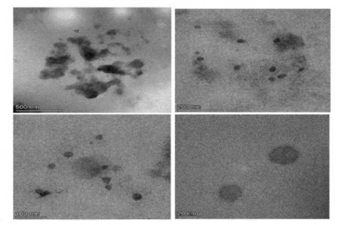
The research on nanoparticles of riksusu leaves produced a less spherical morphology observed using a TEM instrument. TEM results can be seen in Figure 1.
Figure 1. Transmission electron microscope results of Riksusu leaf nanoparticles with magnifications of 500 nm, 200 nm, 100 nm, and 20 nm
The morphology of the riksusu leaf extract nanoparticles shown in Figure 1 tends to be spherical or imperfectly round. Spherical nanoparticles have a greater cellular distribution compared to rod-shaped ones. The less spherical shape of the nanoparticles will facilitate contact between particles, leading to aggregation [26]. According to study [27], the dark-coloured part of the nanoparticles indicates the presence of extracts trapped in the nanoparticle matrix, thus, the riksusu leaf nanoparticles were successfully encapsulated or trapped in chitosan and NaTPP bonds. Research on riksusu leaf nanoparticles produced a less spherical morphology observed using a TEM instrument. This is likely due to the polymer viscosity being too low, resulting in less strong cross-linking bonds, making them easy to shrink and uneven [28]. According to study [29], the shape of less spherical particles can facilitate interactions between particles so that aggregation occurs, which results in the particle size getting larger.
3.4 Anti-inflammatory activity test
The anti-inflammatory activity test in this study used male Wistar rats as test animals. Rats were chosen because they are similar to humans in physiology, anatomy, nutrition, pathology, and metabolism. Male rats were chosen because they have a more stable body condition and do not experience menstruation and pregnancy [30]. The rats that will be used as experimental animals are 2-3 months old and weigh 150-200 grams. The test animals were divided into 2 groups, where each group consisted of 8 male Wistar rats. Group one is the P1 treatment group that will receive a dose of 250 mg/kgBW extract, and group 2 is P2, which will receive a dose of 250 mg/kgBW riksusu leaf extract nanoparticles. The rats were adapted to an environment with room temperature, adequate lighting, and ventilation for 7 days in a cage and were given food and drink [31]. Before being treated, each rat was fasted for 8 hours. This aims to avoid the influence of food on the content of active compounds in riksusu leaf extract and nanoparticles that can affect the anti-inflammatory effects caused. The test requires that the rats be weighed to determine their body weight so that the dose to be given can be estimated. The rats are then measured for inflammation volume and recorded as the initial volume of the rat’s paw (V0). Rats were injected using 1% carrageenan as much as 0.2 mL [32]. Intraplantar injection of 1% carrageenan as much as 0.2 ml on the sole of the rats’ paw can cause inflammation within 5-6 hours and will heal itself because the dose of carrageenan has been reduced [33]. In the study, there was an increase in the volume of rat feet, where, before the administration of carrageenan and 1 hour after the administration of carrageenan, in group P1 the average volume at V0 was 0.448 ± 0.015 to 1.350 ± 0.177, and in group P2 was 0.448 ± 0.015 to 1.400 ± 0.151. The average volume of rat foot edema, percentage of inflammation (%). The average percentage of inhibition for 360 minutes can be seen in Tables 3, 4, and 5.
Table 3. Average volume of rat paw edema
|
Group |
Average Edema Volume (mL)/ and SD Every 1 Hour During 7 Hours of Observation |
|||||||
|
V0 |
Vt |
Vt1 (60) |
Vt2 (120) |
Vt3 (180) |
Vt4 (240) |
Vt5 (300) |
Vt6 (360) |
|
|
P1 |
0.453 ± 0.015 |
1.350 ± 0.093 |
0.883 ± 0.052 |
0.760 ± 0.067 |
0.648 ± 0.062 |
0.640 ± 0.068 |
0.525 ± 0.046 |
0.503 ± 0.002 |
|
P2 |
0.460 ± 0.011 |
1.375 ± 0.017 |
0.878 ± 0.042 |
0.725 ± 0.051 |
0.640 ± 0.032 |
0.618 ± 0.017 |
0.513 ± 0.018 |
0.470 ± 0.015 |
Note: V0 = Time before treatment.
Vt = Time after carrageenan induction for one hour (60 minutes).
Vt1,2,3,4,5,6 = Time after dosing in groups P1 and P2.
P1 = Group of riksusu leaf extract, dose 250 mg/kgBW (only given riksusu leaf extract and fasted during dosing).
P2 = Group of riksusu leaf nanoparticles, dose 250 mg/kgBW (only given riksusu leaf nanoparticles and fasted during dosing).
Table 4. Average percentage of inflammation (%)
|
Group |
Average Inflammation Percentage (%) and SD Every 1 Hour for 6 Hours |
||||||
|
Vt |
Vt1 (60) |
Vt2 (120) |
Vt3 (180) |
Vt4 (240) |
Vt5 (300) |
Vt6 (360) |
|
|
P1 |
1.987 ± 0.242 |
0.952 ± 0.133 |
0.683 ± 0.188 |
0.434 ± 0.171 |
0.418 ± 0.181 |
0.162 ± 0.125 |
0.111 ± 0.042 |
|
P2 |
1.991 ± 0.170 |
0.908 ± 0.094 |
0.575 ± 0.094 |
0.393 ± 0.096 |
0.343 ± 0.048 |
0.115 ± 0.060 |
0.022 ± 0.023 |
Table 5. Average percentage of inhibition over 360 minutes
|
Group |
Average Inhibition Percentage (%) and SD Every 1 Hour for 6 Hours |
|||||
|
Vt1 (60) |
Vt2 (120) |
Vt3 (180) |
Vt4 (240) |
Vt5 (300) |
Vt6 (360) |
|
|
P1 |
0.522 ± 0.012 |
0.659 ± 0.066 |
0.786 ± 0.065 |
0.796 ± 0.071 |
0.921 ± 0.055 |
0.054 ± 0.017 |
|
P2 |
0.540 ± 0.070 |
0.708 ± 0.060 |
0.803 ± 0.040 |
0.828 ± 0.020 |
0.943 ± 0.027 |
0.989 ± 0.012 |
Note: Vt = Time after carrageenan induction for one hour (60 minutes).
Vt1,2,3,4,5,6 = Time after dosing in groups P1 and P2.
P1 = Riksusu leaf extract group dose, 250mg/kgBW (only given riksusu leaf extract and fasted during dosing).
P2 = Riksusu leaf nanoparticles group dose, 250mg/kgBW (only given riksusu leaf nanoparticles and fasted during dosing).
Table 3 indicates that there was an increase in the volume of rat feet, where before the administration of carrageenan and 1 hour after the administration of carrageenan, in group P1, the average volume at V0 was 0.453 ± 0.015 to 1.350 ± 0.093, and in group P2 was 0.460 ± 0.011 to 1.375 ± 0.017. This increase in the volume of rat feet is due to the reaction of the injected carrageenan, which will increase COX-2 so that the formation of oedema occurs quickly, and the purpose of injecting carrageenan which will be increase the soles of the rat's feet [34]. The volume of rat feet decreased from the 60th minute to the 360th minute. During 6 hours of observation between group P1 and group P2, there was a decrease in the volume of inflammation. Based on the results of the study and the data presented in Table 3, the average percentage of inflammation from the pre-test (Vt) was 199% (1.987 ± 0.242) in group P1 and 199% (1.991 ± 0.170) in group P2. This volume size was due to carrageenan-induced inflammation in mice. According to study [15], carrageenan can release inflammatory mediators such as histamine and serotonin that which can cause oedema due to the presence of antibodies in test animals that react to antigens to fight the effects of the antigen; this event triggers inflammation in the test animal group.
The decrease in the percentage of inflammation from the P1 group, 199% (1.987 ± 0.242) to 23% (0.111 ± 0.042), was due to the presence of secondary metabolite compounds that act as anti-inflammatories, including flavonoid compounds. Flavonoids inhibit inflammation by inhibiting pro-inflammatory enzymes such as cyclooxygenase (COX) and lipoxygenase (LOX). When a tissue is injured, arachidonic acid will release prostaglandins with the help of cyclooxygenase and lipoxygenase enzymes. If both enzymes are inhibited, arachidonic acid cannot release prostaglandins and leukotrienes, which are inflammatory mediators [35]. Flavonoids can also inhibit neutrophil degranulation, thereby reducing the release of arachidonic acid by neutrophils, thus reducing the prostaglandins and leukotrienes produced. In addition, flavonoids can also inhibit the release of histamine by mast cells and reduce the number of leukocytes [36]. The decrease in inflammation percentage from group P2 199% (1.991 ± 0.170) decreased to 3% (0.022 ± 0.023) at the 360th minute explaining that riksusu leaf nanoparticles will be maximal as anti-inflammatory if made in the form of nanoparticles and from the results of this decrease in inflammation percentage shows that the dose of riksusu leaf nanoparticles has good anti-inflammatory ability and shows increased efficacy with the use of drug delivery materials in the form of nanochitosan. The high decrease in inflammation in the riksusu leaf nanoparticle group is not only because the extract was successfully encapsulated by chitosan and NaTPP, but is due to the nature of chitosan which can act as an anti-inflammatory and the active period of carrageenan which has decreased so that the ability of riksusu leaf nanoparticles continues to increase. According to study [37], chitosan can act as an anti-inflammatory material and can stimulate cell proliferation and remodelling in the wound-healing process. In wound healing, chitosan has also been shown to activate inflammatory cells such as macrophages, polymorphonuclear (PMN) fibroblasts, and angioendothelial cells [38]. In the inflammatory phase, chitosan can regulate the activity of inflammatory cells, release proinflammatory factors, and provide a good microenvironment for the wound healing process. The level of inflammation percentage reduction can be seen in Table 4.
The instability of riksusu leaf extract in reducing inflammation over 360 minutes may be due to the inability of the extract dose to sustain or enhance its anti-inflammatory effect. According to study [39], a test material is said to have an anti-inflammatory effect if the inhibition of inflammation reaches 50% or more than 50%. Based on the results of the study, it was found that riksusu leaf extract showed an inhibition percentage of more than 50% in the 60th minute of 52% (0.522 ± 0.012) to the 300th minute of 92% (0.921 ± 0.055), while in the 360th minute, the inhibition percentage decreased to 5% (0.054 ± 0.017). In the nanoparticle group, the results of the data analysis of the inhibition percentage were more than 50% at the 60th minute of 54% (0.540 ± 0.070), and continued to increase until the 360th minute of 98.9% (0.989 ± 0.012). The increase in inhibition percentage between P1 and P2 can be seen in Table 5.
If we look at Table 5 comparison of the percentage of inhibition between the riksusu leaf extract group (P1) and the riksusu leaf nanoparticle group (P2) every 60 minutes, the nanoparticles show a percentage of inhibition that continues to increase, it can be said that the riksusu leaf nanoparticles are better than the riksusu leaf extract.
The results of the analysis in Table 4, Pre Test (Vt) of the riksusu leaf extract treatment group (P1) were 199% (1.987 ± 0.242) and Post Test (Vt 1,2,3,4,5,6) for 6 hours (360 minutes) riksusu leaf extract (P1) can inhibit inflammation by 62.30% (0.451 ± 0.086), while in the riksusu leaf nanoparticle group (P2) obtained Pre Test 199% (1.991 ± 0.170) and Post Test (Vt 1,2,3,4,5,6) for 6 hours (360 minutes) riksusu leaf nanoparticles (P2) can inhibit by 80.13% (0.802 ± 0.038). The results of the Pre Test (Vt) analysis and the average Post Test (Vt 1,2,3,4,5,6) for 6 hours at P1 and P2 stated that if the average inhibition percentage of P1 is smaller than the average inhibition percentage of P2, nanoparticles (P2) have greater anti-inflammatory potential compared to riksusu leaf extract (P1). Based on the results of the difference test, there was a significant difference between the posttest edema volume of riksusu extract and the posttest edema volume of nanoparticles (p<0.05). Seen from the average posttest edema volume of nanoparticles is smaller than the pretest edema volume of riksusu extract, so it can be concluded that the posttest nanoparticle treatment reduced the edema volume more than the posttest riksusu extract. Ethanol extract of riksusu leaves and nanoparticles of riksusu leaves (Wrightia pubescens) contain flavonoids, which are thought to be effective as anti-inflammatories. Flavonoids are one of the largest natural phenol compounds. Flavonoids are found in all seed plants, so it is certain that these compounds are found in plant extracts [40]. In general, flavonoids are soluble in polar solvents such as methanol, acetone, water, and ethanol. Flavonoid compounds are specifically able to stop the formation and release of substances that cause inflammation due to allergic reactions. Compounds included in the flavonoid group have different effects in overcoming inflammation. According to study [41], the anti-inflammatory mechanism produced by flavonoids can occur through several pathways, one of which is by directly inhibiting the activity of the COX and lipoxygenase enzymes, which causes inhibition of prostaglandin and leukotriene biosynthesis, which are the end products of the COX and lipoxygenase pathways. Administration of flavonoids can reduce the number of leukocytes and reduce complement activation, thereby reducing leukocyte adhesion to the endothelium, resulting in a decrease in the body's inflammatory response. According to study [42], in addition, it is known that other flavonoid mechanisms in inhibiting inflammation are by inhibiting the release of arachidonic acid, secretion of lysosomal enzymes from neutrophils and endothelial cells, and inhibiting the exudation phase and proliferation phase of the inflammation process. Inhibition of the release of arachidonic acid will cause a decrease in the amount of arachidonic substrate entering the cyclooxygenase pathway and the lipoxygenase pathway, so that in the end there will be a decrease in the amount and suppression of the production of prostaglandins, prostacyclins, endoperoxides, thromboxanes on one side and hydroperoxides, and leukotrienes on the other side [40].
The present study demonstrated that the chitosan-TPP nano-formulated extract of Wrightia pubescens R.Br leaves exhibited significantly enhanced anti-inflammatory activity in the carrageenan-induced paw oedema model in Wistar rats. Compared to the crude leaf extract, the nanoformulation achieved a higher maximum inhibition of oedema (98.9% vs. 92%, p < 0.05) and a greater average inhibition for six hours (80.13% vs. 62.30%, p < 0.05), suggesting improved bioavailability and sustained release of active compounds such as luteolin, kaempferol, and morin.
These findings indicate that chitosan-TPP nanoparticles can serve as an effective delivery system to potentiate the anti-inflammatory effects of Wrightia pubescens and hold promise for further development as a plant-based anti-inflammatory therapeutic agent. Therefore, further pilot randomized controlled trials need to be carried out to gain approval for humans.
All test animals involved in this study were approved by the Animal Ethics Committees of the Faculty of Veterinary Medicine at Udayana University, with reference No.: B/147/UN14.2.9/PT.01.04/2024.
The authors express their gratitude to the Master of Chemistry program at Udayana University and all those who contributed to this research.
[1] Zigterman, B.G.R., Dubois, L. (2022). Ontsteking en infectie: De cellulaire en biochemische processen [Inflammation and infection: Cellular and biochemical processes]. Ned Tijdschr Tandheelkd, 129(3): 125-129. https://doi.org/10.5177/ntvt.2022.03.21138
[2] Riadh, H, Duha, H. (2024). The impact of inflammation on pathological processes. Academic International Journal of Medical Sciences, 1(2): 34-43. https://doi.org/10.59675/m126
[3] Soliman, A.M., Barreda, D.R. (2022). Acute inflammation in tissue healing. International Journal of Molecular Sciences, 24(1): 641. https://doi.org/10.3390/ijms24010641
[4] Liu, W., Liu, Y.C., Xiang, Z. (2023). Editorial: Traditional medicine and chronic inflammatory diseases. Frontiers in Pharmacology, 14: 1202976. https://doi.org/10.3389/fphar.2023.1202976
[5] Balamurugan, J., Lakshmanan, M. (2021). Non-steroidal anti-inflammatory medicines. In: Paul, A., Anandabaskar, N., Mathaiyan, J., Raj, G.M. (eds) Introduction to Basics of Pharmacology and Toxicology. Springer, Singapore. https://doi.org/10.1007/978-981-33-6009-9_19
[6] Caner, G., Bilge, S.S. (2018). Effects of nonsteroidal anti-inflammatory drugs at the molecular level. The Eurasian Journal of Medicine, 50(2): 116-121. https://doi.org/10.5152/EURASIANJMED.2018.0010
[7] Gonfa, Y.H., Tessema, F.B., Bachheti, A., Rai, N., Tadesse, M.G., Nasser Singab, A., Chaubey, K.K., Bachheti, R.K. (2022). Anti-inflammatory activity of phytochemicals from medicinal plants and their nanoparticles: A review. Current Research in Biotechnology, 6: 100152. https://doi.org/10.1016/j.crbiot.2023.100152
[8] Gori, Y.M., Prawirohardjono, W. (2018). Antiinflammatory effect test of 70% ethanol extract of riksusu (Wrightia Pubescens R.Br.) leaves on male white rats (rattus norvegicus) in induction of egg white. Indonesia Natural Research Pharmaceutical Journal, 3(2): 95-101.
[9] Hanutami, B., Budiman, A. (2017). The use of nanotechnology in herbal medicine formulation. Pharmaka, 15(2): 29-39. https://doi.org/10.24198/jf.v15i2.12947
[10] Agnihotri, S.A., Mallikarjuna, N.N., Aminabhavi, T.M. (2004). Recent advances on chitosan-based micro- and nanoparticles in drug delivery. Journal of Controlled Release, 100(1): 5-28. https://doi.org/10.1016/J.JCONREL.2004.08.010
[11] Van Bavel, N., Issler, T., Pang, L., Anikovskiy, M., Prenner, E.J. (2022). A simple method for synthesis of chitosan nanoparticles with ionic gelation and homogenization. Molecules, 28(11): 4328. https://doi.org/10.3390/molecules28114328
[12] Karthikeyan, A., Senthil, N., Min, T. (2020). Nanocurcumin: A promising candidate for therapeutic applications. Frontiers in pharmacology, 11: 487. https://doi.org/10.3389/fphar.2020.00487
[13] Kamal, A., Zaki, S., Shokry, H., Abd-El-Haleem, D. (2020). Using ginger extract for synthesis of metallic nanoparticles and their applications in water treatment. Journal of Pure and Applied Microbiology, 14(2): 1227-1236. https://doi.org/10.22207/JPAM.14.2.17
[14] Jafernik, K., Ładniak, A., Blicharska, E., Czarnek, K., Ekiert, H., Wiącek, A.E., Szopa, A. (2023). Chitosan-based nanoparticles as effective drug delivery systems-A review. Molecules (Basel, Switzerland), 28(4): 1963. https://doi.org/10.3390/molecules28041963
[15] Suryandari, S.S., De Q.E., Datu, O.S.(2021). Anti-inflammatory activity test of ethanol extract of sesewanua leaves (Clerodendrum squamatum Vahl.) on white rats (Rattus norvegicus L.) induced by carrageenan. Pharmacon, 10(3): 1025-1032.
[16] Sharma, S.H., Kumar, J.S., Chellappan, D.R., Nagarajan, S. (2018). Molecular chemopreventionby morin–A plant flavonoid that targets nuclear factor kappa B in experimental colon cancer. Biomedicine Pharmacotherapy, 100: 367-373.
[17] Tjahyanto, T., Eldy, E., Felicia, C., Jessica, K.J.K., Larissa, O. (2022). Antiviral activity of polyphenols as potential prophylaxis and therapy in handling Covid-19. Journal Health Science, 3(2): 311-330. https://doi.org/10.46799/jhs.v3i2.428
[18] Candra, G.N.H., Wijaya, I.M.A.P. (2021). Molecular docking of kaempferol as an anti-inflammatory agent in atherosclerosis in silico. Scientific Journal of Medicine, 7(1): 13-18. https://doi.org/10.36733/medicamento.v7i1.1497
[19] Fadzillah, S.N., Hernawati, D., Putra, R.R. (2024). Anti-inflammatory activity and toxicity prediction of secondary metabolite compounds of ciplukan leaves (Physalis peruviana L.). Bioscientist, 2(1): 517-539. https://dx.doi.org/10.33394/bioscientist.v12i1.9592
[20] Ntalouka, F., Tsirivakou, A. (2023). Luteolin: A promising natural agent in management of pain in chronic conditions. Frontiers in Pain Research, 4: 1114428. https://doi.org/10.3389/fpain.2023.1114428
[21] Maurya, A.K., Agarwal, K., Gupta, A.C., Saxena, A., Nooreen, Z., Tandon, S., Bawankule, D.U. (2018). Synthesis of eugenol derivatives and its anti-inflammatory activity against skin inflammation. Natural Product Research, 34(2): 251-260. https://doi.org/10.1080/14786419.2018.1528585
[22] Salsabila, B.A.A., Yusuf, A.F.N., Gading, A.C.R., Prabuningrat, A., Andanalusia, M. (2023). Eugenol potential in cloves as an analgesic literature review. Journal of Tropical Biology, 23(1): 169-173. https://doi.org/10.29303/jbt.v23i1.4561
[23] Eid, A.M.M., Elmarzugi, N.A., El-Enshasy, H.A. (2013). Preparation and evaluation of olive oil nanoemulsion using sucrose monoester. International Journal of Pharmacy Pharmaceutical Science, 5(3): 434-440.
[24] Feng, Y., Kilker, S.R., Lee, Y. (2020). Surface charge (zeta-potential) of nanoencapsulated food ingredients. Characterization of Nanoencapsulated Food Ingredients, 213-241. https://doi.org/10.1016/B978-0-12-815667-4.00007-9
[25] Makadia, H.K., Siegel, S.J. (2011). Poly Lactic-co-Glycolic Acid (PLGA) as biodegradable controlled drug delivery carrier. Polymers, 3(3): 1377-1397. https://doi.org/10.3390/polym3031377
[26] Oksal, E., Pangestika, I., Muhammad, T.S.T., Mohamad, H., Amir, H., Kassım, M.N.I., Andriani, Y. (2020). In vitro and in vivo studies of nanoparticles of chitosan-Pandanus tectorius fruit extract as new alternative treatment for hypercholesterolemia via Scavenger Receptor Class B type I pathway. Saudi Pharmaceutical Journal,l 28(10): 1263-1275. https://doi.org/10.1016/j.jsps.2020.08.017
[27] Taurina, W., Martien, R., Ismail, H. (2013). Preparation of gamavuton-0 nanoparticles using short chain chitosan and tripolyphosphate as cross linker. Jurnal Ilmiah Farmasi, 10(2): 60-68. https://doi.org/10.20885/jif.vol10.iss2.art4
[28] Kohane, D.S. (2007). Microparticles and nanoparticles for drug delivery. Biotechnology and Bioengineering, 96(2): 203-209. https://doi.org/10.1002/bit.21301
[29] Ikra, N., Esti, M., Rachmat, R.(2020). Phytosome penetration test of ethanol extract of sembung leaves and antioxidant activity test using DPPH method. Syntax Literate, 5(11): 1384-1394. https://doi.org/10.36418/syntax-literate.v5i11.1767
[30] Almunia, J.A., Munesue, Y., Kawasaki, H., Takano, K., Kayahara, C., Noma, S., Morikawa, N., Niida, S., Ogiso, N. (2024). Hematological and biochemical characterization of aging farm male rat strains in the national center for geriatrics and gerontology. Experimental Animals, 74(1): 66-82. https://doi.org/10.1538/expanim.24-0028
[31] Sintowati, R., Handayani, A. P., Aisyah, R.(2017). The Effectiveness of 70% methanolic extract of avocado leaf (persea americana mill) in decreasing blood sugar levels in male rats (rattus norvegicus) wistar strain induced alloxan. Biomedika, 8(1): 15-22. https://doi.org/10.23917/biomedika.v8i1.3018
[32] Yuda, P.E.S.K., Sasmita, G.A.P.Y., Cahyaningsih, E. (2022). Anti-inflammatory activity of traditional instant parem from bali usada ingredients in carrageenan-induced inflammation rats. Jurnal Ilmu Kefarmasian Indonesia, 20(2): 142-149. https://doi.org/10.35814/jifi.v20i2.1076
[33] Pranata, C., Darmirani, Y., Limbing, R. (2024). Test of the anti-inflammatory effectiveness of ethanol extract of cat’s whiskers leaves (orthisiphon aristatus) on paw edema in male mice (mus musculus). Jurnal Farmasimed, 7(1): 41-47. https://doi.org/10.35451/jfm.v7i1.2289
[34] Isrul, M., Dewi, C., Wahdini, V. (2020). Anti-inflammatory effect test of red spinach leaf infusion (Amaranthus tricolor L.) on white rats (Rattus norvegicus) Yang Diinduksi Karagenan. Jurnal Mandala Pharmacon Indonesia, 6(2): 97-103. https://doi.org/10.35311/jmpi.v6i1.61
[35] Nijveldt, R.J., Nood, E.V., Hoorn, D.E.C., Boelens, P.G., Norren, K.V., Leeuwen, P.A.M. (2001). Flavonoids: A review of probable mechanisms of action and potential application. The American Journal of Clinical Nutrition, 74(4): 418-425. https://doi.org/10.1093/ajcn/74.4.418
[36] Sargsyan, S.A., Shearer, A.J., Ritchie, A.M., Burgoon, M.P., Anderson, S., Hemmer, B., Stadelmann, C., Gattenlöhner, S., Owens, G.P., Gilden, D., Bennett, J.L. (2010). Absence of Epstein-Barr virus in the brain and CSF of patients with multiple sclerosis. Neurology, 74(14): 1127-1135. https://doi.org/10.1212/WNL.0b013e3181d865a1
[37] Hartomo, B.T., Firdaus, F.G. (2019). Utilization of chitosan biomaterials in the field of oral surgery. B-Dent: Jurnal Kedokteran Gigi Universitas Baiturrahmah, 6(1): 63-70. https://doi.org/10.33854/jbd.v6i1.82
[38] Evi, K., Naufal, R.P. (2019). Potential of chitosan biopolymer in wound treatment. MEDULA, 9(3): 459-464.
[39] Latief, M., Fisesa, A.T., Sari, P.M., Tarigan, I.L. (2021). Anti inflammatory activity of sungkai leaves (Peronema canescens Jack) ethanol extract in carrageenan induced mice. Jurnal Farmasi Sains, 7(2): 144-153. https://doi.org/10.31603/pharmacy.v7i2.4532
[40] Maulidiyah, M., Hartanto, T.P., Putri, S.N.D., San Sabhira, A., Mukarromah, I.W., Putri, R.A., Ningsih, A.W. (2023). Review article: Phytochemical and pharmacological study of moringa plant (moringa oleifera lam). The Journal General Health and Pharmaceutical Sciences Research, 1(4): 45-52. https://doi.org/10.57213/tjghpsr.v1i4.110
[41] Meilina, R., Maghlisa, U.I., Dhirah, U.H. (2022). Anti-inflammatory ethanol extract of button flower (Gomphrena globosa L.) in rats (Rattus novergicus). Journal of Healthcare Technology and Medicine, 8(2): 648-657. https://doi.org/10.33143/jhtm.v8i2.2337
[42] Widarto, M., Khuluq, H., Rahayu, T.P. (2021). Study of anti-inflammatory activity test of 70% ethanol extract of melinjo leaves (Gnetum Gnemon L.) on wistar strain white rats that carrageenan induced. Prosiding University Research Colloquium, 918-931.