Kiran Napte| Anurag Mahajan*
Special Issue: Technology Innovations and AI Technology in Healthcare
© 2022 IIETA. This article is published by IIETA and is licensed under the CC BY 4.0 license (http://creativecommons.org/licenses/by/4.0/).
OPEN ACCESS
Tremendous advancement takes place in the field of medical science. With this advancement, it is possible to support diagnosis and treatment planning for various diseases related to the abdominal organ. The liver is one of the adnominal organs, a common site for developing tumors. Liver disease is one of the main causes of death. Due to its complex and heterogeneous nature and shape, it is challenging to segment the liver and its tumor. There are numerous methods available for liver segmentation. Some are handcrafted, semi-automatic, and fully automatic. Image segmentation using deep learning techniques is becoming a very robust tool nowadays. There are many methods of liver segmentation which uses Deep Learning. This article provides the survey of the various liver segmentation schemes based on Artificial Neural Network (ANN), Convolution Neural network (CNN), Deep Belief network (DBN), Auto Encoder, Deep Feed-forward neural Network (DFNN), etc based on the architecture details, methodology, performance metrics and dataset details. Researchers are continuously putting efforts into improving these segmentation techniques. So this article give out a comprehensive review of deep learning-based liver segmentation techniques and highlights the advantages of the deep learning segmentation schemes over the traditional segmentation techniques.
liver segmentation, deep learning, review, CNN, algorithm
The liver is very important abdominal organ of our body. Liver disorders are a major cause of death globally. Computer-aided techniques play a significant role in the clinical diagnosis as well as treatment of liver disease. Precise and accurate segmentation of the liver, its vessels, and tumors are required in the disease diagnosis. However, because of the shape of the liver, the intensity of homogeneity inside the liver, low contrast, presence of adjacent abdominal organs, it becomes a challenging task for accurate liver segmentation. Different medical imaging methods, such as computed tomography (CT), ultrasound (US), magnetic resonance imaging (MRI), etc., can be used to diagnose liver problems. Many methods have been formulated like graph cut, Region growing, statistical shape model (SSM), clustering, threshold-based, deformable models, level set, statistical shape-based, support vector machine-based Active contour, neural network-based methods, etc. [1, 2].
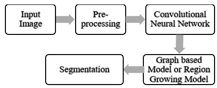
Figure 1. Block diagram of liver segmentation
Recently Deep Convolution neural network has made good progress in medical image processing. The deep learning algorithms have attracted the medical researcher’s attention because of its high attribute representation of the bio-medical modality. It has shown superior feature discrimination, applicable for larger size dataset, higher accuracy, applicable for time series, images and categorical dataset. Using these neural networks, one can have a semi-automatic or fully automatic segmentation method for the liver. These neural networks are part of deep learning, a new field division of machine learning. Figure 1 shows the flow of liver segmentation. Initially, the Input Image is taken and passed through pre-processing. Once pre-processing is done, it is applied to Convolution neural network. Ultimately segmentation is done through any method like a graph-based model or Region growing technique.
Deep learning [3, 4] attempts to build and simulate the learning process of a neural network. There are certain advantages of deep learning in medical image processing and analysis. It can find the features from the given dataset automatically. There are several applications in image detection, registration, segmentation, and classification.
In todays era, various deep learning based frameworks has been employed for the liver segmentation to improve the performance of liver tumor detection. The deep learning frameworks consider various types of medical images such as CT images, ultra-sound images and MRI images.
This paper presents the widespread survey of the recent deep learning based liver segmentation techniques. It focuses on the architectural overview, methodology, dataset, evaluation metrics, constraints and challenges of the current research on liver segmentation. The survey provides the research challenges and constraints that open the future direction for the improvement in liver segmentation performance.
The work in this paper is divided as follows. The subsequent section will explain Deep Learning Architecture. The section 3 describes the Literature survey. Section 4 will tell performance analysis, and the last research paper is concluded in the conclusion section.
This article introduces supervised and unsupervised learning using deep learning architectures. Deep learning isn't just one method; rather, it's a group of topologies and algorithms that can be used to solve a variety of issues. In order to build these deep learning structures, deep learning relies on training neural networks with example data and rewarding them based on their success. Deep learning employs a wide variety of structures and methodologies. The aim that needs to be predicted is specifically identified in the training data when learning is being done under supervision. While unsupervised learning refers to the issue domain where the training data does not contain any target labels.
Modern neural network-based technology called "Deep Learning" aims to replicate the way the human cortex operates. The vast majority of the deep learning architecture is made up of deep/neural networks with various topologies. An input layer, which includes raw data, hidden layers, which gather and process input data, and an output layer are the fundamental parts of neural networks (it produces the outcome: result, estimation, forecast, etc.). Below is an explanation of several Deep Learning architecture types. A number of well-known deep learning designs are also introduced in this article, including the Artificial Neural Network, Deep Feed Forward Network, Stacked Auto Encoders, Deep Belief Network, and Convolution Neural Network.
2.1 Artificial neural network
In 1980 Yegnanarayana [5], the artificial neural network was first developed. Deep learning's central component is an artificial neural network (ANN). Many other algorithms have been developed based on ANN. By adopting a condensed set of concepts from the biological brain system, artificial neural networks mimic biological neural networks. Biological neural networks, which make up the structure of the human brain, are the basis for most artificial neural networks. Artificial neural networks also feature neurons that are coupled to one another in different levels of the networks, similar to how neurons in a real brain are connected to one another. Nodes are the name given to these neurons.
In particular, ANN models mimic the brain and nervous system's electrical activity. Connected to other processing units are components known as neurodes or perceptrons. In most cases, the neurodes are stacked in layers or vectors, with the output of one layer feeding into the input of the next and possibly higher levels. In order to mimic the synaptic connections found in the brain, a neurode may connect to some or all of the neurodes in the layer below. ANN is a computational model comprised of a large number of nodes. Each of its nodes represents some output function, and the connection between these nodes means the ability to the transmission of data between these nodes. The output function is known as the activation function. The output of the network mainly depends on activation functions. ANN can be divided into two types according to connections made as feedforward neural network and feedback neural network. For many years’ vast progress has been made in pattern recognition, robotics, the health sector, etc.
2.2 Deep feed forward network
An artificial neural network with circular node connections is known as a feed-forward neural network. In contrast to feed-forward neural networks, which cycle through many courses, recurrent neural networks only follow one path at a time. The feed-forward model is the simplest form of neural network because it only processes input in one direction. The easiest way to examine a feed-forward neural network is with a single perceptron. A number of inputs will be sent into the layer and multiplied by various weights. The final step is to add up each value to obtain the sum of the weighted input values [1, 2].
Data constantly advances; it never moves backward or in the opposite direction. It is also known as a feed-forward neural network. The purpose of this network is to approximate the corresponding objective function. It includes the input layer, middle layer, and output layer in this network flow of data only in one direction. Data cannot reverse back. So, this deep forward network is a primitive architecture of deep learning [3].
2.3 Stacked auto encoders
An input layer, a hidden layer, and an output layer are the three layers of an autoencoder, a sort of unsupervised learning structure. The two steps in an autoencoder training process are encoder and decoder. An encoder is used to map input data into a hidden representation, while a decoder is used to reconstruct input data from the hidden representation [6].
To efficiently code unlabeled input, an artificial neural network known as an auto encoder is used. Trying to regenerate the input from the encoding serves to verify and enhance the encoding. By teaching the network to reject irrelevant input, the auto encoder creates a representation for a set of data, generally for dimensionality reduction. An input layer, middle layer, and output layer are present in a feed-forward neural network, for instance [6, 7]. Encoder and decoder are separate components of this auto encoder.
2.4 deep belief network
The Deep Belief Network (DBN), a subclass of deep neural network, is constructed from layers of constrained boltzmann machines (RBMs). DBN can be used to complete supervised learning tasks to create classification or regression models as well as unsupervised learning activities to lower the dimensionality of feature space. There are two processes involved in training a DBN: layer-by-layer training and fine-tuning [8, 9].
A deep belief network (DBN) is a type of deep learning architecture made up of many layers of latent variables connected only between units on different levels of the network. A generative graphical model underlies it. A DBN can learn to probabilistically reproduce its inputs.
A deep belief network is a neural network [10] with several Boltzmann machine layers. The output of one layer in this network serves as the input for the next layer. A hierarchical retraining is given to each Boltzmann machine. The supervised learning model's initial input is made up of the results.
2.5 Convolution neural network
A multi-layer neural network with biological cues from the visual cortex of animals is known as a convolution neural network. Image processing applications in particular benefit from the design. The convolution neural network is comparable to a feed-forward neural network [10, 11]. The structure of CNN is shown in Figure 2. Early layers in a deep network discover features (such edges), and later layers reassemble this data into higher-level input quality.
Figure 2. The structure of a CNN [12]
A Convolutional Neural Network (CNN) may distinguish between different objects and elements in an input image by assigning them various values (learnable weights and biases). The convolutional layer is the top layer of a convolutional network. Convolutional layers, further convolutional layers, or pooling layers might come after the fully connected layer, but they do not make up the final layer. With each layer, CNN gets more resilient and is able to recognise bigger portions of the image. Basic features like colours and borders are highlighted in the early layers. As When the visual data progresses through the CNN layers, beginning with identification of the object's primary features or shapes, the target item is finally recognised. Comparatively speaking, a ConvNet requires substantially less pre-processing than other classification methods. ConvNets are able to learn these filters and their characteristics, in contrast to older systems where filters had to be hand-engineered. It's widely used in recognition-intensive activities like document recognition and image classification. In this layer near them, two networks are connected in two different ways. Convolution layer and Pooling layer are two of the CNN's kernel layers. Using the convolution layer, a little neural patch is examined. When provided inputs for visual, speech, or audio signals, convolutional neural networks perform better than other neural networks [13].
The weights of neural connection and kernel are reduced during the procedure of back propagation during the training phase.
Recently in 2019, Seminal contributions have been made in [13] with a new augmentation strategy for image processing using a statistical shape model. They preferred 3 Dimensional Convolution neural network and U Net in their approach. In the experiment, they compared performance with some existing models like random rigid transformation technique and random elastic augmentation. In a further study, they are generating texture of 3D images and introducing GAN for texture generation. Again in the same year, the researcher extended the 2D Convolution neural network to a 3D network [14] with a super vector machine. This was specifically designed for liver tumor differentiation. In their work, they remove preprocessing. Also, volumetric features are adequate in their experiment. But classification accuracy was slightly reduced as compared to other existing techniques. In future scope, they were thinking about a noninvasive CAD solution for more accuracy.
This has been explored in the study [15] that liver semantic segmentation algorithm using adversarial networks work well with weighted loss functions. It has been applied to the LiTS dataset. In the experimentation, it is observed a higher segmentation index with stable convergence performance. In their further study, they are going to optimize the algorithm. Knowledge-aided Convolution networks [16] have been proposed for small organ segmentation. The authors used a graph-based group-wise image registration method. After a demonstration on ISBI 2015, they understand that it avoids over-segmentation with an excellent average dice coefficient for small organs. But localization time is more. In further research, they will reduce localization time. In the study [17] various deep learning schemes for liver segmentation has beem studied and highlighted the challenges such as network complexity, high segmentation time, and semantic gap in segmentation. The study [18] are well documented, and it is also acknowledged that attention hybrid Connection network, i.e., AHCNet with joint dice loss function for Liver Tumor Segmentation in CT volumes, has excellent results For their study 3DIRCADb data set. After experimentation, they realized that training speed was high with faster network convergence. In further research, they will improve the accuracy of lesion segmentation by different silhouettes. Liver extraction is proposed [19] using a residual convolution neural network. This extraction process is accurate. Also, it can remove the noise. They also benefitted from structure preservation and residual mapping, but Poisson noise affects the performance. They will extend work for texture preservation within the liver investigated [20] liver segmentation methods uses three views of CT images on clinical CT images. They did not require to choose a seed point for this. They observed that segmentation is improved. In 2018, Several authors recommended [21] abdomen CT multi-organ segmentation using dense V networks. In this, they use a registration-free segmentation algorithm. It was applied to the BTCV dataset. They analyzed that it can segment multiple organisms with improved boundary accuracy. Studies were conducted [22] on feature learning probability boosting tree initialization and CNN-ASM Refinement. They used Convolution neural network approach. They improved optimal boundary searching, but at the same time, they realized that it has lower performance than a fully connected network. In a further study, they have decided that the boundary model has to be improved by CNN architecture-DenseUNet presented in the study [23] for liver and tumor segmentation with context algorithm. It is applied on MICCAI 2017 and observed competitive results. It does not apply to small liver tumors.
Research provides evidence [24] on multitasking convolution neural networks, for instance, liver tumor segmentation. It is analyzed on the BraTS 2016 dataset. It can be applied to different sizes and shapes of liver tumors. Sometimes data preparation is complex. In the future, they will work on the Discriminator network. Designers in the study [25] have ultrasound image segmentation with the help of a full Convolution neural network. They preferred MICCAI 2011 IVUS dataset. It helps break boundaries. Research [26] focuses on efficient clinical-oriented 3D liver segmentation. It is applied to MICCAI 2007 dataset. It has contour insensitivity and less processing time.
The ability to handle diverse datasets has been observed in active contour parameters by Assaf Hoogi et al. [27] which is based on Convolution neural network. Extension to 3D is required with a complete automatic solution in the future. Laplacian mesh optimization has been developed in for liver segmentation. For effectiveness analysis SLIVER 07 data set is used. It is observed excellent approximation of liver shape with volumetric overlap decrement. But, the volume difference is not improving significantly. A few times, manual segmentation was required [28]. Araújo et al. [29] presented cascaded DCNN for the liver segmentation that can deal with the variable liver size in CT images. It has shown a DICE score of 95,64% on the LiTS dataset. Ahmad et al. [30] explored patch-based stacked autoencoder (SAE) for liver segmentation to overcome the problem of fuzzy boundaries that leads to poor segmentation. The method achieved a Dice score of 96.47 % on the MICCAI-Sliver’07. The performance of the proposed approach may be limited to a more extensive dataset. Zou et al. [31] investigated domain adapted segmentation network (APA2SegNet) for liver segmentation in abdominal CT images. It provided an average transformation parameter error of 22.74mm, showing superiority over exiting techniques.
The research paper discussed in the literature survey is compared based on technique, data set and results achieved in the Table 1. The performance of the many methods is evaluated on the public LiTS, MICCIA, BraTS, and Silver dataset. The performance analysis of the segmentation is mostly analyzed using Volume Overlapping Error (VOE), Relative Volume Difference (RDV), Dice Score (DS), and segmentation accuracy. The VOE and RVD indicates the volume and relative error between ground truth and segmentation results and expected to be minimum. The Ideal value of VOE and RVD is 0. The DS indicates the precision in the segmentation and better segmentation is indicated by value 1. Accuracy provides the quantitative accuracy of the segmentation. It is observed that the deep learningbased liver segmentation results in lower VOE, lower RVD, better DS and accuracy over the traditional machine learning based segmentation techniques.
Table 1. Performance evaluation
|
Ref .No |
Yea r |
Proposed Method |
Dataset |
Results Achieved |
|
[13] |
2019 |
Image augmentation strategy. |
NCI-ISBI 2013, MICCAI18 |
Dice score 81.41% to 89.03% accuracy of liver image segmentation was increased from 88.12% to 90.89% by |
|
[14] |
2019 |
3D CNN for tissue classification, super vector machine |
DW-MRI data, |
classification performance results, 83% (3D) vs 69.6% and 65.2% (2D), |
|
[15] |
2019 |
DeepLab-v3 framework, Pix2pix network, Generative Adversarial Networks model, |
DeepLesion data set, LiTS data-set |
Dice 0.97, VOE 0.079, RVD 1.92, ASSD 0.006, MSSD 35.58 |
|
[16] |
2019 |
Graph-based GroupWise image registration method, knowledge aided convolution neural network |
ISBI 2015 VISCERAL |
average Dice |
|
[17] |
2019 |
Automatic feature learning algorithm based on the deep belief network (DBN-CNN), automatic active contour method for post processing. |
MICCAI-Sliver07, 3Dircadb01 |
94.80% Dice similarity coefficient |
|
[18] |
2019 |
Attention Hybrid Connection Network architecture, joint dice loss function |
LiTS dataset, 3DIRCADb |
Dice score for tumor segmentation is 59.1%, 11.6% and 7.2% improvement on dice global |
|
[19] |
2019 |
Residual convolutional neural networks (LER- CN), |
MICCAI Sliver07 datasets. |
Dice Similarity Coefficient 96.5_1.8%, Volumetric Overlap Error 4.30_0.58%, Average Symmetric surface Distance 1.4_0.5mm |
|
[20] |
2019 |
The confidence connected liver segmentation method |
Clinical CT data sets |
overall true positive rate (TPR) of 0.97., TPR increases by 3.22 |
|
[21] |
2018 |
Registration-free deep learning-based segmentation algorithm |
BTCV data set |
Dice scores 0.87 |
|
[22] |
2018 |
Active shape model |
MICCAI PROMISE12 |
Dice score of 84%, standard deviation of 4% |
|
[23] |
2018 |
Hybrid densely connected UNet, auto-context algorithm |
MICCAI 2017, 3DIRCADb |
98.2% and 93.7% on Dice |
|
[24] |
2018 |
Instance semantic Segmentation method based on deep neural networks (DNNs) |
LiTS-2017, BraTS-2016 |
Dice is 0.82 for 2016 BraTS, 0.74 for LiTS-2017. |
|
[25] |
2018 |
Full Convolution neural network |
MICCAI 2011 IVUS |
Dice index value of 0.91 |
|
[26] |
2017 |
Region Growin Algorithm, Greyscale model, |
MICCAI2007. |
VOE 7.76%, RVD 3.44% |
|
[27] |
2017 |
CNN based and active contour techniques. |
MRI and CT Images. |
average Dice improvement of 0.27 |
|
[28] |
2017 |
Laplacian mesh optimization scheme |
SLIVER07 |
VOE 5.1% for CT,7.6 % for MRI |
|
[29] |
2021 |
Cascaded DCNN |
LiTs-2017 |
Dice 95.64%, RVD 0.41%, VOE 8.28% |
|
[30] |
2022 |
Stacked Autoencoder |
MICCAI2007. |
Dice 96.47% |
|
[31] |
2021 |
adapted segmentation network (APA2SegNet) |
LiTS |
Average transformation parameter error of 22.74mm |
Different deep learning techniques are analyzed for liver segmentation from Table 1, and it is clear that highest segmentation accuracy in terms of Dice Score is achieved through Hybrid densely connected UNet is 98.2% [23]. lowest volumetric overlap error i.e VOE in Generative Adversarial Networks is 0.079 [15] and Relative volume difference i.e RVD for Cascaded CNN is 0.41 [29].
In the above literature review, various liver segmentation techniques have been discussed. Along with these results, limitations and future scope are also discussed. Several questions remain regarding liver segmentation remain to be addressed. Most studies relied on the challenge, constraints, and performance parameters. Most of the techniques are semiautomatic and thus require manual segmentation [22, 26, 28]. Few of the methods apply to the smaller database. Sometimes the problem of over and under segmentation is observed. Some of the ways have less accurate and time-consuming [14, 24]. Very few works have been done on tumor classification after segmentation and on robustness observed in case of noise and resolution as well. Most of them cannot segment the liver and tumor's fine vessels [22, 26].
Although results appeared consistent with prior research, they seemed inconsistent in over-dependency on the database. The previous section points out that, most of the time, segmentation is done with weaker boundaries. It is observed that in a few cases, low SNR is consistent, and it requires more computational power. However, many studies have limited research on high volumetric errors. The problem of selecting an appropriate universal method for liver segmentation is rarely analyzed [27, 28].
Hence Deep learning-based methodology to detect Liver and Liver Tumor segmentation is possible with an Analysis of the Methodology in terms of Accuracy and Statistical Measure. Liver lobe, and vessels tumor burden analysis can be done with the deep learning-based algorithm. The major challenges and constraints of the current deep learning based liver segmentation techniques are summarized as follow [28-31].
This paper presents the survey if generalized deep learning framework of liver segmentation with a literature review. It is being observed that many challenges are there as far as Liver segmentation is concerned. Many Constraints and gaps have been identified. The current liver segmentation techniques are often subjected to the under-segmentation and over-segmentation because of poor contrast, poor vessel segmentation, inferior boundary detection and background segmentation due to noise. Hence a new approach is required to detect Liver and Liver Tumor segmentation based on liver segmentation. It is possible with Analysis of Methodology in Accuracy and Statistical Measure. One of the main challenges in this domain is liver lobe and vessel tumor burden analysis. Availability of larger public dataset for the research is major challenge that limits the performance validation of various algorithms on wide variety of dataset. The traditional deep learning frameworks need larger hyper-parameter tuning, extensive trainable parameters, larger training and segmentation time that limit the hardware design of system.
In future, more concentration can be given on the improving the explainability of the deep learning features, generation of synthetic dataset using deep learning based data augmentation techniques, efficient segmentation using deep learning based approaches, hyper-parameter tuning, and design of lightweight deep learning frameworks for the liver segmentation [32].
[1] Priyadarsini, S., Selvathi, D. (2012). Survey on segmentation of liver from CT images. In 2012 IEEE international conference on advanced communication control and computing technologies (ICACCCT), Ramanathapuram, India, pp. 234-238. http://dx.doi.org/10.1109/ICACCCT.2012.6320777
[2] Campadelli, P., Casiraghi, E., Esposito, A. (2009). Liver segmentation from computed tomography scans: a survey and a new algorithm. Artificial intelligence in medicine, 45(2-3): 185-196. http://dx.doi.org/10.1016/j.artmed.2008.07.020
[3] Bengio, Y., Lamblin, P., Popovici, D., Larochelle, H. (2006). Greedy layer-wise training of deep networks. Advances in Neural Information Processing Systems.
[4] Bengio, Y. (2009). Learning deep architectures for AI. Foundations and trends® in Machine Learning, 2(1): 1-127. http://dx.doi.org/10.1561/2200000006
[5] Yegnanarayana, B. (2009). Artificial neural networks. PHI Learning Pvt. Ltd.
[6] Bourlard, H., Kamp, Y. (1988). Auto-association by multilayer perceptrons and singular value decomposition. Biological Cybernetics, 59(4): 291-294. https://doi.org/10.1007/BF00332918
[7] Alain, G., Bengio, Y. (2014). What regularized auto-encoders learn from the data-generating distribution. The Journal of Machine Learning Research, 15(1): 3563-3593.
[8] Hinton, G.E., Osindero, S., Teh, Y.W. (2006). A fast learning algorithm for deep belief nets. Neural computation, 18(7): 1527-1554. https://doi.org/10.1162/neco.2006.18.7.1527
[9] Lee, H., Grosse, R., Ranganath, R., Ng, A.Y. (2011). Unsupervised learning of hierarchical representations with convolutional deep belief networks. Communications of the ACM, 54(10): 95-103. https://doi.org/10.1145/2001269.2001295
[10] LeCun, Y., Bengio, Y. (1995). Convolutional networks for images, speech, and time series. The handbook of brain theory and neural networks, 3361(10): 1995-1995.
[11] Springenberg, J.T., Dosovitskiy, A., Brox, T., Riedmiller, M. (2014). Striving for simplicity: The all convolutional net. arXiv preprint arXiv:1412.6806.
[12] Commandeur, F., Goeller, M., Betancur, J., Cadet, S., Doris, M., Chen, X. (2018). Deep learning for quantification of epicardial and thoracic adipose tissue from non-contrast CT. IEEE transactions on medical imaging, 37(8): 1835-1846. https://doi.org/10.1109/TMI.2018.2804799
[13] Tang, Z., Chen, K., Pan, M., Wang, M., Song, Z. (2019). An augmentation strategy for medical image processing based on statistical shape model and 3D thin plate spline for deep learning. IEEE Access, 7: 133111-133121. https://doi.org/10.1109/ACCESS.2019.2941154
[14] Trivizakis, E., Manikis, G.C., Nikiforaki, K., Drevelegas, K., Constantinides, M., Drevelegas, A., Marias, K. (2018). Extending 2-D convolutional neural networks to 3-D for advancing deep learning cancer classification with application to MRI liver tumor differentiation. IEEE journal of biomedical and health informatics, 23(3): 923-930. https://doi.org/10.1109/JBHI.2018.2886276
[15] Xia, K., Yin, H., Qian, P., Jiang, Y., Wang, S. (2019). Liver semantic segmentation algorithm based on improved deep adversarial networks in combination of weighted loss function on abdominal CT images. IEEE Access, 7: 96349-96358. https://doi.org/10.1109/ACCESS.2019.2929270
[16] Zhao, Y., Li, H., Wan, S., Sekuboyina, A., Hu, X., Tetteh, G. (2019). Knowledge-aided convolutional neural network for small organ segmentation. IEEE journal of biomedical and health informatics, 23(4): 1363-1373. https://doi.org/10.1109/JBHI.2019.2891526
[17] Rehman, A., ButtiD, M.A., Zaman, M., (2022), Liver lesion segmentation using deep learning models. Acadlore Transactions on AI and Machine Learning, 1(1): 61-67. https://doi.org/10.56578/ataiml010108
[18] Jiang, H., Shi, T., Bai, Z., Huang, L. (2019). Ahcnet: An application of attention mechanism and hybrid connection for liver tumor segmentation in ct volumes. Ieee Access, 7: 24898-24909. https://doi.org/10.1109/ACCESS.2019.2899608
[19] Cheema, M.N., Nazir, A., Sheng, B., Li, P., Qin, J., Feng, D.D. (2019). Liver extraction using residual convolution neural networks from low-dose CT images. IEEE Transactions on Biomedical Engineering, 66(9): 2641-2650. https://doi.org/10.1109/TBME.2019.2894123
[20] Song, X., Deng, G., Zhuang, Y., Zeng, N. (2019). An improved confidence connected liver segmentation method based on three views of CT images. IEEE Access, 7: 58429-58434. https://doi.org/10.1109/ACCESS.2019.2914856
[21] Gibson, E., Giganti, F., Hu, Y., Bonmati, E., Bandula, S., Gurusamy, K. (2018). Automatic multi-organ segmentation on abdominal CT with dense V-networks. IEEE transactions on medical imaging, 37(8): 1822-1834. https://doi.org/10.1109/TMI.2018.2806309
[22] He, B., Xiao, D., Hu, Q., Jia, F. (2017). Automatic magnetic resonance image prostate segmentation based on adaptive feature learning probability boosting tree initialization and CNN-ASM refinement. IEEE Access, 6: 2005-2015. https://doi.org/10.1109/ACCESS.2017.2781278
[23] Li, X., Chen, H., Qi, X., Dou, Q., Fu, C.W., Heng, P.A. (2018). H-DenseUNet: hybrid densely connected UNet for liver and tumor segmentation from CT volumes. IEEE transactions on medical imaging, 37(12): 2663-2674. https://doi.org/10.1109/TMI.2018.2845918
[24] Rezaei, M., Yang, H., Meinel, C. (2018). Instance tumor segmentation using multitask convolutional neural network. In 2018 International Joint Conference on Neural Networks (IJCNN), Rio de Janeiro, Brazil, pp. 1-8. https://doi.org/10.1109/IJCNN.2018.8489105
[25] Mishra, D., Chaudhury, S., Sarkar, M., Soin, A.S. (2018). Ultrasound image segmentation: A deeply supervised network with attention to boundaries. IEEE Transactions on Biomedical Engineering, 66(6): 1637-1648. https://doi.org/10.1109/TBME.2018.2877577
[26] Zhang, Q., Fan, Y., Wan, J., Liu, Y. (2017). An efficient and clinical-oriented 3D liver segmentation method. IEEE Access, 5: 18737-18744. https://doi.org/10.1109/ACCESS.2017.2754298
[27] Hoogi, A., Subramaniam, A., Veerapaneni, R., Rubin, D. L. (2016). Adaptive estimation of active contour parameters using convolutional neural networks and texture analysis. IEEE transactions on medical imaging, 36(3): 781-791. https://doi.org/10.1109/TMI.2016.2628084
[28] Chartrand, G., Cresson, T., Chav, R., Gotra, A., Tang, A., De Guise, J.A. (2016). Liver segmentation on CT and MR using Laplacian mesh optimization. IEEE Transactions on Biomedical Engineering, 64(9): 2110-2121. https://doi.org/10.1109/TBME.2016.2631139
[29] Araújo, J.D. L., da Cruz, L.B., Diniz, J.O.B., Ferreira, J.L., Silva, A.C., de Paiva, A.C. (2022). Liver segmentation from computed tomography images using cascade deep learning. Computers in Biology and Medicine, 140: 105095-105095. https://doi.org/10.1016/j.compbiomed.2021.105095
[30] Ahmad, M., Qadri, S.F., Ashraf, M.U., Subhi, K., Khan, S., Zareen, S.S., Qadri, S. (2022). Efficient liver segmentation from computed tomography images using deep learning. Computational Intelligence and Neuroscience. https://doi.org/10.1155/2022/2665283
[31] Zhou, B., Augenfeld, Z., Chapiro, J., Zhou, S.K., Liu, C., Duncan, J.S. (2021). Anatomy-guided multimodal registration by learning segmentation without ground truth: Application to intraprocedural CBCT/MR liver segmentation and registration. Medical image analysis, 71: 102041-102041. https://doi.org/10.1016/j.media.2021.102041
[32] Wawage, P., Deshpande, Y. (2022). Real-time prediction of car driver's emotions using facial expression with a convolutional neural network-based intelligent system. Acadlore Transactions on AI and Machine Learning, 1(1): 22-29. https://doi.org/10.56578/ataiml010104