Sabeeha A.J. Beden![]() | Hassan I. Dambos
| Hassan I. Dambos![]() | Rana K. Abdulnabi
| Rana K. Abdulnabi![]() | Mohanad Kadhim Mejbel*
| Mohanad Kadhim Mejbel*![]() | Nabil Kadhim Taieh
| Nabil Kadhim Taieh![]()
© 2023 IIETA. This article is published by IIETA and is licensed under the CC BY 4.0 license (http://creativecommons.org/licenses/by/4.0/).
OPEN ACCESS
In this paper we demonstrate physical method (Gamma irradiation technique) for synthesizing Ag-NPs when there is no reduction agent or heat treatment as useful in antimicrobial applications and medical devices. As a strong support, stabilizer, and silver precursor, Chitosan (CS) and AgNO3 were utilized. Ag/Cs characteristics were investigated as a function of gamma dose and -irradiation periods in recent published paper. For conducting the research in Ag-CS-Isopropanol (IPA) by gamma Irradiation method, here we used UV-vis spectroscopy for examining energy gap and Plasmon resonance of surface (SPR) of the produced silver nanoparticles (Ag-NPs). Manufactured Ag-NP UV-vis spectroscopy revealed as the gamma dose increased, the highest wavelength intensity of the plasmon peaks rose. and with energy gap in rang (2.1-2.4) eV. The effect of Ag/CS-APA solutions has examined due on human blood properties and antibacterial activity to water samples selected from local riverbank in Iraq. Overall, the optical characteristic of Ag-CS-APA nanocomposite solution make them promising material for optical thin films and as antibacterial agent in health sector.
Ag-NPs, gamma-irradiation, optical properties, health sector
The chemical and physical and features of Nobel metals such as gold, silver and platinum can drastically improve or changed when applied to nanotechnology methods to reduce particle size to nanoscale [1-3]. CS is a chitin derivative of which is the second predominant naturally occurring polymer after celluslose.it has aroused a lot of interest since it may degrade in the body to form the building block of the body’s tissues [4]. The polymer are excellent hosts for metal nanoparticles since they may act as reducing agent participating in NP synthesis. The rise in the interest in polymer/ Ag metal nanocomposites is due to bring biological, optical, electronic, and magnetic useful.
The noble metal nanoparticles, including silver and gold may show size-dependent electrical, optical and biological features [5, 6].
There are many developed approaches such as chemical [7], electrochemical [8] and decreasing photochemical [9], ultrasound [10], microwave [11], and irradiations of both gamma [12] and electron to [13-19] synthesize Ag-NPs by various stabilizers [20-23]. Ag-NPs depending on sizes and forms utilized in biosensors materials, cosmetics, antimicrobial uses, conducting materials and electrical constituents.
In recent years, silver nanoparticles have increasing interest due to exhibiting antibacterial activity [24], so silver nanocomposites showed extra efficiency against microbial infections than silver alone does [25].
In 1889, Lee was the first to develop colloidal silver for medical applications such as disinfectant in first world war for wound dressing, but today, nano silver is wide spread worldwide in various sciences especially in electronics devices due optical, electrical and thermal compared with other metals. In health and medicine field, studies attempt for additional data on the destruction effects of nano silver particles on tissues and cell of flesh. Various toxins and could have effects on the immune system and blood cells as a disadvantage for health body. Most toxin is in the body and enters the liver for detoxificating and this work investigates the Ag NPs-CS optical properties and its effect on human blood serum. The bactericidal nano material examination significance is due to the new resistance bacteria strains against antibiotics. It seems that silver ions particles are useful and effective in bactericidal uses including water treatment [26, 27]. The silver ion action of bactericidal and nano colloidal silver particles (20-140) nm spread in various medium is common [28, 29]. There has been a use of a solution with Ag as antimicrobial agent in different applications due to their growth and inhibition against microorganisms. Yet, silver nanoparticle helped touching Ag surface with microorganisms to grow and release Ag ions slowly. Despite the fact that the Ag nanoparticles kill many microorganisms such as virus, fungus and bacterium due to its non-toxicity with no skin irritations [30, 31] and Ag-NPs-CS can work as anticontamination for water samples from selected location from the sides of Gherait of Dyala and Tigris River.
In this research novel, clean, easy, nearly to be fast, low cost, green process and no waste final products Photo reduction method used for synthesis nano structures silver particles presented and establishes in Iraqi labs within radiation safety standards. Silver nobel metal is known to kill 650 microorganisms due to nano size which offer large surface area per unit mass, which improves the activities in low concentration compared with other photo reduction method such as UV and optical light related to higher energy of gamma radiation source. The prepared Nano Silver/CS Solutions based chitosan hybrid nanocomposite is safely used for human or environment subjects in some biomedical or anticontamination applications due to effectiveness and easy to be mixed with liquid material or used alone for coating, sputtering, surface penetration or blended with other solutions.
2.1 Materials and chemicals
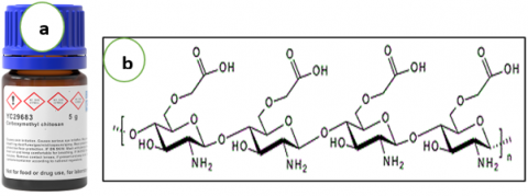
Analytical grades are to all reagents with no additional purification when received. The silver nitrate (AgNO3; 99.98 percent) is selected as silver ion precursor which Merck, Germany provides. Nanoparticles stabilizers used the Carboxymethyl CS which is low molecular weights from Sigma Aldrich in America which has an isopropanol solvent, glacial acetic acid (HAC, 99 percent which the next figure shows. It is a double-distilled water (DD-water) for making every aqueous solution.
Figure 1. (a) Carboxymethyl CS materials and (b)The molecular structure of Carboxymethyl CS
2.2 Methods
To create a stock solution of CS, we dissolved 3 grams of CS in 150 ml of acetic acid using 14:1 distilled water, and left it for 8 hours. Next, we added 0.6 grams of silver nitrate to 20 ml of deionized water and mixed it with the CS solution in a glass ampoule. To prevent the formation of free radicals, we added 23 grams of isopropanol, as shown in Figure 1, and deaerated the solution by bubbling pure nitrogen for 20 minutes. The ampoule was then sealed and exposed to Co-60 1/5 -ray sources. Then 23 g of isopropanol was added as a free radical scavenger as shown in Figure 1 and deaerated with bubbling pure N2 for 20 minutes. It then sealed to irradiate with Co-60 1/5 γ-ray sources. The RD is 8 Gy\hr regulating its total doses when conducting the experiment through adjusting the irradiation time as in Table 1.
The outcome viscous solution, manufactured by AgNPs showed color shift from yellow to brown (Figure 2) because silver salt through reduces by dropping of the compounds in the CM CS extract.
So, Ag ions concentration is (1.76 x10⁻⁵ mol/ml), the –NH2 and the concentration of the groups is (9.35x10⁻⁵ mol/ml) and that of the isopropanol reaches (1.9x10⁻³ mol/ml). The solution pH reaches (3.0). We used diluted irradiation-degrade AgNPs/CS solutions for fixing the made silver particles from radiation–reduction. The RD rates and Times (hr.) to Ag-NPs /CM-CS - isopropanol is seen in the table below with additional physical features.
Table 1. Gamma dose and time of irradiation and properties
|
1 |
2 |
3 |
|
|
Sample Code |
A1 |
A2 |
A3 |
|
RD (Gy) |
768 |
1151 |
1920 |
|
Irradiation Time (hr.) |
96 |
144 |
240 |
|
PH. |
5 |
4.8 |
5 |
|
Con.% |
0.182 |
0.19 |
0.264 |
|
T℃ |
27 |
26 |
26 |
Figure 2. a) Preparation of AgNO3, b) AgNO3 sample before, c) after gamma irradiation Treatment
There were many sprays of silver solutions on glass substrates to the distribution of the size which AFM (Angstrom dvancedtypeAA3000) determines. X-ray diffraction were used for the silver thin films samples for crystalline structures [32-38]. For the influence of AgNPs on human blood; the experiment was conducted on six donner mal health adult. We labeled the samples and the controls (B1-B6) and examined pre and post nano silver particles injecting to blood, urea, blood creatinine, and phosphates of alkaline test by Uv-Vis spectrophotometer (SP-3000 Optima-plus- Chapan) [36, 39-55]. The former was red upper level and yellow color serum lower level. We established the tests for both with injecting 0.1 ml (volume ratio) of sample A3 of AgNPs solution besides the controls.
To evaluate the antimicrobial efficiency of AgNPs solutions for river water treatment, we considered the following;
i) We collected five water samples from different locations along the Dyala River, which flows primarily through Iraq. In addition, we collected one water sample from the Al-Gherait riverbank, and each sample was labeled and placed into standard containers that had been treated with AgNPs solutions (sample A3). These samples were selected due to their contamination status.
ii) The samples above evaluate as antimicrobial efficiency for achieving through the standard approach to inspect water inspections in the aerobic nutrient agar for whole bacteria inspections and macconky broth counts tocoliform and to the testing of the fecal coliform bacterias [56].
3.1 Structure properties
Ag-NPs / (CM CS – isopropanol) is from reducing AgNO3 into ions (Ag+) of positive silver helped by fragmenting large Ag-NPs in γ-irradiation [12]. The figure below shows the manufactured Ag-NPs. The Ag-NPs/(CM CS-isopropanol color intensity rose step by step. The A0 sample with not ray shows a clear yellow solution (as Figure 3 shows). The colloidal solution turned dark brown which is the -rays strengthened (samples A1-A3), yet the AgNO3 solution had no color as in Figure 3). The Ag-NPs/CM CS-isopropanol change the pure yellow colloidal solution from light to dark brown after gamma irradiations (samples (A0-A3). This confirms Hanan et al. [57], Shriram et al. [58], and M.S.N. Salleh et al. [23] to form AgNPs by the reduction of AgNO3.
Figure 3. Feature shifts in the color of colloidal solutions which can be Ag-NPs /(CM CS - isopropanol)) after being exposed to (0, 768, 1151, 1920 Gy) doses (A0–A3, respectively)
Peng Chen Briefly reported photo-induced fragmenting Ag-NPs [19, 59]:
$(A g)_n+2 h v \rightarrow(A g)_n^{+}+e_{a q}^{-}$ (1)
$(A g)_n^{+}+e_{a q}^{-} \rightarrow(A g)_n$ (2)
$(A g)_n^{+} \rightarrow(A g)_{n-1}+A g^{+}(A g)_n^{+} \rightarrow(A g)_{n-1}+A g^{+}$ (3)
Here: (Ag)n = The silver nanocluster with (n) silver atoms
e − = The aqueous electrons
On gamma -irradiation on aqueous suspending CS/ silver nitrate (Cts/AgNO3). In addition to the, the aqueous electrons decrease the positive silver ions into silver.
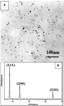
Figure 4. (a)TEM and (b) XRD results of Ag /CM-CSs sample A3
Figure 4 is the x-ray diffraction of the silver samples for studying their structure showing the crystalline which as patterned with 2Ө (33.0337) of highest positions along the high intensities (100%) to plane (111) and second peak positions 2Ө (43.9817) with relative intensities (52%) to plane (200), (220) band for face-centered cubics (fcc) structure of Ag-NPs [24, 58].
The XRD shows the most suitable gamma dose for gaining silver nano particle because the Co-60 is 1920 Gy over 240 hr [25]. The produced AgNPs morphology and particle size distributions were examined by Transmission Electron Microscopy (TEM) imaging. Also the figure below is a typical sample A3olution TEM image with dispersed well Ag-NPs in the (CM-CS-isopropanol) matrix.
Figure 5. The size distribution (a) and (b) AFM image of AgNPs (A3) sample
The AgNP TEM images and size distributions shows the average diameter of the nanoparticles in sample A3 ranging 40 and 50 nm along with the cubic phases. Yet, that of the A1 and A2 is larger than 100 nm.
Figure 5 is the spherical AgNPs (A3) particle AFM made by gamma irradiation with (40-100) nm distribution.
3.2 UV–visible spectroscopy analysis
This research included studying the optical properties of the samples prepared by irradiation method using cobalt (Co 60) source as finding the optical energy gaps for the permissible direct electronic transitions, by recording the absorption and transmittance spectra of the prepared solutions within the wavelength range (400-1100) nm.
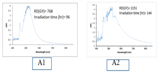
UV–seen absorbing spectra are sensitive to form Ag-NPs as Ag-NPs show intense absorption peak because of the SPR. Figures 6 is UV–vis silver nanoparticle spectra at constant CM-CS and it is also at silver nitrate concentrations yet at other gamma irradiation doses (768, 1151, 1920 Gy) to (96, 144, and 240 hr), in the same order.
All spectra show an absorption band (370–470 nm). It is a Ag-NPs SPR band. Weak peaks around 330 and 560 nm indicate the nanoparticle aggregations. One strong peak with a highest of d 430 nm was seen in the UV–vis spectra corresponding to the specific SPR of electrons from the of Ag-NPs surface. The nanoparticle synthesis efficiency rises gamma irradiation doses rise (768, 1151, 1920 Gy) because oxidating hydroxyl set of CM-CS by silver ions rises. Also, in producing nanoparticle rise by monitoring silver nitrate when gamma irradiation doses increase, and Figure 6 displays their respective spectra. The absorption of the solution intensity rose when the gamma irradiation doses increased, in particular by A3 subjected a 1920Gy gamma dose of irradiations sample A3:
Figure 6. UV– silver nanoparticle visible absorption spectra of CM -CS- isopropanol by various gamma irradiation doses: (AI) at 768Gy dose for 96hr, (A2) at 1151Gy dose for 144 and (A3) at 1920Gy dose for 240 hr
Figure 7. (αhv) versus hv for Ag-NPs stabilized within CM -CS- isopropanol using various gamma irradiation doses
Figure 6 is specific UV-visible absorption spectra to the silver solutions. It reveals the highest absorbance in a set of spectra band. There is a dominate SP absorption peak (420-430 nm).
This research founds the optical energy gap value for the permissible direct electronic transitions, by recording the absorption and transmittance spectra of the prepared solutions within the wavelength range (400-1100) nm by plotting the graph between (hƲ) on the X-axis and (αhƲ)2 on the Y-axis and drawing the tangent, we can extract the value of the energy gap. The optical energy gaps are fulfilled by scheming (αhν) against (hν) as shown in Figure 7. It increases from the values of the energy gap of the films grown at (96, 144 and 240) hr were (1.9, 2.32 and 2.33) eV. Respectively.
The variation of the optical energy with correlated to the change of the radiation rate, as shown in the Table 2 [60].
Table 2. Cs/Ag nanocomposite solution energy gaps with Irradiation Gamma dose rate
|
Sample Code |
A1 |
A2 |
A3 |
|
RD (Gy) |
768 |
1151 |
1920 |
|
Irradiation Time (hr.) |
96 |
144 |
240 |
|
PH. |
5 |
4.8 |
5 |
|
Con.% |
0.182 |
0.19 |
0.264 |
|
T℃ |
27 |
26 |
26 |
|
Eg (ev) |
1.9 |
2.33 |
2.34 |
Figure 8 shows the optical transmission ratio behavior for the three Samples (A1-A3). Sample A3 show the highest ratio of transmission light in a range of 489-789 nm due to its particle nano size than the others.
Figure 8. The Transmitions optical features (T%) versus wave length (180-1100) nm for Ag-NPs stabilized (CM -CS- isopropanol) using various gamma irradiation doses
It is obvious that the optical energy gap and light transmission percent widely depended on gamma irradiation energy dose rate.
3.3 Applying Ag-NPs /CM CS – isopropanol.
I) Effect of AgNPS on human blood
Nanosilver solution prepared by gamma RDrate (sample A3- 1920 Gy) using gamma source Co-60 within CS agent. Sample (A3) with particle size about 40 nm with 0.63 ppm of concentration inserted into second group (B1-B6). We measured the concentration of the Serum concentration through biochemical spectrophotometer and biochemical devices and calculated the Urean, Alkaline phosphate and blood crentinine. Tables 3, 4, 5 are the examinations of to six blood sample outcomes.
Table 3. Blood urea results
|
Sample |
No. |
B1 |
B2 |
B3 |
B4 |
B5 |
B6 |
|
|
B.Urea |
Befor AgNPs |
Ingection |
50.8 |
37.7 |
28 |
46 |
31 |
31 |
|
B.Urea |
after AgNPs Ingection |
upper level |
27 |
26 |
14 |
31 |
21 |
23 |
|
B.Urea |
after AgNPs Ingection |
lower level |
49 |
36.3 |
27 |
44 |
31 |
29 |
Table 4. Blood crentinine result
|
Sample No. |
B1 |
B2 |
B3 |
B4 |
B5 |
|
B.Crentinine befor AgNPs ingection |
1.27 |
0.94 |
0.7 |
1.1 |
0.7 |
|
B.Crentinine after AgNPs ingection upper level |
0.67 |
0.65 |
0.3 |
0.7 |
0.5 |
|
B.Crentinine after AgNPs ingection lower level |
1.22 |
0.9 |
0.7 |
1.1 |
0.7 |
Table 5. Alkaline phosphates examinations
|
Sample |
No. |
B1 |
B2 |
B3 |
B4 |
||
|
A.Phos. |
before AgNPs |
injection |
8.2 |
15.1 |
9 |
10 |
|
|
A.Phos. |
after AgNPs |
Injection |
upper level |
10.7 |
11.4 |
8 |
7 |
|
A.Phos. |
after AgNPs |
Injection |
lower level |
8.4 |
14.6 |
9 |
10 |
The group (B1-B6) contained the control identical to the treated samples to the compared methods. Tables 3, 4, 5 are the mean Urea, phosphate and crentinine of the six samples for two blood levels, the upper of which is red cells group and the lower is yellow. The results make it clear that the nanosilver particle impact (sample A3) contradictorily on upper (red one) with no effect on the yellow. Reducing urean, alkaline, phosphate and blood crentinine could is possibly treated by the aggregating influence of nanosilver particles on blood cells. The influence on nanosilver particles (Figure 9) in the blood cells cause reduces parameters injected in the liver kidney factors [60, 61].
II) Effect of AgNPS with river water treatment
The Purification of water by silver seems to be wide spread to a long time and traditionally applied in in different fields [62, 63]. Evaluations of antimicrobial effect of AgNPs against contaminated river water was achieved to six samples from Dyala and Al Gherait river and tested by the standard methods for water inspection. Tables 6 and 7 is one good activity obtained by using AgNPs concentration (5.3 - 33.03) ppm as antimicrobial water bacteria.
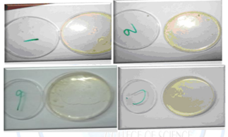
Table 6 and Table 7 show good activity by AgNPs techniques as antimicrobial water bacteria in which the typical images (Figure 10) are the for total water sample count inspecting. In addition, the good agreement between the AgNPS efficiency in each sample of Al_Gherai’t and Dyala is reported regardless of the highly contaminated water in Dyala River water in comparison to Al_Gherai’t, and the various concentration uses where we have Table 6 of silver shows no inhabitation in the bacteriological purification.
Table 6 is the outcomes of the silver (5.3-33.03) ppm concentration to all samples and the inspiring activities. In addition, sample (5) exhibits some aerobic bacteria. The quantity is suitable based on the Iraqi standard for bacteriology water inspections. The Dyala contamination is bigger than that of Al_Gherai’t which encouraged greater silver concentration (33.03) ppm in purifying the sample of the later. Table 7 showed that the in Bridge – Dyala and Join Dyala aerobic bacteria and the Tigris are somehow larger than the rest of the areas because of their high contamination.
Figure 9. Effect of AgNPs: Aggregate morphology of red blood cells (in vitro application)
Table 6. Bacteriology effective treating AgNPS on AL-Gherait side river water
|
Sample No. |
C1 |
C2 |
C3 |
C4 |
C5 |
C6 |
|
AgNPs concentration (ppm) |
5.3 |
7.05 |
12.82 |
14.87 |
28.09 |
33.03 |
|
Total count |
0 |
0 |
0 |
0 |
9 |
0 |
|
coliform |
0 |
0 |
0 |
0 |
0 |
0 |
|
Fecal coliform |
-ve |
-ve |
-ve |
-ve |
-ve |
-ve |
Table 7. Bacteriology effective treating AgNPs on Dyala side river water
|
Site of the sample |
Location 1- Alsdur |
location 2- Baquba |
location 3- Dyala Bridge |
Location 4 new Dyala Bridge |
Location 5- join Dyala river with Tigris river |
|
Water to AgNPs ratio |
03:01 |
03:01 |
03:01 |
03:01 |
03:01 |
|
Total count |
1 |
1 |
2 |
1 |
2 |
|
coliform |
0 |
0 |
0 |
0 |
0 |
|
Fecal coliform |
-ve |
-ve |
-ve |
-ve |
-ve |
Figure 10. Typical picture for total count inspecting water samples
Ag-NPs stabilized in (CM -CS- isopropanol) were prepared by -irradiation method with (40-500) nm particle size distribution form the use of RDrates. The wavelength at the highest absorptions changed to longer with the initial concentration rise of AgNPs ions. A silver nanoparticle smaller size was obtained by the use of CS as stabilizer. The unique shows that the -irradiation is suitable in particular. In producing colloidal and nano Ag-NPs on large scales. The process of producing is through the economic and available methods, tools and materials. This work examined the physiological influence that nanosilver particles (sample A3) at (0.63) ppm concentrations with (0.1) ml volume ratio injected to human blood samples. The study showed reduced urea, blood crentinine and alkaline phosphate values. There is also extreme stability of these nanoparticles at physiological conditions and the blood was compatible for use to more potent antiplatelet agent than aspirin.
There is a successful silver solution development for bacteriological water purification proving their effectiveness at various concentration to various contaminated water samples of. Using various concentration of Ag-NPs in CM -CS- isopropanol for Al_Gherai’t samples (5.3- 33.03) ppm were positive.
[1] Dhawale, D.S., Lokhande, C.D. (2011). Chemical route to synthesis of mesoporous ZnO thin films and their liquefied petroleum gas sensor performance. Journal of Alloys and Compounds, 509(41): 10092-10097. https://doi.org/10.1016/j.jallcom.2011.08.046
[2] Santiago, T.R., Bonatto, C.C., Rossato, M., Lopes, C.A., Lopes, C.A., Mizubuti, E.S., Silva, L.P. (2019). Green synthesis of silver nanoparticles using tomato leaf extract and their entrapment in chitosan nanoparticles to control bacterial wilt. Journal of the Science of Food and Agriculture, 99(9): 4248-4259. https://doi.org/10.1002/jsfa.9656
[3] Calderón-Jiménez, B., Johnson, M.E., Montoro Bustos, A.R., Murphy, K.E., Winchester, M.R., Vega Baudrit, J.R. (2017). Silver nanoparticles: Technological advances, societal impacts, and metrological challenges. Frontiers in Chemistry, 5: 6. https://doi.org/10.3389/fchem.2017.00006
[4] AL‐Shnani, F., Al‐Haddad, T., Karabet, F., Allaf, A.W. (2017). Chitosan loaded with silver nanoparticles, CS‐AgNPs, using thymus syriacus, wild mint, and rosemary essential oil extracts as reducing and capping agents. Journal of Physical Organic Chemistry, 30(11): e3680. https://doi.org/10.1002/poc.3680
[5] Wessels, J.M., Nothofer, H.G., Ford, W.E., von Wrochem, F., Scholz, F., Vossmeyer, T., Schroedter, A., Weller, H., Yasuda, A. (2004). Optical and electrical properties of three-dimensional interlinked gold nanoparticle assemblies. Journal of the American Chemical Society, 126(10): 3349-3356. https://doi.org/10.1021/ja0377605
[6] Heilmann, A., Kiesow, A., Gruner, M., Kreibig, U. (1999). Optical and electrical properties of embedded silver nanoparticles at low temperatures. Thin Solid Films, 343: 175-178. https://doi.org/10.1016/S0040-6090(98)01599-5
[7] Mansoor, S., Zahoor, I., Baba, T.R., Padder, S.A., Bhat, Z.A., Koul, A.M., Jiang, L. (2021). Fabrication of silver nanoparticles against fungal pathogens. Frontiers in Nanotechnology, 3: 679358. https://doi.org/10.3389/fnano.2021.679358
[8] Zhang, Z., Zhao, B., Hu, L. (1996). PVP protective mechanism of ultrafine silver powder synthesized by chemical reduction processes. Journal of Solid State Chemistry, 121(1): 105-110. https://doi.org/10.1006/jssc.1996.0015
[9] Chou, K.S., Ren, C.Y. (2000). Synthesis of nanosized silver particles by chemical reduction method. Materials Chemistry and Physics, 64(3): 241-246. https://doi.org/10.1016/S0254-0584(00)00223-6
[10] Nersisyan, H.H., Lee, J.H., Son, H.T., Won, C.W., Maeng, D.Y. (2003). A new and effective chemical reduction method for preparation of nanosized silver powder and colloid dispersion. Materials Research Bulletin, 38(6): 949-956. https://doi.org/10.1016/S0025-5408(03)00078-3
[11] Sondi, I., Goia, D.V., Matijević, E. (2003). Preparation of highly concentrated stable dispersions of uniform silver nanoparticles. Journal of Colloid and Interface Science, 260(1): 75-81. https://doi.org/10.1016/S0021-9797(02)00205-9
[12] Beden, S.A., Ati, A.A., Fayadh Alimarah, K.A., Saihood, S.K., Abdulnabi, R.K., Mejbel, M.K. (2022). Green synthesis and characterization of silver nanoparticles/CM-chitosan-isopropanol by gamma irradiations method. Revue des Composites et des Matériaux Avancés-Journal of Composite and Advanced Materials, 32(4): 173-180. https://doi.org/10.18280/rcma.320402
[13] Yin, B., Ma, H., Wang, S., Chen, S. (2003). Electrochemical synthesis of silver nanoparticles under protection of poly (N-vinylpyrrolidone). The Journal of Physical Chemistry B, 107(34): 8898-8904. https://doi.org/10.1021/jp0349031
[14] Huang, H.H., Ni, X.P., Loy, G.L., Chew, C.H., Tan, K.L., Loh, F.C., Deng, J.F., Xu, G.Q. (1996). Photochemical formation of silver nanoparticles in poly (N-vinylpyrrolidone). Langmuir, 12(4): 909-912. https://doi.org/10.1021/la950435d
[15] Ye, X., Zhou, Y., Chen, J., Sun, Y. (2007). Deposition of silver nanoparticles on silica spheres via ultrasound irradiation. Applied Surface Science, 253(14): 6264-6267. https://doi.org/10.1016/j.apsusc.2007.01.111
[16] Jiang, H., Moon, K.S., Zhang, Z., Pothukuchi, S., Wong, C.P. (2006). Variable frequency microwave synthesis of silver nanoparticles. Journal of Nanoparticle Research, 8: 117-124. https://doi.org/10.1007/s11051-005-7522-6
[17] Henglein, A., Giersig, M. (1999). Formation of colloidal silver nanoparticles: capping action of citrate. The Journal of Physical Chemistry B, 103(44): 9533-9539. https://doi.org/10.1021/jp9925334
[18] Shin, H.S., Yang, H.J., Kim, S.B., Lee, M.S. (2004). Mechanism of growth of colloidal silver nanoparticles stabilized by polyvinyl pyrrolidone in γ-irradiated silver nitrate solution. Journal of colloid and interface science, 274(1): 89-94. https://doi.org/10.1016/j.jcis.2004.02.084
[19] Chen, P., Song, L., Liu, Y., Fang, Y.E. (2007). Synthesis of silver nanoparticles by γ-ray irradiation in acetic water solution containing chitosan. Radiation Physics and Chemistry, 76(7): 1165-1168. https://doi.org/10.1016/j.radphyschem.2006.11.012
[20] Li, T., Park, H.G., Choi, S.H. (2007). γ-Irradiation-induced preparation of Ag and Au nanoparticles and their characterizations. Materials Chemistry and Physics, 105(2-3): 325-330. https://doi.org/10.1016/j.matchemphys.2007.04.069
[21] Hornebecq, V., Antonietti, M., Cardinal, T., Treguer-Delapierre, M. (2003). Stable silver nanoparticles immobilized in mesoporous silica. Chemistry of Materials, 15(10): 1993-1999. https://doi.org/10.1021/cm021353v
[22] Bogle, K.A., Dhole, S.D., Bhoraskar, V.N. (2006). Silver nanoparticles: Synthesis and size control by electron irradiation. Nanotechnology, 17(13): 3204. https://doi.org/10.1088/0957-4484/17/13/021
[23] Salleh, M.S.N., Ali, R.R., Shameli, K., Hamzah, M.Y., Chan, J.Z. (2020). Silver nanoparticles on pullulan derived via gamma irradiation method: A preliminary analysis. In IOP Conference Series: Materials Science and Engineering, 808(1): 012030. https://doi.org/10.1088/1757-899X/808/1/012030
[24] Barot, T., Rawtani, D., Kulkarni, P. (2020). Physicochemical and biological assessment of silver nanoparticles immobilized Halloysite nanotubes-based resin composite for dental applications. Heliyon, 6(3): e03601. https://doi.org/10.1016/j.heliyon.2020.e03601
[25] Kaur, A., Preet, S., Kumar, V., Kumar, R., Kumar, R. (2019). Synergetic effect of vancomycin loaded silver nanoparticles for enhanced antibacterial activity. Colloids and Surfaces B: Biointerfaces, 176: 62-69. https://doi.org/10.1016/j.colsurfb.2018.12.043
[26] Morones, J.R., Elechiguerra, J.L., Camacho, A., Holt, K., Kouri, J.B., Ramírez, J.T., Yacaman, M.J. (2005). The bactericidal effect of silver nanoparticles. Nanotechnology, 16(10): 2346. https://doi.org/10.1088/0957-4484/16/10/059
[27] Kim, J.S., Kuk, E., Yu, K.N., et al. (2007). Antimicrobial effects of silver nanoparticles. Nanomedicine: Nanotechnology, Biology and Medicine, 3(1): 95-101. https://doi.org/10.1016/j.nano.2006.12.001
[28] Elechiguerra, J.L., Burt, J.L., Morones, J.R., Camacho-Bragado, A., Gao, X., Lara, H.H., Yacaman, M.J. (2005). Interaction of silver nanoparticles with HIV-1. Journal of Nanobiotechnology, 3: 1-10. https://doi.org/10.1186/1477-3155-3-6
[29] Pal, S., Tak, Y.K., Song, J.M. (2007). Does the antibacterial activity of silver nanoparticles depend on the shape of the nanoparticle? A study of the gram-negative bacterium Escherichia coli. Applied and Environmental Microbiology, 73(6): 1712-1720. https://doi.org/10.1128/AEM.02218-06
[30] Vermerie, N., Malbrunot, C., Azar, M., Arnaud, P. (1997). Stability of nystatin in mouthrinses; effect of pH, temperature, concentration and colloidal silver addition, studied using an in vitro antifungal activity. Pharmacy World and Science, 19: 197-201. https://doi.org/10.1023/A:1008664917377
[31] Lee, H.J., Yeo, S.Y., Jeong, S.H. (2003). Antibacterial effect of nanosized silver colloidal solution on textile fabrics. Journal of Materials Science, 38: 2199-2204. https://doi.org/10.1023/A:1023736416361
[32] Aziz, W.J., Abid, M.A., Kadhim, D.A., Mejbel, M.K. (2020). Synthesis of iron oxide (β-fe2o3) nanoparticles from Iraqi grapes extract and its biomedical application. In IOP Conference Series: Materials Science and Engineering, 881(1): 012099. https://doi.org/10.1088/1757-899X/881/1/012099
[33] Mezher, S.J., Kadhim, KJ., Abdulmunem, O.M., Mejbel, M.K. (2020). Microwave properties of Mg–Zn ferrite deposited by the thermal evaporation technique. Vacuum, 173: 109114. https://doi.org/10.1016/j.vacuum.2019.109114
[34] Ghazi, A.K., Muhmmed, A.A., Taieh, N.K., Mejbel, M.K. (2022). Tribological and mechanical performance of epoxy reinforced by fish scales powder. Revue des Composites et des Matériaux Avancés-Journal of Composite and Advanced Materials, 32(3): 149-155. https://doi.org/10.18280/rcma.320306
[35] Mezher, S.J., Dawood, M.O., Abdulmunem, O.M., Mejbel, M.K. (2020). Copper doped nickel oxide gas sensor. Vacuum, 172: 109074. https://doi.org/10.1016/j.vacuum.2019.109074
[36] Al-Saadi, T.H.A., Mohammad, S.H., Daway, E.G., Mejbel, M.K. (2021). Synthesis of intumescent materials by alkali activation of glass waste using intercalated graphite additions. Materials Today: Proceedings, 42: 1889-1900. https://doi.org/10.1016/j.matpr.2020.12.228
[37] Mezher, S.J., Dawood, M.O., Beddai, A.A., Mejbel, M.K. (2020). NiO nanostructure by RF sputtering for gas sensing applications. Materials Technology, 35(1): 60-68. https://doi.org/10.1080/10667857.2019.1653595
[38] Mikhlif, H.M., Dawood, M.O., Abdulmunem, O.M., Mejbel, M.K. (2021). Preparation of High-Performance Room Temperature ZnO Nanostructures Gas Sensor. Acta Physica Polonica, A., 140(4): 320-326. http://doi.org/10.12693/APhysPolA.140.320
[39] Mejbel, M.K., Allawi, M.K., Oudah, M.H. (2019). Effects of WC, SiC, iron and glass fillers and their high percentage content on adhesive bond strength of an aluminium alloy butt joint: An experimental study. Journal of Mechanical Engineering Research and Developments (JMERD), 42(5): 224-231. http://doi.org/10.26480/jmerd.05.2019.224.231
[40] Jawad, L.K., Beddai, A.A., Nasser, M.A., Mejbel, M.K. (2022). Scrutinizing the physical and strength properties of fabricated date palm frond leaves particleboard. Materials Today: Proceedings, 57: 980-988. https://doi.org/10.1016/j.matpr.2022.03.396
[41] Mejbel, M.K., Abdullah, I.T., Taieh, N.K. (2022). Thin wall manufacturing improvement using novel simultaneous double-sided cutter milling technique. International Journal of Automotive and Mechanical Engineering, 19(1): 9519-9529. https://doi.org/10.15282/ijame.19.1.2022.15.0734
[42] Moosa, A.U., Hernández-Nava, E., Mejbel, M.K., Todd, I. (2022). The joining of CP-vanadium and Ti–6Al–4V using the Electron Beam Melting Additive Manufacturing method. Advances in Industrial and Manufacturing Engineering, 5: 100102. https://doi.org/10.1016/j.aime.2022.100102
[43] Al-Saadi, T.H.A., Daway, E.G., Mohammad, S.H., Mejbel, M.K. (2020). Effect of graphite additions on the intumescent behaviour of alkali-activated materials based on glass waste. Journal of Materials Research and Technology, 9(6): 14338-14349. https://doi.org/10.1016/j.jmrt.2020.10.035
[44] Mejbel, A.M.K.M.K., Khalaf, M.M., Kwad, A.M. (2021). Improving the machined surface of AISI H11 tool steel in milling process. Journal of Mechanical Engineering Research and Developments, 44(4): 58-68.
[45] Allawi, M.K., Oudah, M.H., Mejbel, M.K. (2019). Analysis of exhaust manifold of spark-ignition engine by using computational fluid dynamics (CFD). Journal of Mechanical Engineering Research and Developments (JMERD), 42(5): 211-215. http://doi.org/10.26480/jmerd.05.2019.211.215
[46] Allawi, M.K., Mejbel, M.K., Oudah, M.H. (2021). Variable valve timing (VVT) modelling by Lotus engine simulation software. International Journal of Automotive and Mechanical Engineering, 17(4): 8397-8410. https://doi.org/10.15282/ijame.17.4.2020.15.0635
[47] Beddai, A.A., Badday, B.A., Al-Yaqoobi, A.M., Mejbel, M.K., Al Hachim, Z.S., Mohammed, M.K. (2022). Color removal of textile wastewater using electrochemical batch recirculation tubular upflow cell. International Journal of Chemical Engineering, 2022: 4713399. https://doi.org/10.1155/2022/4713399
[48] Muhmmed, A.A., Hussain, M.K., Khudadad, A.R., Mahdi, H.H., Mejbel, M.K. (2021). Mechanical behavior of laser engraved single lap joints adhered by polymeric material. International Review of Mechanical Engineering, 15(12): 622-628. https://doi.org/10.15866/ireme.v15i12.21278
[49] Allawi, M.K., Mejbel, M.K., Younis, Y.M., Mezher, S.J. (2020). A simulation of the effect of Iraqi diesel fuel cetane number on the performance of a compression ignition engine. International Review of Mechanical Engineering, 14(3): 151-159. https://doi.org/10.15866/ireme.v14i3.18137
[50] Allawi, M.K., Mejbel, M.K., Oudah, M.H. (2020). Iraqi gasoline performance at low engine speeds. In IOP Conference Series: Materials Science and Engineering, 881(1): 012065. https://doi.org/10.1088/1757-899X/881/1/012065
[51] Allawi, M.K., Oudah, H., Mejbel, M.K. (2019). Experimental investigation of exhaust emissions from spark-ignition engine using the different types of fuels. International Journal of Mechanical Engineering & Technology, 10(1): 2109-2113.
[52] Mejbel, M.K., Atwan, H.R., Abdullah, I.T. (2021). Void formation in friction stir welding of AA5052 butt joining. Journal of Mechanical Engineering Research and Developments, 44(5): 318-332.
[53] Baqer, A.R., Beddai, A.A., Farhan, M.M., Badday, B.A., Mejbel, M.K. (2021). Efficient coating of titanium composite electrodes with various metal oxides for electrochemical removal of ammonia. Results in Engineering, 9: 100199. https://doi.org/10.1016/j.rineng.2020.100199
[54] Abood AL-Saadi, T.H., Abdulnabi, R.K., Ismael, M.N., Hassan, H.F., Mejbel, M.K. (2022). Glass waste based geopolymers and their characteristics. Revue des Composites et des Matériaux Avancés, 32(1): 17-23. https://doi.org/10.18280/rcma.320103
[55] Oudah, M.H., Mejbel, M.K., Allawi, M.K. (2021). R134a flow boiling heat transfer (FBHT) characteristics in a refrigeration system. Journal of Mechanical Engineering Research and Development, 44(4): 69-83.
[56] Lorian, V. (Ed.). (2005). Antibiotics in Laboratory Medicine. Lippincott Williams & Wilkins.
[57] Ghetas, H.A., Abdel-Razek, N., Shakweer, M.S., Abotaleb, M.M., Paray, B.A., Ali, S., Eldessouki, E.A., Dawood, M.A.O., Khalil, R.H. (2022). Antimicrobial activity of chemically and biologically synthesized silver nanoparticles against some fish pathogens. Saudi Journal of Biological Sciences, 29(3): 1298-1305. https://doi.org/10.1016/j.sjbs.2021.11.015
[58] Mirajkar, S., Rathod, P., Pawar, B., Penna, S., Dalvi, S. (2021). γ-irradiated chitosan mediates enhanced synthesis and antimicrobial properties of chitosan–silver (Ag) nanocomposites. ACS Omega, 6(50): 34812-34822. https://doi.org/10.1021/acsomega.1c05358
[59] Chen, P., Wang, Z., Zong, S., Chen, H., Zhu, D., Zhong, Y., Cui, Y. (2014). A wide range optical pH sensor for living cells using Au@ Ag nanoparticles functionalized carbon nanotubes based on SERS signals. Analytical and Bioanalytical Chemistry, 406: 6337-6346. https://doi.org/10.1007/s00216-014-8064-5
[60] Al-Baker, A.A., Al-Kshab, A.A., Ismail, H.K., Ashwaq, A.H. (2020). Effect of silver nanoparticles on some blood parameters in rats. Iraqi Journal of Veterinary Sciences, 34(2): 389-395.
[61] Kumar, K.P., Paul, W., Sharma, C.P. (2012). Green synthesis of silver nanoparticles with Zingiber officinale extract and study of its blood compatibility. BioNanoScience, 2: 144-152. https://doi.org/10.1007/s12668-012-0044-7
[62] Lv, Y., Liu, H., Wang, Z., Liu, S., Hao, L., Sang, Y., Liu, D., Wang, J., Boughton, R.I. (2009). Silver nanoparticle-decorated porous ceramic composite for water treatment. Journal of Membrane Science, 331(1-2): 50-56. https://doi.org/10.1016/j.memsci.2009.01.007
[63] Gadkari, R.R., Ali, S.W., Alagirusamy, R., Das, A. (2018). Silver nanoparticles in water purification: opportunities and challenges. In: Oves, M., Zain Khan, M., M.I. Ismail, I. (eds) Modern Age Environmental Problems and their Remediation. Springer, Cham. https://doi.org/10.1007/978-3-319-64501-8_13