Sabeeha A.J. Beden | Ali A. Ati | Kifah A. Fayadh Alimarah | Suhailah K. Saihood | Rana K. Abdulnabi | Mohanad Kadhim Mejbel*
© 2022 IIETA. This article is published by IIETA and is licensed under the CC BY 4.0 license (http://creativecommons.org/licenses/by/4.0/).
OPEN ACCESS
The present study demonstrates a green process for the synthesis of spherical-shaped and stabilized silver nanoparticles Ag NPs using gamma irradiated conditions. This method has certain advantages over conventional methods since it can manage the particle size and structure and generates totally reduced and extremely pure nanoparticles free from by-products or chemical reducing agents. Silver nanoparticles composite (Ag NPs / CM) is prepared from an aqueous solution of silver nitrate, chitosan and isopropanol, at room temperature using gamma irradiation doses to induce reduction and cross-linking to formation in situ Ag NPs/ CM-chitosan-isopropanol solutions. The production and homogenous distribution of silver nanoparticles in the hydrogel matrix were characterized using transmission electron microscopy, XRD, Zeta potential and UV-Vis spectrophotometer analysis. The silver/ CM-chitosan-isopropanol matrixes possessed different gamma doses using a Co- 60 gamma source. Through the comprehensive results of IR- Ag NPs /(CM- chitosan isopropanol solutions due to antibacterial activity test, the results show the prepared Ag NPs /CM-chitosan-isopropanol could be used as an antibacterial agent. The obtained Ag nanoparticles were stable for over 3 months at room temperature. the analysis outcomes indicate that the colloidal Solution (silver nanoparticles / (CM chitosan - isopropanol)) has a promising ability to be used in antibacterial applications in the future.
Ag-NPs, gamma radiation, CM-chitosan -isopropanol, antibacterial agent
Nanotechnology is an emerging field of scientific research. It is thriving due to the advent of nanomaterials, like inorganic nanoparticles (NPs) and nanostructures [1]. Nanoparticles Researchers from a wide range of fields are interested in studying this topic. Because of their extraordinary features, which include distinct functionalities and size-dependent physicochemical characteristics that vary greatly from their bulk counterparts [2]. Which can develop nanostructures with a large surface area to volume ratio.
Nanoparticles may be classified into organic and inorganic varieties [3-6]. Throughout the globe, inorganic nanoparticles have been used in nanomedicine, medication delivery, cosmetics, electronics, and the energy sector [7, 8]. Among metal nanoparticles, silver nanoparticles (Ag NPs) have received attention not only in the researches field. It is also for industrial applications, and different fields of science, as it has chemical and biological characteristics that may be of interest [9]. Depending on the silver nanoparticles size and form, they may be used in biosensor materials, cosmetic items, antimicrobial applications, conducting materials, and electrical components [10]. Researchers have been interested in silver nanoparticles because of their unique properties, such as their optical, antibacterial, and electrical capabilities (NPs) [11]. Silver nanoparticles can be synthesized by different techniques, such as physical, chemical, and biological method [12].
Documentation of many methods for synthesizing silver nanoparticles are: Electron and gamma (γ)-irradiation, artificial and biological procedures, microwave and ultrasonic processing, laser ablation, photochemical reduction, and so forth [13-15].
Chitosan (Cts) is one of the most efficient and effective biomaterials to synthesize Ag-NPs in our study because of its substantial antibacterial properties against Gram-negative and Gram-positive bacteria and low toxicity toward mammalian cells [16, 17].
A stabilizer is usually added to prevent agglomeration and control the formation and stabilize the NPs. Additionally, the NPs produced by chemical synthesis will be sedimented with chemicals and are not applicable for biomedical application [18].
Indeed, when comparing such a method that uses ionizing radiation (gamma and electron) to other approaches, it has several advantages [19].
Mass production carried out at a comparatively reasonable cost, the particles size is controlled by varying the concentration of silver ion, colloidal silver nanoparticles prepared without silver ion residue and excessive reductant contamination, reaction rate controlled by varying irradiation time, reducing agents may be found in the irradiated solution in a very consistent manner, and the process is carried out at room temperature.
The majority of the chemical mentioned techniques include toxic reducing and stabilizing chemicals, which result in non-biodegradable compounds with high environmental toxicity. AgNPs have been shown in numerous studies to be harmful to human health. Nano-silver particles may trigger genes linked to cell cycle progression and DNA damage, as well as mitochondrial death when they interact with individual cells. Alternative green synthesis methods have evolved in this regard as cost-effective, non-toxic, ecologically friendly, scalable in bulk synthesis, and do not involve the use of harmful compounds [20]. In addition, silver nanoparticle production may be classed as either green or non-green based on the synthesis methods used [21]. To create and stabilize nano-silver, green synthetic methods employ sugars, plant extracts and microbes and fungus. During nanoparticle production, it is crucial to monitor the concentration of nano silver, as well as its size, shape, surface charge and crystal structure [21-23].
Recently, there has been a lot of interest in improving antimicrobials' efficiency and stability by building composites with other antibacterial compounds [24]. The manufacture of chitosan metal nanocomposites using diverse processes (chemical or physical) has recently become a prominent focus for future antibacterial activity improvement. Silver nanoparticles have shown significant improvements in antibacterial and antifungal activities against a potential microbial species (Staphylococcus aureus, Escherichia coli, Mycobacterium, etc.). Therefore, Silver nanocomposites are more efficient against microbial infections than silver nanoparticles or chitosan alone [25, 26]. Research on using chitosan for artificial skin, bone replacements, and food packaging film has focused on its nontoxicity, biocompatibility, biodegradability, bioactivity, and multifunctional groups' solubility in aqueous media [27, 28].
The use of gamma irradiation in the presence of chitosan to decrease ions of silver in the solution has been previously documented [29, 30] among the numerous methods utilized for silver and chitosan nanocomposites production. However, we believe that there are no studies on employing gamma irradiation to produce silver nanoparticles for antimicrobial applications (silver nanoparticles/(CM chitosan- isopropanol)). The silver ions are in situ reduced by gamma-ray irradiation and protected by the chitosan fragments. The resulting silver nanoparticles are even and densely dispersed in the solution. Beside the synthesis method, the concentration of silver and the irradiation dose also affect the size of NPs. It was revealed that as the concentration of silver or the irradiation dose is increased, the size of the NPs increased as well due to an increase in the reduction of Ag+ ions and formation of Nps [31].
In a study by Nhien et al. [32], synthesis of Ag-NPs/Chitosan by $\gamma-$ Irradiation from (8 to 28 kGy) inhibited the growth of Corynespora cassiicola on rubber-leaf extract media. In contrast, Ag chitosan NPs were created by (25 kGy) -ray irradiation in a work by Yoksan and Chirachanchai [33], and demonstrated inhibitory action against S. aureus and E. coli.
In the present study, (IR- Ag NPs / (CM- chitosan- isopropanol) were synthesized using (the thermal reduction) method for the chemical materials and followed with a physical method by irradiating the samples with different gamma doses using a (Co- 60) gamma source. Irradiation with ɣ-rays brings to an in-situ silver ions reduction, which are then protected by the chitosan fragments.
The production and homogenous distribution of silver nanoparticles in the matrix were determined using transmission electron microscopy, XRD, FTIR, Zeta potential and UV-Vis spectrophotometer analysis. After 15 weeks of preparation, the XRD and TEM, Zeta potential tests for the samples, and the PH values are relatively stable. An effective and reliable technology to be taken into consideration for the synthesis of Ag-NPs is gamma irradiation. This method is regarded as a "green method" since it produces metal NPs without the use of a reducing agent, making it more environmentally friendly.
2.1 Materials and chemicals
All reagents were analytical grade and utilized without further purification once received. The silver nitrate (99.98% AgNO3) choosen as a silver ion precursor, supplied by Merck, Germany. Nanoparticle’s stabilizer utilized the Chitosan (low molecular weight from Sigma Aldrich, USA). Sigma-Aldrich in the United States provided the isopropanol solvent, glacial acetic acid (HAC, 99%), as shown in Figure 1. Double-distilled water (DD-water) to make all aqueous solutions.
Figure 1. Where (a) The experimental part of the mixing system preparation of AgNO3 solution and (b): (silver nitrate /CM chitosan) solution samples before irradiation
2.2 Methods
Referred to the reports of the researchers Chen et al. [30] and Nhien et al. [32] the irradiation–reduction procedure to fabricate silver particles is the following:
Dried chitosan (3 gm) is dissolved in (150 ml) acetic acid solution with distilled water (14:1) to get CM chitosan solution (acid: water). The CM chitosan-acid mixture stirrers until dissolved. Dissolve (0.6 gm) silver nitrate in (20 ml) deionized water and then homogenized with the chitosan solution. Once adding AgNO3 solution, the final mixture gets denser by progressively dropping silver nitrate into the CM chitosan-acetic acid solution, followed by adding (23 g) of isopropanol as a free radical scavenger (a colloidal solution) as shown in Figure 2.
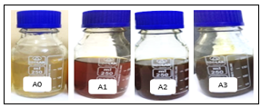
Figure 2. Characteristic changes in the colour of colloidal Solution
The resulting viscous solution, manufacturing AgNPs revealed a colour change from yellow to brown (Figure 2) owing to silver salt reduction by reducing compounds in the extraction of CM chitosan. The color change was a result of the excitation of the surface plasmon resonance and SPR band which both play an important role in the confirmation of silver nanoparticles formation [34].
As a result, solution’s pH is approximately (3.0), the Ag ions concentration in the solution is (1.76 x10⁻⁵ mol/ml), the isopropanol concentration is (1.9x10⁻³ mol/ml), and the –NH2 group concentration is (9.35x10⁻⁵ mol/ml).
Finally, irradiate the three (AgNPS/CM chitosan - isopropanol)) mixtures on the gamma irradiator, with a Co-60 1/5 source of γ-ray. The dose rate of irradiation sets at 8 Gy\hr and regulates the total dose of irradiation in the experiment by adjusting the time of irradiation. The irradiation dose rate and irradiation time (hr.) for the prepared (silver nanoparticles /CM-chitosan - isopropanol) solution as predicated in Table 1. Table 2 shows Other important parameters for the prepared (silver nanoparticles /CM-chitosan - isopropanol) solution. In a dilute irradiation-degrade AgNPs/chitosan solution, the produced silver particles from radiation–reduction method were stable.
Table 1. Irradiation dose rate and Irradiation Time (hr.) for the prepared (silver nanoparticles /CM-chitosan - isopropanol) solution
|
Sample Code |
Irradiation Dose (Gy) |
Irradiation Time (hr.) |
|
A1 |
768 |
96 |
|
A2 |
1151 |
144 |
|
A3 |
1920 |
240 |
Table 2. The important parameters for the prepared (silver nanoparticles /CM-chitosan - isopropanol) solution
|
Test |
A1 |
A2 |
A3 |
|
PH |
5.0 |
4.8 |
5 |
|
CON. (%) |
0.182 |
0.19 |
0.264 |
2.3 Characterization of (Ag /CM-chitosan) solution
The stabilized silver nanoparticles in the (CM-chitosan- isopropanol) solution analyze by ultraviolet-visible (UV–Vis) absorbance spectroscopy (at room temperature in the wavelength range of 340–900 nm) using the model (H.UV.1650 PC, Shimadzu), Japan [35-39]. A transmission electron microscopy (TEM) and UV–vis spectrophotometer was used to measure the solution’s absorption spectrum at 420 nm, which resulted in the confirmation of the nanoparticles production. The particle size was determined using a Hitachi H7100 transmission electron microscopy (TEM) [40-43] and (UTHSCSghA Image Tool) version 3.00 application to calculate the distributions [44-46].
X-ray diffraction (XRD) measurement for obtained silver nanoparticles (stabilized in CM-chitosan) carried by an X-ray diffractometer [47-52]. Also, the Ag(CM-chitosan- isopropanol) solution structure investigate through Powder X-Ray Diffraction (model PXRD-6000 SHIMADZU, Japan. As a result, these observations were at a wavelength of 0.15418 nm at a scan speed of 2° min. The PXRD patterns were obtained when the instrument operated at 0.388 min. scan rate, current of 30 mA, and 40 kV.
Colloidal solution (silver nanoparticles / (CM chitosan - isopropanol)) prepared by AgNO3 reduction into positive silver ions (Ag+) and assisted with the fragmentation of large Ag-NPs during γ-irradiation [14, 29]. Figure 2 illustrates the synthesized silver nanoparticles samples. The colour intensity of the (silver nanoparticles/(CM chitosan-isopropanol)) samples gradually increased. The colloidal solution (sample A0) without γ-ray exhibits a clear yellow solution (Figure 2). The colloidal solution became dark brown as the -rays intensified (samples A1-A3), while the AgNO3 solution was colourless (see Figure 1). The (silver nanoparticles/(CM chitosan-isopropanol)) alter the color of the clear yellow colloidal solution after gamma irradiation from light brown to brown to darker brown (samples (A0-A3) .
These results agree with the results reported by Hanan et al. [53], Shriram et al. [14], and M.S.N. Salleh et al. [54] for the formation of AgNPs by the reduction of AgNO3.
3.1 UV–visible spectroscopy analysis
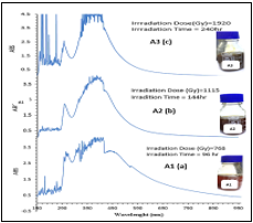
Because silver nanoparticles display a high absorption peak owing to surface plasmon resonance, the UV–visible absorption spectra that were obtained are extremely sensitive to the production of silver nanoparticles (SPR). Figure 3 show the UV–vis silver nanoparticles spectra prepared at constant CM-chitosan and silver nitrate concentrations but with different gamma irradiation doses (768, 1151, 1920 Gy) for (96, 144, and 240 hr), respectively. Every spectrum displays an absorption band with a wavelength range of 370–470 nm, which corresponds to a typical silver nanoparticles plasmon resonance band. Some faint peaks may be seen at about 330 and 560 nm. These peaks are an indication that some of the nanoparticle aggregations do exist. The usual conducting electrons SPR emerging from the silver nanoparticles surface is represented in the UV–vis spectra by a single, prominent peak with a maximum of about 430 nm. This peak was detected in the UV–vis spectra. The efficiency of nanoparticle synthesis increases with increased gamma irradiation doses (768, 1151, 1920 Gy) as a result of silver ions' ability to more effectively oxidize the hydroxyl groups found in CM-chitosan. Further, Increasing the gamma irradiation dosages allowed for the synthesis nanoparticles monitoring from silver nitrate, and the relevant spectra of these nanoparticles are shown in Figure 3. The higher the dosages of gamma irradiation that were applied, the greater the degree to which the solutions we're able to absorb the radiation, especially with the A3 sample exposed to a 1920Gy dose of gamma irradiation (Figure 3 c (sample A3)).
Figure 3. Silver nanoparticles stabilized UV–visible absorption spectra
3.2 Silver nanoparticles X-Ray analysis
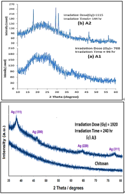
The XRD approach was selected to analyze the green-generated silver nanoparticles' crystal structure and establish the presence of currently synthesized silver nanoparticles. The XRD pattern of CM chitosan and silver nanoparticles (CM-chitosan-isopropanol) is shown in Figure 4. In Figure 4c, there was only one peak at 20° for chitosan (see Figures (4a and 4b)), but four Brag reflections with 2θ values of 77.21, 64.5, 43.95, and 38.25 for silver nanoparticles/(CM-chitosan-isopropanol)) were seen, corresponding to the (311), (220), (200), and (111) sets, respectively, which may be referred to as the band for silver nanoparticles with a face-centred cubic (FCC) structure [54, 55].
Figure 4. Silver nanoparticles and chitosan XRD patterns
The (111) plane's peak is the most intense of the group. The influence of nanoparticles is mostly responsible for the widening of these peaks. The XRD pattern for (sample A3) There were no substantial indications of other peak phases in their crystalline cubic phase, which suggested the dominant silver. A further consideration is the presence of a nanoparticle of colloidal solution (silver nanoparticles/(CM chitosan - isopropanol)) that exhibited gamma irradiation at 1929Gy doses contributes to the formation of the varied and distinct XRD pattern peaks [56] more than sample A1 and A2. From the XRD pattern, one can conclude the most appropriate gamma dose to get silver nanoparticles due to Co-60 is 1920 Gy and for 240 hr. irradiation dose time [56].
3.3 TEM, XRD and Zeta potential after weeks of storage
The produced AgNPs particle size distribution and morphology were evaluated using TEM imaging. Figure 5 displays a typical TEM image of sample A3 in the solution that there is a good distribution of silver nanoparticles throughout the (CM-chitosan-isopropanol) matrix.
Figure 5. (a)TEM and (b)XRD pattern test of Ag /CM-chitosans sample A3 after week from preparation time and storage at room temperature and in a dark place
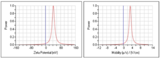
According to size distributions and TEM images of AgNPs, the nanoparticle's mean diameter in sample A3 is generally between 40 and 50 nm with a cubic phase. While particle size distribution of A1 and A2 seems bigger than 100 nm. After gamma-irradiation on aqueous suspensions of chitosan/ silver nitrate (Cts/AgNO3), consider aqueous electrons create and reduce the positive silver ions into silver. Then, the shielded silver clusters by the chitosan as unique structure silver atoms nucleates and develops into silver clusters rather than being oxidized back into silver ions [57]. After 15 weeks of preparation, neither the XRD and TEM, Zeta potential tests for the samples, nor the PH values, as indicated in Table 3 and Figure 7, have altered.
Figure (5 b) displays the X-ray diffraction patterns of sample A3 of chitosan – nano silver after a week of storage at room temperature. It showed the peaks at about 20 degrees with the other two peaks which indicate that the Nano silver particles are blended with chitosan fragment. This testified that the nano silver particles have cubic crystal patterns. The figure of Ag-CS indicates a notable peak of most 2theta is 19.68, which determines the synthesized Ag-NPs crystalline structure. For Zeta potential of nano silver particles distribution in the matrix, PH value of solutions determination as in Table 3 and Zeta potential analysis applied for the synthesis samples. Table 3 indicates the PH values almost stability of the Ag solutions with storage time at room temperature and in a dark place. For 15 weeks, the PH values of Ag solution do not exceed 5.2 which leads to quite a stability of silver ions with ±40 mV zeta potential with nearly ionic mobility as shown in Figure 6.
Table 3. The PH value of the prepared sample with storage time and room temperature
|
No. |
Time |
A1-PH |
A2-PH |
A3-PH |
Temperature℃ |
|
1 |
Week-1 |
5.1 |
4.8 |
5 |
27.3 |
|
2 |
Week -2 |
5 |
4.9 |
5.1 |
27.0 |
|
3 |
Week-3 |
5 |
4.8 |
5.1 |
27.1 |
|
4 |
Week-4 |
5.1 |
4.8 |
5.1 |
27 |
|
5 |
Week-5 |
5.2 |
5 |
5.1 |
27 |
|
6 |
Week-6 |
5 |
5 |
4.9 |
29 |
|
7` |
Week-7 |
5.2 |
5.1 |
5 |
27 |
|
8 |
Week-8 |
5 |
4.9 |
5 |
26 |
|
9 |
Week-9 |
5 |
5 |
5.1 |
26 |
|
10 |
Week-10 |
5.1 |
5 |
5.1 |
25 |
|
11 |
Week-11 |
5 |
4.9 |
5 |
25 |
|
12 |
Week12 |
5.1 |
5 |
5.1 |
18 |
|
13 |
Week-13 |
5 |
5.1 |
5 |
17 |
|
14 |
Week-14 |
5 |
5 |
4.9 |
17 |
|
15 |
Week-15 |
5 |
4.9 |
5 |
18 |
(A1)
(A2)
(A3)
Figure 6. The Zeta potential and Mobility of ionic Nano silver particles of samples (A1, A2, and A3) after 15 weeks of storage at room temperature
3.4 FT-IR spectroscopy
The FTIR analysis assesses the molecular interaction of silver nanoparticles with chitosan at the molecular level (Figure 7). The FTIR spectra of pure chitosan display bands at 1037 cm-1 (C-O stretching), 1600 cm-1 (NH2 bending), and 3327 cm-1 O-H and N-H stretching are the FTIR spectra of pure chitosan [58]. The FTIR spectra of silver nanoparticles capped with chitosan revealed a wider peak at 3327 cm-1 indicating that the O-H group is more prominent since the N-H group is essential in silver metal binding.
Figure 7. FT-IR spectra of (a) A1, Irradiation dose (768 Gy); (b) A2, Irradiation dose (1151 Gy); (c) A3, Irradiation dose (1920 Gy)
While compared to the chemical reduction approach, the radiation approach is one of the most important ways to create metal clusters under ambient settings. Because the metal reduction is carried out by radical species that are produced as a result of the interaction of ionizing radiation with the solvent, the addition of reducing agents is not required in this process, and the complete process may conduct at room temperature.
The growing degree to which the reduction is controlled by the dosage absorbed, as well as the reduction rate, which is controlled by the dose rate (Figure 8). At a bit high doses, an instantaneous distribution of atoms obtaining through the solution. The varying dose rate of the irradiation controlled the number of zerovalent nuclei in the solution [59], therefore, Gamma irradiation-induced strategy offered silver nanoparticles with a narrower sized distribution.
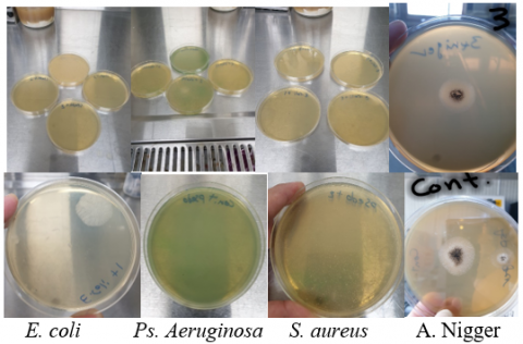
Figure 8. Test of antibacterial activity
3.5 Antimicrobial Activity of (silver nanoparticles/CM chitosan – isopropanol)
The (silver nanoparticles/(CM chitosan-isopropanol)) solution tested for antibacterial activity; against (gram-positive , Staphylococcus aureus) and (gram-negative, Escherichia coli) bacteria. The disc diffusion technique was used to examine the antibacterial activity of the produced AgNPs by measuring the inhibition zone against two bacteria and fungus, Aspergillus niger vs. control.
After 24 hours of being kept in an incubator at a temperature of 37℃, the diameter (in mm) of the inhibitory zone was measured as shown in Figure 8. The results show that the silver nanoparticles and (CM chitosan-isopropanol)) for samples (A2) and (A3) appeared to have an inhibition zone against S.aureus and E.coli.
Table 4 shows the effectiveness of various gamma irradiation doses (768, 1151, 1920 Gy) of antibacterial activity of biologically synthesized (silver nanoparticles / (CM chitosan - isopropanol)) in (Gram-positive), Staphylococcus aureus (S.aureus), and Gram-negative (Escherichia coils)as relative to the control sample. From Table 4 one can find that the microbe resistance was less effective for lower gamma rays. In short, the colloidal Solution (silver nanoparticles /(CM chitosan - isopropanol)) when coupled with silver nanoparticles, an inhibitory effect was seen against S. aureus. It is anticipated that the silver nanoparticles, which are responsible for rupturing the cell wall membrane of bacteria, will be the source of this antibacterial property. When the cell membrane of the bacteria is damaged, the respiration of the germs is immediately hampered, which brings an immediate halt to the activity of the bacteria [60, 61]. The analysis outcomes indicate that the colloidal Solution (silver nanoparticles / (CM chitosan - isopropanol)) has a promising ability to be used in antibacterial applications.
Table 4. Test results of Antibacterial activity for (silver nanoparticles / (CM chitosan - isopropanol)) solutions
|
SAMPLE |
Sauries |
E.coli |
Ps.aeruginosa |
A.Nigger, Growth diameter (mm) |
|
A1 |
60 |
40 |
138X102 |
19 |
|
A2 |
40 |
250 |
70X10 |
20 |
|
A3 |
NILL |
70 |
450 |
17 |
|
CONTROL |
Hi |
550 |
50X104 |
22 |
The present study demonstrates a green process for the synthesis of spherical-shaped and stabilized silver nanoparticles Ag NPs using gamma irradiated conditions. The findings of this work indicate that, in comparison to the control sample, the generated Ag NPs/CM-chitosan-isopropanol might be employed as an antibacterial agent in (Gram-positive), Staphylococcus aureus (S. aureus), and Gram-negative (Escherichia coils) bacteria.
Irradiation with gamma rays was used to successfully produce silver nanoparticles in the interlaminar space of chitosan. No reducing agent or heat treatment was used in the production of these nanoparticles. Chitosan molecules were shown to be responsible for the stabilization of the Ag-NPs over 15 weeks at room temperature. The dosage rate and irradiation periods both affect the size distributions of the Ag-NPs. According to the findings of this investigation, an increase in the amount of irradiation time led to a reduction in particle size along with stability in the distribution of silver ions. The silver nanoparticles that were synthetically produced are stable in aqueous solutions and the analysis outcomes indicate that the colloidal Solution (silver nanoparticles / (CM chitosan - isopropanol)) has a promising ability to be used in antibacterial applications in the future.
[1] Mohseniazar, M., Barin, M., Zarredar, H., Alizadeh, S., Shanehbandi, D. (2011). Potential of microalgae and lactobacilli in biosynthesis of silver nanoparticles. Bioimpacts, 1(3): 149-152. https://doi.org/10.5681/bi.2011.020
[2] Haider, A., Kang, I.K. (2015). Preparation of silver nanoparticles and their industrial and biomedical applications: A comprehensive review. Adv. Mater. Sci. Eng., 2015: 165257. https://doi.org/10.1155/2015/165257
[3] Khan, I., Saeed, K., Khan, I. (2019). Nanoparticles: Properties, applications and toxicities. Arab. J. Chem., 12(7): 908-931. https://doi.org/10.1016/j.arabjc.2017.05.011
[4] Aziz, W.J., Abid, M.A., Kadhim, D.A., Mejbel, M.K. (2020). Synthesis of iron oxide (β-fe2o3) nanoparticles from Iraqi grapes extract and its biomedical application. IOP Conf. Ser. Mater. Sci. Eng., 881: 12099. https://doi.org/10.1088/1757-899x/881/1/012099
[5] Mikhlif, H., Dawood, M., Abdulmunem, O., Mejbel, M.K. (2021). Preparation of high-performance room temperature ZnO nanostructures gas sensor. ACTA Phys. Pol. A, 140(4): 320-326. https://doi.org/10.12693/APhysPolA.140.320
[6] Mezher, S.J., Dawood, M.O., Beddai, A.A., Mejbel, M.K. (2020). NiO nanostructure by RF sputtering for gas sensing applications. Mater. Technol., 35(1): 60-68. https://doi.org/10.1080/10667857.2019.1653595
[7] Mansoor, S., Zahoor, I., Baba, T.R., Padder, S.A., Bhat, Z.A., Koul, A., Jiang, L.H. (2021). Fabrication of silver nanoparticles against fungal pathogens. Frontiers in Nanotechnology, 3. https://doi.org/10.3389/fnano.2021.679358
[8] Ghosh Chaudhuri, R., Paria, S. (2012). Core/Shell nanoparticles: Classes, properties, synthesis mechanisms, characterization, and applications. Chem. Rev., 112(4): 2373-2433. https://doi.org/10.1021/cr100449n
[9] Siddiqi, K.S., Husen, A., Rao, R.A.K. (2018). A review on biosynthesis of silver nanoparticles and their biocidal properties. J. Nanobiotechnology, 16(1): 14. https://doi.org/10.1186/s12951-018-0334-5
[10] Maaz, K. (Ed.) (2018). Silver Nanoparticles - Fabrication, Characterization and Applications. London, United Kingdom, IntechOpen. https://doi.org/10.5772/intechopen.71247
[11] Iravani, B.Z.S., Korbekandi, H., Mirmohammadi, S.V. (2014). Synthesis of silver nanoparticles: chemical, physical and biological methods. Res. Pharm. Sci., 9(6): 385-406.
[12] Zhang, X.F., Liu, Z.G., Shen, W., Gurunathan, S. (2016). Silver nanoparticles: Synthesis, characterization, properties, applications, and therapeutic approaches. International Journal of Molecular Sciences, 17(9): 1534. https://doi.org/10.3390/ijms17091534.
[13] Güzel, R. (2018). Synthesis of Silver Nanoparticles. G. E. E.-K. Maaz, Ed., Rijeka: IntechOpen. https://doi.org/10.5772/intechopen.75363
[14] Mirajkar, S., Rathod, P., Pawar, B., Penna, S., Dalvi, S. (2021). γ-irradiated chitosan mediates enhanced synthesis and antimicrobial properties of chitosan–silver (Ag) nanocomposites. ACS Omega, 6(50): 34812-34822. https://doi.org/10.1021/acsomega.1c05358
[15] Galindo, D.O.O., Utrera, O.H., Mejia, R.M., Aranda, M.A.S. (2016). Silver nanoparticles by laser ablation confined in alcohol using an Argon gas environment. J. Laser Micro Nanoeng., 11(2): 158-163. https://doi.org/10.2961/jlmn.2016.02.0004
[16] Palem, R.R., Saha, N., Shimoga, G.D., Kronekova, Z., Sláviková, M., Saha, P. (2018). Chitosan–silver nanocomposites: New functional biomaterial for health-care applications. Int. J. Polym. Mater. Polym. Biomater., 67(1): 1-10. https://doi.org/10.1080/00914037.2017.1291516
[17] Latif, U., Al-Rubeaan, K., Saeb, A.T.M. (2015). A review on antimicrobial chitosan-silver nanocomposites: A roadmap toward pathogen targeted synthesis. Int. J. Polym. Mater. Polym. Biomater., 64(9): 448-458. https://doi.org/10.1080/00914037.2014.958834
[18] Jyoti, K., Baunthiyal, M., Singh, A. (2016). Characterization of silver nanoparticles synthesized using Urtica dioica Linn. leaves and their synergistic effects with antibiotics. J. Radiat. Res. Appl. Sci., 9(3): 217-227. https://doi.org/10.1016/j.jrras.2015.10.002
[19] Du, B.D., Van Phu, D., Duy, N.N., Lan, N.T.K., Lang, V.T.K., Thanh, N.V.K., Phong, N.T.P., Hien, N.Q. (2008). Preparation of colloidal silver nanoparticles in poly(N-vinylpyrrolidone) by γ-irradiation. J. Exp. Nanosci., 3(3): 207-213. https://doi.org/10.1080/17458080802353527.
[20] Pirtarighat, S., Ghannadnia, M., Baghshahi, S. (2019). Green synthesis of silver nanoparticles using the plant extract of Salvia spinosa grown in vitro and their antibacterial activity assessment. J. Nanostructure Chem., 9(1): 1-9. https://doi.org/10.1007/s40097-018-0291-4
[21] Malassis, L., Dreyfus, R., Murphy, R.J., Hough, L.A., Donnio, B., Murray, C.B. (2016). One-step green synthesis of gold and silver nanoparticles with ascorbic acid and their versatile surface post-functionalization. RSC Adv., 6(39): 33092-33100. https://doi.org/10.1039/C6RA00194G
[22] Divya, K., Kurian, L.C., Vijayan, S., Manakulam Shaikmoideen, J. (2016). Green synthesis of silver nanoparticles by Escherichia coli : Analysis of antibacterial activity. J. Water Environ. Nanotechnol., 1(1): 63-74. https://doi.org/10.7508/jwent.2016.01.008
[23] Ulug, B., Haluk Turkdemir, M., Cicek, A., Mete, A. (2015). Role of irradiation in the green synthesis of silver nanoparticles mediated by fig (Ficus carica) leaf extract. Spectrochim. Acta Part A Mol. Biomol. Spectrosc., 135: 153-161. https://doi.org/10.1016/j.saa.2014.06.142
[24] Yu, J., Wang, D., Geetha, N., Khawar, K.M., Jogaiah, S., Mujtaba, M. (2021). Current trends and challenges in the synthesis and applications of chitosan-based nanocomposites for plants: A review. Carbohydr. Polym., 261: 117904. https://doi.org/10.1016/j.carbpol.2021.117904
[25] Affes, S., Maalej, H., Aranaz, I., Kchaou, H., Acosta, N., Heras, Á., Nasri, M. (2020). Controlled size green synthesis of bioactive silver nanoparticles assisted by chitosan and its derivatives and their application in biofilm preparation. Carbohydr. Polym., 236: 116063. https://doi.org/10.1016/j.carbpol.2020.116063
[26] Li, J.H., Zhuang, S.L. (2020). Antibacterial activity of chitosan and its derivatives and their interaction mechanism with bacteria: Current state and perspectives. Eur. Polym. J., 138: 109984. https://doi.org/10.1016/j.eurpolymj.2020.109984
[27] Jiménez-Gómez, C.P., Cecilia, J.A. (2020). Chitosan: A natural biopolymer with a wide and varied range of applications. Molecules, 25(17): 3981. https://doi.org/10.3390/molecules25173981
[28] Martău, G.A., Mihai, M., Vodnar, D.C. (2019). The Use of chitosan, alginate, and pectin in the biomedical and food sector—biocompatibility, bioadhesiveness, and biodegradability. Polymers, 11(11): 1837. https://doi.org/10.3390/polym11111837
[29] Van Phu, D., Lang, V.T.K., Lan, N.T.K., Duy, N.N., Chau, N.D., Du, B.D., Cam, B.D., Hien, N.Q. (2010). Synthesis and antimicrobial effects of colloidal silver nanoparticles in chitosan by γ-irradiation. J. Exp. Nanosci., 5(2): 169-179. https://doi.org/10.1080/17458080903383324
[30] Chen, P., Song, L.Y., Liu, Y.K., Fang, Y.E. (2007). Synthesis of silver nanoparticles by γ-ray irradiation in acetic water solution containing chitosan. Radiat. Phys. Chem., 76(7): 1165-1168. https://doi.org/10.1016/j.radphyschem.2006.11.012
[31] Mossa, S., Shameli, K. (2021). Gamma irradiation-assisted synthesis of silver nanoparticle and their antimicrobial applications: A review. J. Res. Nanosci. Nanotechnol., 3(1): 53-75. https://doi.org/10.37934/jrnn.3.1.5375
[32] Nhien, L.T.A., Luong, N.D., Tien, L.T.T., Luan, L.Q. (2018). Radiation synthesis of silver nanoparticles/chitosan for controlling leaf fall disease on rubber trees causing by Corynespora Cassiicola. J. Nanomater., 2018: 7121549. https://doi.org/10.1155/2018/7121549
[33] Yoksan, R., Chirachanchai, S. (2009). Silver nanoparticles dispersing in chitosan solution: Preparation by γ-ray irradiation and their antimicrobial activities. Mater. Chem. Phys., 115(1): 296-302. https://doi.org/10.1016/j.matchemphys.2008.12.001
[34] Vanaja, M., Gnanajobitha, G., Paulkumar, K., Rajeshkumar, S., Malarkodi, C., Annadurai, G. (2013). Phytosynthesis of silver nanoparticles by Cissus quadrangularis: influence of physicochemical factors. J. Nanostructure Chem., 3(1): 17. https://doi.org/10.1186/2193-8865-3-17
[35] Beddai, A.A., Badday, B.A., Al-Yaqoobi, A.M., Mejbel, M.K., Al Hachim, Z.S., Mohammed, M.K.A. (2022). Color removal of textile wastewater using electrochemical batch recirculation tubular upflow cell. Int. J. Chem. Eng., 2022: 4713399. https://doi.org/10.1155/2022/4713399
[36] Allawi, M.K., Mejbel, M.K., Younis, Y.M., Mezher, S.J. (2020). A simulation of the effect of Iraqi diesel fuel cetane number on the performance of a compression ignition engine. Int. Rev. Mech. Eng., 14(3): 151-159. https://doi.org/10.15866/ireme.v14i3.18137
[37] Allawi, M.K., Mejbel, M.K., Oudah, M.H. (2020). Iraqi gasoline performance at low engine speeds. IOP Conf. Ser. Mater. Sci. Eng., 881: 12065. https://doi.org/10.1088/1757-899x/881/1/012065
[38] Oudah, M.H., Mejbel, M.K., Allawi, M.K. (2021). R134a flow boiling heat transfer (FBHT) characteristics in a refrigeration system. J. Mech. Eng. Res. Dev., 44(4): 69-83. https://jmerd.net/Paper/Vol.44,No.4(2021)/69-83.pdf.
[39] Allawi, M., Mejbel, M., Oudah, M. (2021). Variable valve timing (VVT) modelling by lotus engine simulation software. Int. J. Automot. Mech. Eng., 17(4): 8397-8410. https://doi.org/10.15282/ijame.17.4.2020.15.0635
[40] Mejbel, M.K., Abdullah, I.I., Taieh, N.K. (2022). Thin wall manufacturing improvement using novel simultaneous double-sided cutter milling technique. Int. J. Automot. Mech. Eng., 19(1): 6519-6529. https://doi.org/10.15282/ijame.19.1.2022.15.0734
[41] Kadhim Jawad, L., Beddai, A.A., Ali Nasser, M., Kadhim Mejbel, M. (2022). Scrutinizing the physical and strength properties of fabricated date palm frond leaves particleboard. Mater. Today Proc., 57(2): 980-988. https://doi.org/10.1016/j.matpr.2022.03.396
[42] Mejbel, A.M.K.M.K., Khalaf, M.M., Kwad, A.M. (2021). Improving the machined surface of AISI H11 tool steel in milling process. J. Mech. Eng. Res. Dev., 44(4): 58-68. https://jmerd.net/Paper/Vol.44,No.4(2021)/58-68.pdf.
[43] Mejbel, M.K., Allawi, M.K., Oudah, M.H. (2019). Effects of WC, SiC, iron and glass fillers and their high percentage content on adhesive bond strength of an aluminium alloy butt joint: An experimental study. J. Mech. Eng. Res. Dev., 42(5): 224-231. https://doi.org/10.26480/jmerd.05.2019.224.231
[44] Allawi, M.K., Oudah, M.H., Mejbel, M.K. (2019). Analysis of exhaust manifold of spark-ignition engine by using computational fluid dynamics (CFD). J. Mech. Eng. Res. Dev., 42(5): 211-215. https://doi.org/10.26480/jmerd.05.2019.211.215
[45] Mezher, S.J., Kadhim, K.J., Abdulmunem, O.M., Mejbel, M.K. (2020). Microwave properties of Mg–Zn ferrite deposited by the thermal evaporation technique. Vacuum, 173: 109114. https://doi.org/10.1016/j.vacuum.2019.109114
[46] Mezher, S.J., Dawood, M.O., Abdulmunem, O.M., Mejbel, M.K. (2020). Copper doped nickel oxide gas sensor. Vacuum, 172: 109074. https://doi.org/10.1016/j.vacuum.2019.109074
[47] Mejbel, M.K., Atwan, H.R., Abdullah, I.T. (2021). Void formation in friction stir welding of AA5052 butt joining. J. Mech. Eng. Res. Dev., 44(5): 318-332. https://jmerd.net/Paper/Vol.44,No.5(2021)/318-332.pdf.
[48] Muhmmed, A.A., Hussain, M.K., Khudadad, A.R., Mahdi, H.H., Mejbel, M.K. (2021). Mechanical behavior of laser engraved single lap joints adhered by polymeric material. Int. Rev. Mech. Eng., 15(12): 622-628. https://doi.org/10.15866/ireme.v15i12.21278
[49] Al-Saadi, T.H.A., Mohammad, S.H., Daway, E.G., Mejbel, M.K. (2021). Synthesis of intumescent materials by alkali activation of glass waste using intercalated graphite additions. Mater. Today Proc., 42(5): 1889-1900. https://doi.org/10.1016/j.matpr.2020.12.228
[50] AL-Saadi, T.H.A., Abdulnabi, R.K., Ismael, M.N., Hassan, H.F., Mejbel, M.K. (2022). Glass waste based geopolymers and their characteristics. Revue des Composites et des Matériaux Avancés-Journal of Composite and Advanced Materials, 32(1): 17-23. https://doi.org/10.18280/rcma.320103
[51] Abood Al-Saadi, T.H., Daway, E.G., Mohammad, S.H., Mejbel, M.K. (2020). Effect of graphite additions on the intumescent behaviour of alkali-activated materials based on glass waste. J. Mater. Res. Technol., 9(6): 14338-14349. https://doi.org/10.1016/j.jmrt.2020.10.035
[52] Baqer, A.R., Beddai, A.A., Farhan, M.M., Badday, B.A., Mejbel, M.K. (2021). Efficient coating of titanium composite electrodes with various metal oxides for electrochemical removal of ammonia. Results Eng., 9: 100199. https://doi.org/10.1016/j.rineng.2020.100199
[53] Ghetas, H.A., Abdel-Razek, N., Shakweer, M.S., Abotaleb, M.M., Paray, B.A., Ali, S., Eldessouki, E.A., Dawood, M.A.O., Khalil, R.H. (2022). Antimicrobial activity of chemically and biologically synthesized silver nanoparticles against some fish pathogens. Saudi J. Biol. Sci., 29(3): 1298-1305. https://doi.org/10.1016/j.sjbs.2021.11.015
[54] Salleh, M.S.N., Rasit Ali, R., Shameli, K., Hamzah, M.Y., Chan, J.Z. (2020). Silver nanoparticles on pullulan derived via gamma irradiation method: A preliminary analysis. IOP Conf. Ser. Mater. Sci. Eng., 808(1): 12030. https://doi.org/10.1088/1757-899x/808/1/012030
[55] Barot, T., Rawtani, D., Kulkarni, P. (2020). Physicochemical and biological assessment of silver nanoparticles immobilized Halloysite nanotubes-based resin composite for dental applications. Heliyon, 6(3): e03601. https://doi.org/10.1016/j.heliyon.2020.e03601
[56] Kaur, A., Preet, S., Kumar, V., Kumar, R., Kumar, R. (2019). Synergetic effect of vancomycin loaded silver nanoparticles for enhanced antibacterial activity. Colloids Surfaces B Biointerfaces, 176: 62-69. https://doi.org/10.1016/j.colsurfb.2018.12.043
[57] Ahmad, M.B., Shameli, K., Darroudi, M., Yunus, W.M.Z.W., Ibrahim, N.A. (2009). Synthesis and characterization of silver/clay/chitosan bionanocomposites by UV-irradiation method. Am. J. Appl. Sci., 6(12): 2030-2035. https://doi.org/10.3844/ajassp.2009.2030.2035
[58] Nate, Z., Moloto, M.J., Mubiayi, P.K., Sibiya, P.N. (2018). Green synthesis of chitosan capped silver nanoparticles and their antimicrobial activity. MRS Adv., 3(42-43): 2505-2517. https://doi.org/10.1557/adv.2018.368
[59] Eisa, W.H., Abdel-Moneam, Y.K., Shaaban, Y., Abdel-Fattah, A.A., Abou Zeid, A.M. (2011). Gamma-irradiation assisted seeded growth of Ag nanoparticles within PVA matrix. Mater. Chem. Phys., 128(1): 109-113. https://doi.org/10.1016/j.matchemphys.2011.02.076
[60] Salleh, M.S., Ali, R.R., Shameli, K., Hamzah, M.Y., Kasmani, R.M., Nasef, M.M. (2021). Interaction insight of pullulan-mediated gamma-irradiated silver nanoparticle synthesis and its antibacterial activity. Polymers, 13(20): 3578. https://doi.org/10.3390/polym13203578
[61] Wang, X.H., Du, Y.M., Fan, L.H., Liu, H., Hu, Y. (2005). Chitosan- metal complexes as antimicrobial agent: Synthesis, characterization and Structure-activity study. Polym. Bull., 55(1): 105-113. https://doi.org/10.1007/s00289-005-0414-1