Mohammed A. Abd Ali* | Ali Aboud Shareef
© 2022 IIETA. This article is published by IIETA and is licensed under the CC BY 4.0 license (http://creativecommons.org/licenses/by/4.0/).
OPEN ACCESS
The goal of this research is to use dental caries patients’ oral cavity-isolated Enterobacter aerogenes bacterial strains (S1, S2 and S3) to create silver nanoparticles (AgNPs) in an environmentally friendly and cost-effective manner. In addition, the study explores the combination of antibiotics with Streptococcus mitis MDR, which was isolated from patients with dental caries to determine their antibacterial efficiency. Clinical bacterial strains identified from dental caries patients' mouths were all resistant to standard antibiotics. Antibiotics and AgNPs have a synergistic impact, which suggests that antibiotics will make up a larger portion of the diet. It was shown that erythromycin E had the greatest synergistic impact with AgNPs (0.1 mg/ml), but Streptomycin and Tetracycline had only 6 mm inhibitory zones when paired with AgNPs (0.1 mg/ml) in comparison. Antagonizing effects are meant by this. It was revealed that antibiotics such as penicillin P and cephalexin CN had distinct effects on patients. When used in combination with antibiotics, Enterobacter aerogenes AgNPs demonstrated excellent antibacterial efficacy on Streptococcus mitis isolates. As a result, AgNPs in the dental care area have a wide range of applications.
green synthesis, silver nanoparticles, dental, XRD, antagonistic effects
Humans have been searching for new antibiotics since the discovery of penicillin at the turn of the twentieth century, despite the fact that bacteria are developing resistance mechanisms to the antibiotics already in use [1]. In addition to increasing the likelihood of accidents, the rise in antibiotic resistance to conventional antibiotics is an important worldwide health issue [2]. Unique structural size and shape, which are defined by their chemical, biological, and physical properties, nanotechnology has emerged as a rapidly expanding area of nanoscience with crucial applications, including the creation of nanomaterials and their application in medicine and biology [3]. It is common to refer to nanotechnology as the "fourth industrial revolution" in the history of humanity civilization. According to Parthasarathi and Thilagavathi [4], ceramics, metals, polymers, and biomaterials are all included in this category. Antibiotics, sunscreens, and sunblocks are examples of biogenic products that are being developed and improved., it has made a substantial contribution [5]. Since environmentally friendly derivates are used to reduce nanoparticle metals, rather than hazardous chemicals, the green synthesis method is superior to the traditional chemical method [6].
According to numerous studies, nanoparticle products have evolved over time. These nanoparticles have already been used in implant coatings as well as food containers and ointments. The United States of America's Food and Medicine Administration has given clearance to use the drug [7]. According to numerous studies, Nano silver is bactericidal to both gram-positive and gram-negative microorganisms [8].
Microorganisms that live in the mouth cavity form a self-contained ecosystem [9]. Despite its prevalence, dental caries is one of the world's most common chronic infectious diseases. If the enamel is damaged, caries can begin as small areas of demineralization beneath the surface of the tooth's surface and progress through the dentine and into the pulp [10]. An infection caused by bacteria weakens the teeth's enamel, dentin, and cementum and can cause tooth loss. In most developed nations, dental caries affects 60–90% of school-aged children and the vast majority of adults, making it a serious public health issue [11].
Traditional medical infections often include a single infectious agent, and that infectious agent may also be present in isolated animals that are not traditionally hosts [12]. On the other hand, dental caries can be brought on by oral commensal microbes that spread a polymicrobial infection. About 600 different species of microorganisms have been found in the oral microbiome, which is a microbiome that lives inside the human mouth [13].
Cells, water, and bacteria extracellularly form a biofilm, which adheres to the surface of an organism [14]. The formation of biofilms is a result of a variety of environmental factors [15]. As the plankton moves from free-swimming to sessile life in a biofilm, a range of environmental and genetic factors must be taken into account that are specific to the individual species [16].
Periodontal disease is defined as the breakdown of periodontal teeth's supporting tissues in the oral cavity, such as alveolar bone and gingiva, which can be a risk factor for certain systemic conditions [17]. Topcuoglu et al. [18] discovered in a study that red-complex bacteria were the most consistent, with extremely high stages in all groups. Fusobacterium nucleatum was found in high concentrations in all of the samples. With the exception of Aggregatibacterer actinomycomiltas, which was found in all localised competitive periodontitis groups, the green and blue complex bacteria were less numerous than the red and orange complex microorganisms [19].
Dental caries-related bacteria are often detected using based culture procedures that do not yet exclude cultured species. Methods of based culture that do not exclude species that have not yet been grown.
Oriented culture techniques do not exclude species that have never been grown before. For more specific species investigations of bacteria in the oral cavity, including species that aren't currently cultivable, molecular approaches for bacterial identification and enumeration are now commonly used [20].
Oral bacteria, including those that aren't currently cultivable, are increasingly being studied using molecular techniques for species identification and enumeration [20]. Cell communities, rather than pure, isolated cultures, are what distinguish uncultivable species from those that can be propagated in a petri dish or petri dish-less medium. Species and genus names can be assigned using ecological molecular-enabled phylogenetic trees, which now contain creatures that cannot be grown, as is the case in many cases. Bacterial species linked to the oral cavity are increasingly being studied using molecular approaches for bacterial enumeration and identification, including species that are currently uncultivated.
In recent years, the field of nanotechnology has arisen as a rapidly expanding area of nanoscience, with various significant applications, including nanomaterial synthesis and use in medicine and biology [3]. Nanotechnology is the fourth industrial revolution in human history, and it encompasses a wide spectrum of materials, including ceramics, metals, polymers, and biomaterials [4]. Antibiotics, sunscreen, and sunblock are only a few of the biogenic items that have benefited greatly from research and development. The use of environmentally acceptable materials as reducing agents rather than dangerous chemicals makes the green synthesis of nanoparticle metals more advantageous than chemical synthesis.
The purpose of this research is to generate silver nanoparticles (AgNPs) using dental caries patients' oral cavity-isolated Enterobacter aerogenes bacterial strains (S1, S2, and S3) in an environmentally friendly and cost-effective manner As far as the researchers are aware, this is the first study at the Iraqi level to use bacteria isolated from the oral cavity of some dental caries patients in the creation of silver nanoparticles for bactericidal Streptococcus mitis multidrug resistant bacterial isolates.
2.1 Solutions of silver nitrate
Nanotechnology refers to the study of nanometer-scale materials ranging from 1-100 nanometers in size [21]. In this regard, antimicrobial drug under development is to be more effective with free form bacterial resistance it most act at several cellular levels and not specifically like traditional antibiotics. The effect of silver either as a metal or in compounds, is known to be not specific at a single level but to influence many bacterial structures and metabolic processes at the same time Nanoparticles AgNPs were shown to in activate bacterial enzymes.
In order to prevent PHTO-oxidation, silver nitrate was dissolved in DDW in a dark atmosphere. As a substrate, the solution was used to make silver nanoparticles [21].
2.2 Isolation and identifying of bacteria
Specimens were taken from the mouths of patients with dental disease at the Dental Specialist Canter in the Iraqi province of Misan. The isolates were identified using a combination of gram staining and biochemical assays, as described above. Finally, PCR and sequencing were used. The identified cultures were transferred to nutrient agar slant and stored at 4℃ in the refrigerator to ensure their long-term survival.
2.3 Strain identification via molecular sequence
BLAST is a bioinformatics technique and tool for comparing primary biological sequence information, such as protein amino acid sequences or DNA and/or RNA nucleotide sequences. A BLAST search enables a researcher to compare a topic protein or nucleotide sequence (referred to as a query) to a library or database of sequences and to identify database sequences that are more similar to the query sequence than a specified threshold.
For this study, we used the BLAST tool, an online comparison tool, to estimate the degree of similarity between three strains of Enterobacter aerogenes bacteria (S2, S1, S2, S3). These three strains (S1, S2, and S3) were isolated and their 16S rDNA sequenced, extracted, and amplified using PCR.
2.4 Activation of bacterial strains
Ten millilitres of nutritional broth were activated in a serum tube using an aseptic method, and the bacteria were cultured overnight at 37℃.
2.5 Production of biomass
In a shaking incubator, a bacterial isolate (Enterobacter aerogenes) was injected into a 500 ml flask containing 6.5 g/l of nutritious broth and cultivated for 48 hours at 200 rpm.
2.6 Obtaining the cell free medium
Four stages were taken to obtain cell-free supernatant: activation with an inoculum, incubated biomass generation, cultivation, and centrifugation for cell exclusion. The supernatant was collected from the broth culture pellet cells at the bottom of the tubes by centrifugation. After collecting the supernatant in a sterile flask, it is utilized to create silver nanoparticles [22].
In general, bacterial cell lysate supernatant was mixed with 0.1 mM AgNO3 solution and incubated in dark at 35℃ for 24 h. AgNO3 and bacterial cell lysate supernatant without AgNO3 served as controls and were kept under the same conditions. After the incubation period, the mixtures were observed for the presence of brown color, which indicated the positive formation of AgNPs.
2.7 Biogenic synthesis
Enriched Enterobacter aerogenes liquid culture supernatant was combined with 250ml of 1mM silver nitrate solution to produce silver nanoparticles (filtered through a 0.2m pore size). The cells were then grown in an orbital shaker for 5 days at 37℃ in the dark (200 rpm). As a control, a flask with cell-free supernatant missing (AgNO3) was used [23].
2.8 AgNPs characterization
Analytical techniques such as FTIR, XRD, and SEM were employed to investigate the presence of biomolecules in pure AgNPs as well as their structure [24].
2.9 Test for antimicrobial activity
Johan et al. [25] evaluated antimicrobial activity using two methods well and disc diffusion.
Susceptibility to antibiotics was determined using AgNPs in a combination experiment. Bacterial cells were cultured in nutritional broth. The disk-diffusion method was used to determine the antibacterial activity of a combination of antibiotics and biologically produced AgNPs against several bacterial isolates from dental caries and periodontitis. On Muller – Hinton lates, nutrient broth was grown with Streptococcus mitis isolates [26].
On the other hand, synergy between antibiotics is often quantified as the total of the Fractional Inhibitory Concentrations (FIC) of two combinations. FIC is calculated by following for combination A, B:
FIC= FIC of antibacterial A+ FIC of antibacterial B (1)
3.1 Characterization morphologically and biochemically
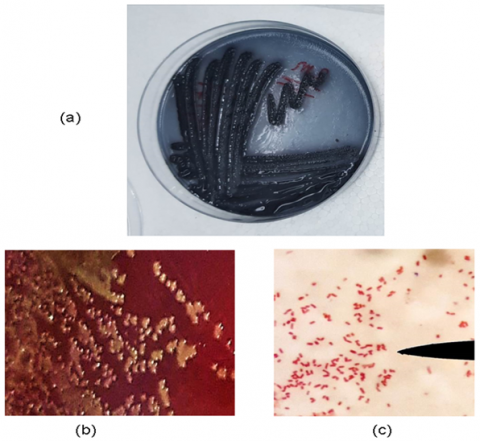
A dental clinic in Misan City, Iraq, was used to collect clinical bacterial strains from the oral cavities of dental illness patients. S2 Enterobacter aerogenes was found to be involved in the synthesis of AgNPs and their biological application in the control of infectious bacteria, and this isolate was identified as a result, as seen in Figure 1.
3.2 Molecular identification
3.2.1 AgNPs characterization (bacterial strains)
In addition to biochemical tests, 16SrRNA gene sequencing, gene extraction, and amplification were used to identify isolates. Three strains (S1, S2, and S3) of Enterobacter aerogenes were identified and one was chosen (S2) as shown in Figure 2. Genomic DNA was extracted from strain IHB B 6843 and amplified using PCR. The sequence most closely related to NR 117547 was IHB B 6843. A new Enterobacter soli ATCC BAA.2102 strain LF7.1 was identified using a 16S rRNA analysis.
3.2.2 Harvesting the cell free medium
Centrifugation was used to extract the broth culture pellet cells' supernatant. The supernatant is then collected in a sterile flask and utilized to generate silver nanoparticles in a subsequent step [22]. Silver nanoparticles were created by mixing 250 ml of cell-free supernatant from an Enterobacter aerogenes 48-hour liquid culture with 250 ml of a 1 mM silver nitrate (AgNO3) solution. The supernatant combination was then cultivated for 5 days at 37°C in the dark on an orbital shaker (200 rpm). The experimenter's control was a flask containing cell-free supernatant free of (AgNO3) [23].
Figure 1. Enterobacter aerogenes bacterial strain cultured on (a) Mitis Salivarius Agar, (b) Blood Agar, and (c) Gram stain of Enterobacter aerogenes bacterial isolate revealed by microscope at 1250x magnification
Figure 2. On a 1% agarose gel at 5vol. cm for 1.15 hours, electrophoresis patterns of genomic DNA extracted from Enterobacter aerogenes strains (S1, S2, and S3) were electrophoresis
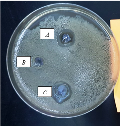
Because of the growing concern about multi-drug resistance (MDR), the study is focusing on developing effective alternatives to resistant antibiotics. Nanoparticles are being researched as a potential replacement for conventional antibiotics. AgNPs have recently been hailed as a promising target for developing a new antibiotic generation [27]. As shown in Figures 3 and 4, AgNPs from the Enterobacter aerogenes strain (S2) were effective against the multidrug-resistant bacterial isolate Streptococcus mitis.
Figure 3. The effects of nanoparticles on multidrug-resistant streptococcus mitis isolated from dental caries patients (a), (b) Control DDW, and (c) AgNO3 antibacterial activity
Figure 4. Antibiotics' effect on streptococcus mitis bacterial pathogens (a), and (b) Nanoparticles AgNPs
3.3 Antibacterial activity
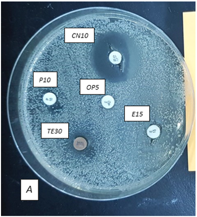
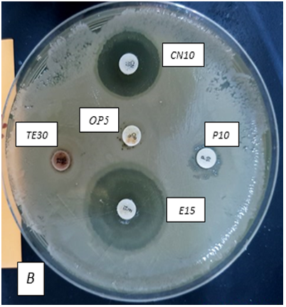
Using a bacterial suspension well and the disc diffusion method, the antimicrobial activity of Optochin OP, Penicillin P, Tetracycline TE, Cefalexin CN, and Streptomycin S antibiotics was evaluated against some pathogenic bacteria isolated from dental patients in Misan province, Iraq, alone and in combination with Biogenic AgNPs synthesised by Enterobacter aerogenes as shown in Figure 5.
Figure 5. Lowest concentrations required to inhibit the growth of bacteria that produce non-tumour necrosis factor (NTF)
Figure 6. Extracellular of AgNPs synthesis
Table 1. Antibiotic-induced inhibition zone against Streptococcus mitis as a percentage
|
Increase in fold area |
Antibiotics With AgNPs Effects B |
Antibiotics Effects A |
Concentration µg̷ disk |
Antibiotics |
No |
|
0.48+ |
7.3 |
6 |
5 |
OP Optochin |
1 |
|
+1.16 |
9.7 |
6.6 |
10 |
P Penicillin |
2 |
|
-0.85 |
6.0 |
15.6 |
30 |
TE Tetracycline |
3 |
|
+16.7 |
30.7 |
7.3 |
15 |
E Erythromycin |
4 |
|
-0.39 |
20.6 |
26.3 |
30 |
Cefalexin CN |
5 |
|
-0.97 |
6.0 |
40.6 |
10 |
S Streptomycin |
6 |
Table 2. The FIC index is used to determine the interaction between antibiotics
|
No |
AgNPs+Antibiotics |
FICI |
Interaction |
|
1 |
Optochin + AgNPs |
_ |
_ |
|
2 |
Penicillin + AgNPs |
1.46 |
Synergistic |
|
3 |
Tetracycline+ AgNPs |
2.00 |
Antagonistic |
|
4 |
Erythromycin+ AgNPs |
0.20 |
Synergistic |
|
5 |
Cefalexin+ AgNPs |
_ |
|
|
6 |
Streptomycin+ AgNPs |
4.11 |
Antagonistic |
Table 3. Lowest inhibitory concentration (MIC) of biosynthesized AgNPs
|
No |
Multi drug Streptococcus miti MIC (mg/ml) |
|
|
1 |
NPs alone |
0.009 |
|
2 |
Optochin + AgNPs |
_ |
|
3 |
Penicillin + AgNPs |
0.0037 |
|
4 |
Tetracycline+ AgNPs |
0.018 |
|
5 |
Erythromycin+ AgNPs |
0.0018 |
|
6 |
Cefalexin+ AgNPs |
_ |
|
7 |
Streptomycin+ AgNPs |
0.039 |
According to the CLSI Standard, it was determined that, at a concentration of 100g, the mixed formulation of AgNPs exhibited antibacterial activity via a variety of antibiotic discs, including optochin, penicillin, tetracycline, erythromycin, cephalexin, and streptomycin as shown in Figure 6.
In this study, only Erythromycin E (30.7 mm) had a synergistic effect with antibiotics, but all other clinical bacterial strains isolated from dental disease patients' oral cavities were resistant to standard antibiotics such as Optochin OP, Penicillin P, and Erythromycin E. Streptococcus mitis, on the other hand, was more responsive to Tetracycline TE, Cefalexin CN, and Streptomycin S. Improved antibiotic effectiveness in combination with AgNPs has been reported (percentage). AgNPs synthesized were tested against multidrug resistant Streptococcus mitis strains using commercially available antibiotics, comparable to Fayaz et al. [28].
When erythromycin E was administered at 30.7 mm, the AgNPs (0.1 mg/ml) showed the strongest synergistic effect. Unlike Streptomycin S (Streptomycin 40mm was Streptomycin with a 6.0-mm AgNPs inhibition zone) and Tetracycline TE (Tetracycline 15.6mm was Tetracycline with a 6-mm AgNPs inhibition zone), antagonistic effects were found. It is clear from Figure 4 and the Tables 1 and 2 that Optochin OP, Penicillin P, and Cefalexin CN did not work.
These results corroborate those of Birla et al. [29], who investigated biologically synthesized AgNPs against gram-negative and gram-positive bacteria using currently available antibiotics. In the presence of silver nanoparticles, AgNPs, the antimicrobial activity of erythromycin, ampicillin, kanamycin, and chloramphenicol was prolonged.
Finally, the inhibitory zone's lowest concentration was identified using the following biosynthesized AgNPs detection concentrations for Enterobacter aeruginosa (0.1, 0.05, 0.025, 0.0125, and 0.0062 mg/ml) as illustrated in Figure 4 and Table 3.
The current study discovered that silver nanoparticles behave similarly to silver ions in that they interact with groups of electron donors containing oxygen, sulfur, or nitrogen atoms that are typically found as phosphates or thiols on amino acids and nucleic acids. According to Kvitek et al. [30], AgNPs interact with the surface of the bacterial cell's membrane via interactions with proteins that continue the sulphur chain. This results in a disruption of the respiratory functions of the cell and a decrease in the permeability of the cell membrane, which ultimately results in cell death.
3.4 Physical characterization
XRD, FT-IR, and UV-vis spectroscopy were used to examine the AgNPs' optical properties. Infectious Enterobacter aerogenes made up the bulk of them (S2 strain). The color shift in the reaction solution from light yellowish brown to dark brown is generated by the excitation vibrations of the integrated AgNPs' surface plasmon, which is the first sign of AgNP synthesis. Changes in color can be seen as an indication that silver nanoparticles are being produced [31].
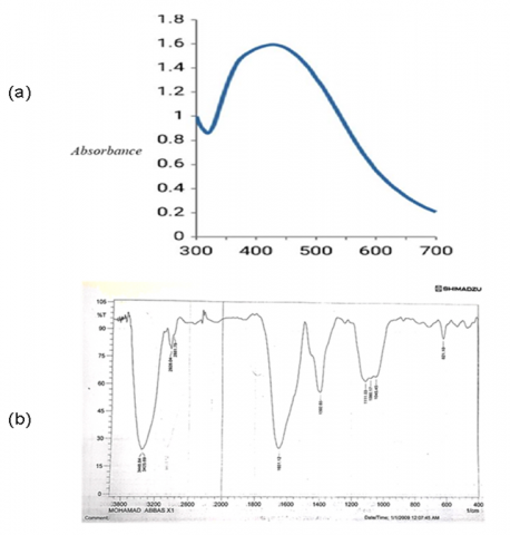
Figure 7. AgNPs synthesized (a) UV- Vis absorption spectrum and (b) FT- IR spectrum
Table 4. Lattice plane
|
Source of AgNPs |
h kl index |
|||
|
|
111 |
200 |
220 |
311 |
|
Enterobacter aerogenes |
38.4213 |
46.5167 |
67.7625 |
77.0906 |
Table 5. XRD results of silver nanoparticles
|
Pos. [°2Th.] |
Height [cts] |
FWHM Left [°2Th.] |
d-spacing [Å] |
Rel. Int. [%] |
Tip Width |
|
38.4213 |
132.40 |
0.5904 |
2.34297 |
4.80 |
0.7085 |
|
46.5167 |
1340.05 |
0.3000 |
1.95073 |
48.56 |
0.3600 |
|
67.7625 |
108.62 |
0.4800 |
1.38177 |
3.94 |
0.5760 |
|
77.0906 |
255.07 |
0.4800 |
1.23617 |
9.24 |
0.5760 |
Figure 8. Scanning electron microcopy(a) and (b) XRD
UV-vis spectroscopy confirmed the strong absorption of this color range. Figure 7 shows that the reaction mixture's absorption peaks were about 420nm (a). As Basavaraja et al. [32] have discovered, the absorption at 420nm is compatible with the reduction of Ag+ to Ag°.
The average crystallization size of AgNPs can be calculated using the Deby–Scherrer formula [33]. The four major peaks in the complete spectrum of (2) 38.42, 46.51, 67.76, and 77.09 are referred to as XRD patterns as shown in Figure 8. According to the Joint Committee Powder Diffraction Standards (JCPDS) Card No.087–0720, these values correspond to the (111), (200), (220), and (311) planes of the face–centered–cubic (FCC) sliver, with a network parameter of 4.08 °A as shown in Table 4. Using scanning electron microscopy, nanoparticles were discovered in a variety of shapes; however, all of the shapes were spherical and ranged in size from 71.56 nm to 71.56 nm. Electron microscopy and XRD pictures of AgNPs formed on a copper surface by Enterobacter aerogenes isolated from the oral cavity of dental caries patients in Misan province's central region. Table 5 shows XRD patterns.
The findings of this study indicate that Enterobacter aerogenes nanoparticles may be beneficial alone or in combination with antibiotics in the treatment of oral infections caused by MDR Streptococcus mitis bacterial isolates, which are frequently used in dentistry to treat a variety of oral cavity illnesses. The current study's findings suggest that the combination of the antibiotic Erythromycin and AgNP nanoparticles had a synergistic effect on Streptococcus mitis isolates from some dental caries patients. Numerous hypotheses were addressed regarding the mechanism of action of these combinations of AgNP nanoparticles and antibiotics. Finally, detailed data is required to clarify the mechanism of the synergistic impact.
[1] Clatworthy, A.E., Pierson, E., Hung, D.T. (2007). Targeting virulence: A new paradigm for antimicrobial therapy. Nature Chemical Biology, 3(9): 541-548. https://doi.org/10.1038/nchembio.2007.24
[2] Manna, P., Jain, S.K. (2015). Obesity, oxidative stress, adipose tissue dysfunction, and the associated health risks: causes and therapeutic strategies. Metabolic Syndrome and Related Disorders, 13(10): 423-444: 246-251. https://doi.org/10.1089/met.2015.0095
[3] Mahasneh, A.M. (2013). Bionanotechnology: The novel nanoparticles based approach for disease therapy. Jordan Journal of Biological Sciences, 6(4).
[4] Parthasarathi, V., Thilagavathi, G. (2011). Synthesis and characterization of zinc oxide nanopartilce and its application on fabrics for microbe resistant defence clothing. International Journal of Pharmacy and Pharmaceutical Sciences, 3(4): 392-398. https://d1wqtxts1xzle7.cloudfront.net/31879772/2690-with-cover-page-v2.pdf.
[5] Raghavan, V., Fan, H.M., Dockery, P., Wheatley, A., Keogh, I., Olivo, M. (2015). Multimodal gold nanoprobes for SERS bioimaging. Journal of Nanomedicine & Nanotechnology, S6: 1. http://dx.doi.org/10.4172/2157-7439.S6-002
[6] Abdeen, S., Geo, S., Praseetha, P.K., Dhanya, R.P. (2014). Biosynthesis of silver nanoparticles from Actinomycetes for therapeutic applications. International Journal of Nano Dimension (IJND), 5(2): 155-162.
[7] Dunn, K., Edwards-Jones, V. (2004). The role of Acticoat™ with nanocrystalline silver in the management of burns. Burns, 30: S1-S9. https://doi.org/10.1016/S0305-4179(04)90000-9
[8] Morones, J.R., Frey, W. (2010). Room temperature synthesis of an optically and thermally responsive hybrid PNIPAM–gold nanoparticle. Journal of Nanoparticle Research, 12(4): 1401-1414. https://doi.org/10.1007/s11051-009-9686-y
[9] Chopde, N., Jawale, B., Pharande, A., Chaudhari, L., Hiremath, V., Redasani, R. (2012). Microbial colonization and their relation with potential cofactors in patients with denture stomatitis. J Contemp Dent Pract, 13(4): 456-459. https://doi.org/10.5005/jp-journals-10024-1168
[10] Petersen, P.E. (2008). World Health Organization global policy for improvement of oral health-World Health Assembly 2007. International Dental Journal, 58(3): 115-121. https://doi.org/10.1111/j.1875-595X.2008.tb00185.x
[11] Petersen, P.E., Bourgeois, D., Ogawa, H., Estupinan-Day, S., Ndiaye, C. (2005). The global burden of oral diseases and risks to oral health. Bulletin of the World Health Organization, 83: 661-669.
[12] Marsh, P.D. (2005). Dental plaque: Biological significance of a biofilm and community life‐style. Journal of Clinical Periodontology, 32: 7-15. https://doi.org/10.1111/j.1600-051X.2005.00790.x
[13] Dewhirst, F.E., Chen, T., Izard, J., et al. (2010). The human oral microbiome. Journal of Bacteriology, 192(19): 5002-5017. https://doi.org/10.1128/JB.00542-10
[14] Sutherland, I.W. (2001). The biofilm matrix–an immobilized but dynamic microbial environment. Trends in Microbiology, 9(5): 222-227. https://doi.org/10.1016/S0966-842X(01)02012-1
[15] O'Toole, G., Kaplan, H.B., Kolter, R. (2000). Biofilm formation as microbial development. Annual Reviews in Microbiology, 54(1): 49-79.
[16] Monds, R.D., O’Toole, G.A. (2009). The developmental model of microbial biofilms: Ten years of a paradigm up for review. Trends in Microbiology, 17(2): 73-87. https://doi.org/10.1016/j.tim.2008.11.001
[17] Agnello, M., Marques, J., Cen, L., et al. (2017). Microbiome associated with severe caries in Canadian first nations children. Journal of Dental Research, 96(12): 1378-1385.
[18] Topçuoğlu, H.S., Üstün, Y., Akpek, F., Aktı, A., Topçuoğlu, G. (2016). Effect of coronal flaring on apical extrusion of debris during root canal instrumentation using single‐file systems. International Endodontic Journal, 49(9): 884-889. https://doi.org/10.1111/iej.12520
[19] Hajishengallis, G. (2015). Periodontitis: from microbial immune subversion to systemic inflammation. Nature Reviews Immunology, 15(1): 30-44. https://doi.org/10.1038/nri3785
[20] Paster, B.J., Olsen, I., Aas, J.A., Dewhirst, F.E. (2006). The breadth of bacterial diversity in the human periodontal pocket and other oral sites. Periodontology 2000, 42(1): 80-87. https://doi.org/10.1111/j.1600-0757.2006.00174.x
[21] Chaudhari, P.R., Masurkar, S.A., Shidore, V.B., Kamble, S.P. (2012). Antimicrobial activity of extracellularly synthesized silver nanoparticles using Lactobacillus species obtained from VIZYLAC capsule. Journal of Applied Pharmaceutical Science, 2(3): 25-29. https://doi.org/10.7324/JAPS.2012.2305
[22] Singh, H., Du, J., Singh, P., Yi, T.H. (2018). Extracellular synthesis of silver nanoparticles by Pseudomonas sp. THG-LS1. 4 and their antimicrobial application. Journal of Pharmaceutical Analysis, 8(4): 258-264. https://doi.org/10.1016/j.jpha.2018.04.004
[23] Saifuddin, N., Wong, C.W., Yasumira, A.A. (2009). Rapid biosynthesis of silver nanoparticles using culture supernatant of bacteria with microwave irradiation. E-Journal of Chemistry, 6(1): 61-70.
[24] Zhou, N., Wong, H.M., McGrath, C. (2019). Oral health and associated factors among preschool children with special healthcare needs. Oral Diseases, 25(4): 1221-1228. https://doi.org/10.1111/odi.13057
[25] Johan, P., Harley, I., Prescott, M. (2003). Laboratory Exercise in Microbiology. McGraw-Hill. USA, 149-1538.
[26] Privett, B.J., Deupree, S.M., Backlund, C.J., Rao, K.S., Johnson, C.B., Coneski, P.N., Schoenfisch, M.H. (2010). Synergy of nitric oxide and silver sulfadiazine against gram-negative, gram-positive, and antibiotic-resistant pathogens. Molecular Pharmaceutics, 7(6): 2289-2296. https://doi.org/10.1021/mp100248e
[27] Rai, M.K., Deshmukh, S.D., Ingle, A.P., Gade, A.K. (2012). Silver nanoparticles: the powerful nanoweapon against multidrug‐resistant bacteria. Journal of Applied Microbiology, 112(5): 841-852. https://doi.org/10.1111/j.1365-2672.2012.05253.x
[28] Fayaz, A.M., Balaji, K., Girilal, M., Yadav, R., Kalaichelvan, P.T., Venketesan, R. (2010). Biogenic synthesis of silver nanoparticles and their synergistic effect with antibiotics: A study against gram-positive and gram-negative bacteria. Nanomedicine: Nanotechnology, Biology and Medicine, 6(1): 103-109. https://doi.org/10.1016/j.nano.2009.04.006
[29] Birla, S.S., Tiwari, V.V., Gade, A.K., Ingle, A.P., Yadav, A.P., Rai, M.K. (2009). Fabrication of silver nanoparticles by Phoma glomerata and its combined effect against Escherichia coli, Pseudomonas aeruginosa and Staphylococcus aureus. Letters in Applied Microbiology, 48(2): 173-179. https://doi.org/10.1111/j.1472-765X.2008.02510.x
[30] Kvítek, L., Panáček, A., Soukupova, J., Kolář, M., Večeřová, R., Prucek, R., Holecová, M., Zbořil, R. (2008). Effect of surfactants and polymers on stability and antibacterial activity of silver nanoparticles (NPs). The Journal of Physical Chemistry C, 112(15): 5825-5834. https://doi.org/10.1021/jp711616v
[31] Kalimuthu, K., Babu, R.S., Venkataraman, D., Bilal, M., Gurunathan, S. (2008). Biosynthesis of silver nanocrystals by Bacillus licheniformis. Colloids and Surfaces B: Biointerfaces, 65(1): 150-153. https://doi.org/10.1016/j.colsurfb.2008.02.018
[32] Basavaraja, C., Pierson, R., Huh, D.S. (2008). Synthesis, characterization, and comparative study of conducting polyaniline/lead titanate and polyaniline–dodecyl benzenesulfonic acid/lead titanate composites. Journal of Applied Polymer Science, 108(2): 1070-1078. https://doi.org/10.1002/app.27582
[33] Allafchian, A., Bahramian, H., Jalali, S.A.H., Ahmadvand, H. (2015). Synthesis, characterization and antibacterial effect of new magnetically core–shell nanocomposites. Journal of Magnetism and Magnetic Materials, 394: 318-324. https://doi.org/10.1016/j.jmmm.2015.06.086