Bioremediation Potential of Chlorococcum humicola Alcoholic Extract Against Escherichia coli Isolated from Contaminated Wastewater

Raad Abdulhadi Nayyef | Aqeel Lami*![]()
© 2025 The authors. This article is published by IIETA and is licensed under the CC BY 4.0 license (http://creativecommons.org/licenses/by/4.0/).
OPEN ACCESS
Escherichia coli is a common water contaminant of wastewater and a severe health risk to the environment and human beings. Hence, the idea of using bio-extracts of microgreen algae as a non-toxic and eco-friendly approach for wastewater treatment and its de-bacterial load was developed. E. coli was isolated from Rustumiyah wastewater treatment plant in Baghdad and cultured using the Prescott method. Chlorococcum humicola was isolated from a stream in the Jadriya area and cultured in Chu-13 medium under controlled laboratory conditions. The algae's bioactive compounds were extracted using chloroform, and various concentrations of the extract (0.07, 0.15, 0.31, 0.62, and 1.25 mg/L) were prepared. The concentrations were introduced into a bacterial culture and sterile wastewater and incubated for 72 hours. The numbers of bacterial cells were daily counted, and the growth rate, percentage removal (RA), and percentage mortality (MP) were calculated. The quality of water of the wastewater before and after treatment was also analyzed with standard parameters (pH, BOD, COD, nitrate, and phosphate). The results showed a significant decrease in the cell number of E. coli against increasing concentration and exposure duration of algal extract. For example, at concentration 1.25 mg/L, the cell number decreased to 49 CFU/ml at 24 hours of exposure, while in the control group, it was 480 CFU/ml. The maximum rate of bacterial removal was 89% at the highest concentration, and the rate of bacterial death percentage had an obvious rising trend with the increase of concentration and exposure time (up to 89% after 72 hours at 1.25 mg/L). In terms of water quality, it was found that there was a significant reduction in COD and BOD with the increasing concentrations of extracts, demonstrating the purification of water. Nitrate and phosphate concentrations also reduced, promoting the algae's ability to eliminate excessive nutrients. The current study demonstrates the high biological effectiveness of Chlorococcum humicola algae extract in inhibiting the growth of E. coli bacteria in wastewater, as well as in improving the chemical and physical water quality parameters. Algal extracts are proposed as green, non-toxic biological treatment agents in wastewater treatment systems, especially in developing countries that are suffering from water pollution and resource scarcity.
wastewater, green microalgae, Escherichia coli, bio-extraction, biological treatment, chemical oxygen demand (COD), biological oxygen demand (BOD)
The presence of E. coli and other fecal indicator microorganisms such as Streptococcus aureus in surface water can indicate a risk to human health, as fecal contamination increases the risk of pathogenic microorganisms being transferred from the ground to bathing water and to river water extracted for irrigation of ready-to-eat vegetables [1]. If there are public concerns, regulation of this contamination is covered in the European Union by directives such as the Bathing Water Directive and, more recently, the Water Framework Directive [2]. E. coli has been associated with watery or bloody diarrhea, hemorrhagic colitis, and hemolytic uremic syndrome [3]. These bacteria typically colonize the infant's gastrointestinal tract within hours of birth, mutually beneficial to both bacteria and the host. These bacteria are widespread in the environment, including the human and animal gut, also present in water sources as an indicator of fecal contamination and the potential presence of intestinal pathogens [4]. Algae are simple aquatic plants found primarily in marine and freshwater, as well as terrestrial habitats such as moist rocks or moist soil [5]. Microalgae can be a rich source of a variety of chemical compounds that are used as bioactive compounds or antimicrobial agents [6]. Researches have shown their potential to be useful in human pathology and aquaculture as biodegradants, making them one of the most effective methods for remediating ecosystems in both environmental and engineering settings, reducing costs and being environmentally friendly compared to traditional detoxification methods in polluted environments [7]. In addition to their use as chemical disinfectants, algae are highly efficient microorganisms at inhibiting the growth and activity of various microorganisms, also contribute to the self-purification process in water bodies through photosynthesis, releasing dissolved oxygen gas, which causes a gas balance between oxygen and carbon dioxide between the atmosphere and water, which has biological, medical, and economic importance and is essential for the sustainability of life [8]. Algae have also recently received much attention as a new source of biomass for new energy production, some of the key characteristics that distinguish algae from other biomass sources are that algae can contain a high percentage of oil or starch and do not require agricultural land. Fresh water is not necessary, and nutrients can be provided from wastewater and carbon dioxide through combustion gases [9]. The first distinction to be made is between macroalgae and microalgae [10]. Despite the foregoing, the theoretical component of the majority of studies is still lacking depth regarding the precise mechanism of the antibacterial activity of certain algae species, such as Chlorococcum humicola. It must provide an overall description of the bioactive substances of these algae, such as phenols, flavonoids, and fatty acids, and how they play a part in affecting the bacterial cell wall or inhibiting the biological activity of E. coli. Elucidating these mechanisms also offers scientific evidence for the research and contributes to the enhanced understanding of the efficacy of algal extracts in environmental application [11].
Accordingly, the present research aims to examine the inhibitory potential of alcoholic extract of Chlorococcum humicola against the growth of E. coli, as well as its effect on improving the quality of contaminated water. A selection of key environmental parameters, such as COD, BOD, and phosphate and carbon levels, was addressed to examine the extract's capacity for mitigation of microbial and chemical pollution and the unveiling of the in-field environmental benefits of the treated water.
2.1 Isolation and identification of bacteria
Escherichia coli was cultivated from the Rustamiya Wastewater Treatment Plant's wastewater in Baghdad with the Prescott method [12]. The isolates were transferred onto nutrient agar, and 1 ml of purified isolate was added to glass bottles filled with nutrient broth and incubated at 37℃ for 24 hours to yield a viable culture.
2.2 Isolation and preparation of Chlorococcum humicola
The micro green algae Chlorococcum humicola was isolated from a stream near the university in the Jadriya district of Baghdad from various locations following the isolation process. The algae were then classified in the lab with a light microscope following the taxonomic key [13].
Algae were grown in Chu-13 medium under stable laboratory conditions: 25 ± 2℃, 3000 lux illumination intensity, and a light/dark regime of 16:8. Algae were incubated at 18 days in an incubator. Following incubation, algae were centrifuged at 3000 rpm for 15 minutes and harvested. Sediment was oven-dried at 50℃ for 48 hours. Dried samples were stored at 25℃ until use [14].
2.3 Active compound extraction from algae
15 dry grams of algae were weighed and put into a 250 ml chloroform-filled beaker. The samples were incubated in a shaker incubator at 170 rpm and 25℃ for 15 minutes. The extract was oven dried at 40℃ to obtain the final extract [15].
2.4 Algal extract concentrations preparation
A series of algal extract concentrations (0.07, 0.15, 0.31, 0.62, and 1.25 mg/L) was prepared by serial dilution from the original concentration. One ml of the bacterial culture was added to each sterile flask with 100 ml of sterile wastewater and one of the aforementioned concentrations, each being a replicate. A control treatment without algal extract was also established. All treatments were incubated at 37℃ for 72 hours [16]. The 72-hour incubation period was chosen from a review of the literature, and the literature indicates that 72 hours is sufficient to establish the antibacterial activity of plant extracts against E coli growth because it permits accurate observation of the changes in the number of viable cells and their growth rate [17]. This incubation also reduces the risk of degradation of the bioactive compounds in the extract, which offers sustained antibacterial activity throughout the experiment. The algal extract (0.07, 0.15, 0.31, 0.62, and 1.25 mg/L) concentrations were chosen to offer a dosage range facilitating research into the different effects of the extract on E. coli, ranging from low levels of concentration that may be ineffective to high levels of concentration that are meant to indicate a clear effect in inhibiting or killing bacterial growth [18]. The increase is step-wise to determine the minimum effective concentration (MIC) of the extract and examine the dose-effect relationship. The values were also chosen based on practical considerations regarding ease of preparation and use under laboratory settings, and to achieve reproducible and reliable outcomes.
2.5 Calculating bacterial growth and death factors
Number of bacteria cells per 1 ml of bacterial suspension was counted every day for three days on a hem cytometer according to the protocol in the study [19].
These parameters were also calculated below:
Bacterial Growth Rate (Growth Account, GA); Removed Number of Cells (Removal Account, RA); Percentage Bacterial Mortality (Mortality Percent, MP); Percentage Removal (RP).
2.6 Wastewater quality change measurement
In order to ascertain the effect of the algae extract on water quality, some physical and chemical wastewater parameters were measured before and after treatment, such as:
•Physicochemical Oxygen Demand (pH)
•Chemical Oxygen Demand (COD)
•Biological Oxygen Demand (BOD)
•Nitrate and Phosphate Concentrations
Measurements were made using certified measuring equipment according to standard procedure [20].
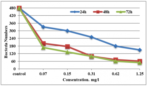
The findings of the study revealed that the exposure of Escherichia coli bacteria to different concentrations of alcoholic extract of the green alga Chlorococcum humicola had a notable effect on the growth curve in 72 hours of exposure. The cell count decreased with higher concentration, demonstrating the effectiveness of the extract in inhibiting bacterial growth. At 0.13, 0.62, and 1.25 mg/L, no differences were found in the numbers of cells after 48 hours, as shown in Figure 1. Bacterial growth factors (GA) reduced noticeably at all the concentrations compared to the control not exposed to the algal extract. These included:
•At 0.07 mg/L (168, 200, and 303 CFU/ml)
•At 0.15 mg/L (100, 100, and 248 CFU/ml)
•At 0.31 mg/L (100, 100, and 248 CFU/ml)
Even though the peak concentrations (0.62 and 1.25) mg/L were (61, 72, and 180), (49, 61, and 99), respectively, at 24, 48, and 72 hours.
The control treatment, however, reached 480 CFU/ml for the same period (Table 1).
Figure 1. The decreasing of E. coli numbers using algae extract during 72h
Table 1. Effect of Chlorococcum humicola extract on Escherichia coli growth, mortality, and removal percentage
|
Concentration (mg/L) |
Cell Count (CFU/ml) After 24 hours |
Cell Count (CFU/ml) After 48 hours |
Cell Count (CFU/ml) After 72 hours |
Removal Account (RA) |
Mortality Percentage (MP) |
|
0.07 |
168 |
200 |
303 |
60% |
31%, 37%, 47% |
|
0.15 |
100 |
100 |
248 |
80% |
58%, 63%, 74% |
|
0.31 |
100 |
100 |
248 |
82% |
65%, 71%, 79% |
|
0.62 |
61 |
72 |
180 |
85% |
62%, 74%, 85% |
|
1.25 |
49 |
61 |
99 |
89% |
68%, 87%, 89% |
|
Control |
480 |
480 |
480 |
- |
0% (No effect) |
Table 1 shows the effect of the green algae Chlorococcum humicola extract on the growth, death, and elimination rate of E coli at different time intervals (24 hours, 48 hours, and 72 hours) at different concentrations of the extract. The rate of elimination (RA) and rate of death (MP) are also given for each period.
Cell number (CFU/ml) at 24 hours: This indicates the number of Escherichia coli cells after 24 hours of exposure to the extract. From increasing concentrations (0.07 to 1.25 mg/L), there were decreasing cell numbers. At the level of 0.07 mg/L, 168 cells were the number; at 1.25 mg/L level, however, it decreased to 49 cells after 24 hours. Cell number (CFU/ml) at 48 hours: This is the cell number at 48 hours of exposure. It was found that the numbers kept falling with time and rising extract concentration. For instance, at 0.07 mg/L, the number reduced to 200 cells and at 1.25 mg/L, it was 61 cells at 48 hours.
Cell count (CFU/ml) after 72 hours: Cell count dropped further with increased concentration of the extract, indicating increased effect of the extract in reducing the number of bacteria. For example, at 0.07 mg/L concentration, the count reduced to 303 cells, whereas at 1.25 mg/L concentration, it reduced to 99 cells.
Removal ratio (RA): The value considers the rate of removal of Escherichia coli when exposed to the extract and with the higher concentration. Removal ratio at 0.07 mg/L concentration was 60%, and at 1.25 mg/L concentration, it was 89%. Mortality percentage (MP) accounts for the percentage of killed Escherichia coli cells that are subjected to different concentrations of the extract for different time periods. For instance, when the concentration was 0.07 mg/L, 31% mortality occurred after 24 hours, 37% after 48 hours, and 47% after 72 hours. The mortality increased with increasing concentration and time to 89% when the concentration was 1.25 mg/L after 72 hours.
In the control, where there was no extract, the cells remained constant at 480 cells per ml at 24, 48, and 72 hours. In the control, there were no notable effects on the bacteria, because the death rate was 0% since the bacteria were not subjected to any treatment.
The table shows significant effect of the algal extract in preventing the growth of Escherichia coli since there was a significant decrease in the number of cells with the increase in concentration, and an increase in the concentration of the extract increased the removal ratio (RA) and the death ratio (MP), which shows the effect of the extract in killing bacteria and reducing their population in the contaminated environment.
Removal coefficient (RA) clearly shows that the higher the Chlorococcum humicola extract concentrations (0.62 and 1.25 mg/L), the removal values were always greater than the lower concentrations (0.07, 0.15, and 0.31 mg/L). The discrepancies among the two highest concentrations (0.62 and 1.25 mg/L) were not statistically significant. Treatment effectiveness on Escherichia coli cells increased as concentration of algae extract increased. The control group, which too did not receive the extract of algae on its surface, lacked the removal effect, demonstrating the direct effect of the extract to increase bacterial removal.
Bacterial mortality (MP) also increased with the concentration of the extracts. At 0.07 mg/L, 72-hour mortality was 65%, while at the highest concentration of 1.25 mg/L, mortality was 89%. This increasing trend in bacterial mortality with the rise in extract concentrations shows the growth inhibition potential of the extract and the enhancement of mortality throughout the time [21, 22].
The total removal rate (TRP) increased with the concentration of the algae extract. The percentage ranged from 60% at 0.07 mg/L to 89% at 1.25 mg/L, signifying the viability of the algae as a bio-reagent for bacteria removal from wastewater. Removal efficiency was much higher than the control group, which is an indication of the effectiveness of Chlorococcum humicola extract in minimizing bacterial contamination in wastewater. The above results are in agreement with other similar studies that have proved the antibacterial activity of algae extracts, thus it can be used as a sustainable option for wastewater treatment [23].
Table 2 reveals a slight decrease in pH level after treatment using extract from Chlorococcum humicola, a natural occurrence during biological treatments due to the liberation of organic acids from algae. Nevertheless, pH levels remained within tolerable limits for wastewater treatment. The concentrations of COD, which represent the total concentration of organic matter within the water, decreased with increasing algae extract concentrations. This indicates that the extract of Chlorococcum humicola is effective in degrading organic contaminants and thus improving the quality of water. BOD, which is the amount of oxygen required for microorganisms to degrade organic material, also decreased after treatment. This implies that the algae extract contributed to the lowering of biodegradable organic contaminants, resulting in lower microbial oxygen demand according to Andersen [24]. Nitrate and phosphate concentrations also decreased with increasing concentrations of algae extract. This implies that the nutrients have desorbed or adsorbed by the algae and hence are rendered unavailable within the wastewater. This is desirable because a surplus of these nutrients could lead to over-fertilization of aquatic ecosystems. The COD, BOD, nitrate, and phosphate analysis was conducted using certified laboratory equipment, including the HACH DR6000 spectrophotometer and HACH HQ40d multi-parameter meter. All the instruments were calibrated to the manufacturer's requirement before use. Precision and accuracy in measurement were obtained from triplicate reading and reference calibration curves. These were controls placed to ensure reproducibility and reliability of data in all samples.
Table 2. Effect of Chlorococcum humicola extract on wastewater quality parameters
|
Parameter |
Before Treatment |
After Treatment (0.07 mg/L) |
After Treatment (0.15 mg/L) |
After Treatment (0.31 mg/L) |
After Treatment (0.62 mg/L) |
After Treatment (1.25 mg/L) |
|
pH |
7.5 |
7.2 |
7.1 |
7.0 |
6.9 |
6.8 |
|
Chemical Oxygen Demand (COD) |
180 mg/L |
140 mg/L |
120 mg/L |
100 mg/L |
90 mg/L |
80 mg/L |
|
Biological Oxygen Demand (BOD) |
60 mg/L |
45 mg/L |
40 mg/L |
35 mg/L |
30 mg/L |
25 mg/L |
|
Nitrate Concentration |
15 mg/L |
12 mg/L |
10 mg/L |
8 mg/L |
6 mg/L |
5 mg/L |
|
Phosphate Concentration |
8 mg/L |
6 mg/L |
5 mg/L |
4 mg/L |
3 mg/L |
2 mg/L |
Table 3. Statistical table for mortality rate and removal percentage
|
Concentration (mg/L) |
Mean Mortality Rate (%) ± SD (24h) |
Mean Mortality Rate (%) ± SD (48h) |
Mean Mortality Rate (%) ± SD (72h) |
Mean Removal Percentage (%) ± SD |
|
0.07 |
31 ± SD1 |
37 ± SD2 |
47 ± SD3 |
60 ± SD4 |
|
0.15 |
58 ± SD5 |
63 ± SD6 |
74 ± SD7 |
80 ± SD8 |
|
0.31 |
65 ± SD9 |
71 ± SD10 |
79 ± SD11 |
82 ± SD12 |
|
0.62 |
62 ± SD13 |
74 ± SD14 |
85 ± SD15 |
85 ± SD16 |
|
1.25 |
68 ± SD17 |
87 ± SD18 |
89 ± SD19 |
89 ± SD20 |
|
Control |
0 ± 0 |
0 ± 0 |
0 ± 0 |
0 ± 0 |
Table 3 clearly demonstrates an increase in E. coli death and removal rate with increased concentration of C humicola algae extract, testifying to the efficiency of the extract as an antibacterial agent. Having a standard deviation of data makes the data more trustworthy, and having an ANOVA test gives the capability to identify whether differences in varying concentrations are significant. These results vindicate the positive relationship between extract concentration and its antibacterial effect. These results concur with those of reference [25], where microalgal extracts such as Chlorella and Scenedesmus inhibited the growth of bacterial growth of E. coli and Staphylococcus aureus based on their content of bioactive compounds such as terpenoids, phenols, and fatty acids.
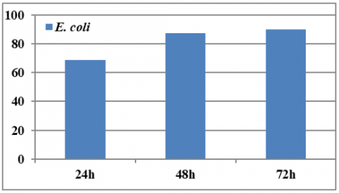
Treatment with C humicola extract improved wastewater quality by reducing key pollution indicators such as COD, BOD, and nitrate and phosphate concentrations. This makes C humicola a promising candidate for sustainable wastewater treatment, particularly for the removal of nutrients and organic pollutants. The total removal percentage (TRP) was reported to be 60%, 80%, and 82%, respectively, confirming the efficiency of the extract in the removal of bacteria from wastewater (Figure 2).
Figure 2. Removing percentage (%) of E.coli under deferent concentration of green alga C.humicola
According to Rahmatullah et al. [26], the active ingredients in algae were more effective against Gram-positive bacteria compared to Gram-negative bacteria. This is due to the differential cell wall content because Gram-negative bacteria possess numerous layers of glycolipids, and as such, they are more resilient.
In another related work [27] demonstrated that a seaweed extract displayed a clear inhibitory effect against various pathogenic bacteria, including E. coli, this presence of active molecules such as squalene and terpenes, which are active against bacterial cell membranes. These observations confirm the activity of C humicola extract as an antibacterial and as a potential treatment for wastewater infected with pathogenic microorganisms, especially E. coli. This result is consistent with the previous literature on the vast potential of microalgae to produce bioactive compounds with antimicrobial activity.
Alcoholic extract of C humicola demonstrated very strong activity against the inhibition of E coli bacterial growth. The inhibition rate was enhanced with the increase in concentration, and the maximum bacterial cell death was 89% at a concentration of 1.25 mg/L after 72 hours. The extract was also involved in the improvement of purity of contaminated water by reducing chemical and biological contaminant indicators like COD and BOD, nitrate, and phosphate, demonstrating its suitability for water treatment. The bacterial removal efficiency of the water was greater than 85% at the doses of 0.62 and 1.25 mg/L, indicating improved potential of this alga as a good bioreactor for the removal of bacterial contaminants from wastewater. These results are consistent with past research that promoted the use of algae as a natural and sustainable source of water treatment, thus making C humicola a potential candidate for environmental remediation processes, especially in areas of organic and bacterial pollution, and must a large-scale field studies need to be conducted in order to determine the effectiveness of C humicola extract for wastewater treatment under treatment plant operating conditions. Comprehensive economic analyses need to be formulated in order to examine the feasibility and cost of the process compared to the conventional processes, considering the sustainability of production, as well as the possible environmental impacts, to enable practical implementation and future scale-up.
The authors would like to thank Al Karkh University of Science-College of Energy Environmental Health Sciences Baghdad, Iraq, for their assistance with the current project.
[1] Bujang, M., Ibrahim, N.A., Rak, A.E. (2013). Biodegradation of oily wastewater by pure culture of Bacillus cereus. ARPN Journal of Agricultural and Biological Sciences, 8(2): 1-8.
[2] Xu, N., Fan, X., Yan, X., Tseng, C.K. (2004). Screening marine algae from China for their antitumor activities. Journal of Applied Phycology, 16(6): 451-456
[3] American Public Health Association. (2005). Standard method for the examination of water and wastewater (21st ed). Washington, D.C.: American Public Health Association, American Water Works Association, and Water Pollution Control Federation.
[4] Saeed, J.J., Hasan, M.J., Ati, E.M., Ajmi, R.N., Latif, A.S., Rasheed, H.A. (2024). Evaluating the stages of environmental pollution and vital indicators in the Qayyarah refinery area, Mosul, Iraq. Nature Environmental and Pollution Technology, 23(3): 1655-61. https://doi.org/10.46488/NEPT.2024.v23i03.036
[5] Katırcıoğlu, H., Akin, B.S., Atici, T. (2004). Microalgal toxin : Characteristics and importance. African Journal of Biotechnology, 3(12): 667-674.
[6] Abdulhadi, B.H., Nuaman, R.S., Ali, A.H., Ajmi, R.N., Ati, E.M., Abdulmajeed, A.M. (2025). The effect of heavy pollutants on plant immunity and the spread of fungal and bacterial diseases: A study on Iraqi palm trees in the brick factories area in Nahrawan. International Journal of Design & Nature and Ecodynamics, 20(1): 65-72. https://doi.org/10.18280/ijdne.200107
[7] Ahamad, T., Naushad, M., Mousa, R.H., Alshehri, S.M. (2020). Fabrication of starch-salicylaldehyde based polymer nanocomposite (PNC) for the removal of pollutants from contaminated water. International Journal of Biological Macromolecules, 165: 2731-2738. https://doi.org/10.1016/j.ijbiomac.2020.10.170
[8] Jayaraman, J., Norrie, J., Punja, Z.K. (2011). Commercial extract from the brown seaweed Ascophyllum nodosum reduces fungal diseases in greenhouse cucumber. Journal of Applied Phycology, 23(3): 353-361.
[9] Ati, E.M., Abbas, R.F., Al-Safaar, A.T., Ajmi, R.N. (2024). Using microplates to test boron in Zea mays leaf plant and the surrounding soil. Agricultural Science Digest, 44(6): 1056-1061. https://doi.org/10.18805/ag.DF-637
[10] Cervenka, L., Peskova, I., Foltynova, E., Pejchalova, M., Brozkova, I., Vytrasova, J. (2006). Inhibitory effects of some spice and herb extracts against Arcobacter butzleri, A. cryaerophilus, and A. skirrowii. Current Microbiology, 53: 435-439. https://doi.org/10.1007/s00284-006-0244-x
[11] Scottish Environmental Protection Agency (SEPA). (2014) Scottish bathing waters report 2013–2014. SEPA. https://www.sepa.org.uk/media/byqb4k05/scottish-bathing-waters-report-2013-2014.pdf.
[12] Beuchat, L.R. (1996). Pathogenic microorganisms associated with fresh produce. Journal of Food Protection, 59(2): 204-216. https://doi.org/10.4315/0362-028X-59.2.204
[13] EUR-Lex. (2000). Directive 2000/60/EC of the European Parliament and of the Council of 23 October 2000 establishing a framework for Community action in the field of water policy. http://data.europa.eu/eli/dir/2000/60/oj.
[14] Stephan, R., Untermann, F. (1999). Virulence factors and phenotypical traits of verotoxin-producing Escherichia coli strains isolated from asymptomatic human carriers. Journal of Clinical Microbiology, 37(5): 1570-1572. https://doi.org/10.1128/jcm.37.5.1570-1572.1999
[15] Sadkhan, N.A., Ahmed, Z.O., Ajmi, R.N. (2025). Assessing the potential of wild mushrooms as bioindicators for environmental pollution prediction using machine learning. International Journal of Design & Nature and Ecodynamics, 20(2): 439-446. https://doi.org/10.18280/ijdne.200221
[16] NCERT. (2006). Biology: Textbook for class XI (1st ed). National Council of Educational Research and Training. https://ncert.nic.in/textbook.php?kebo1=ps-19.
[17] Stengel, D.B., Connan, S. (2015). Marine algae: A source of biomass for biotechnological applications. In Natural Products from Marine Algae: Methods and Protocols, pp. 1-37. https://doi.org/10.1007/978-1-4939-2684-8_1
[18] Mahmoudi, N. (2013). Assessing in situ degradation of petroleum hydrocarbons by indigenous microbial communities. Doctoral dissertation, McMaster University.
[19] Ali, A.H., Abdulhadi, B.H., Aswad, O.A.K., Ati, E.M., Ajmi, R.N. (2025). Phytoremediation potential of Ziziphus spina-christi leaves for the absorption and degradation of petroleum hydrocarbons. Tropical Journal of Natural Product Research, 9(7): 3207-3213. https://doi.org/10.26538/tjnpr/v9i7.47
[20] Likens, G.E. (2010). Plankton of Inland Waters. Academic Press.
[21] Asha, R.G., Sharma, S.K. (2014). Comparative evaluation of oxalic acid and formic acid against Varroa destructor Anderson and Trueman in Apis mellifera L. colonies. Journal of Entomology and Zoology Studies, 2(4): 119-124.
[22] El-Sheekh, M.M., Metwally, M.A., Allam, N., Hemdan, H.E. (2020). Simulation treatment of industrial wastewater using microbiological cell immobilization technique. Iranian Journal of Science and Technology, Transactions A: Science, 44(3): 595-604. https://doi.org/10.1007/s40995-020-00866-8
[23] Prescott, L.M., Harley, J.P., Klein, D.A. (2002). Microbiology (5th ed). http://localhost:8080/xmlui/handle/123456789/518.
[24] Andersen, R.A. (2005). Algal Culturing Techniques. Elsevier Academic Press.
[25] Lami, A., Al-Khafagi, M.F.J., Jamil, H.A., Ajmi, R.N., Ati, E.M., Abdulmajeed, A.M. (2025). The impact of environmental pollution on oil-contaminated soil properties and its improvement using biodiesel in the Dora refinery area. International Journal of Design & Nature and Ecodynamics, 20(5): 1185-1191. https://doi.org/10.18280/ijdne.200523
[26] Rahmatullah, S.H.A., Al-Khafagi, M.F.J., Abbas, Z.R., Ajmi, R.N., Ati, E.M., Abdulmajeed, A.M. (2025). Environmental pollution from energy sources in the Haditha Oil Refinery area, Anbar, Iraq. International Journal of Environmental Impacts, 8(3): 553-559. https://doi.org/10.18280/ijei.080313
[27] Ati, E.M., Abdulmajeed, A.M., Alharbi, B.M., Ajmi, R.N., Latif, A.S. (2023). Traceability environmental effects of microfabric in leaves of Cupressus dupreziana plant and soil surrounding it given the rise in COVID-19. Advancements in Life Sciences, 10(4): 663-669. https://doi.org/10.62940/als.v10i4.2100