Fatima Hadi Kareem
© 2026 The author. This article is published by IIETA and is licensed under the CC BY 4.0 license (http://creativecommons.org/licenses/by/4.0/).
OPEN ACCESS
Growing broad bean (Vicia faba L.) in the Babylon Governorate of Iraq is threatened by root rot and damping off due to fungal infection that occurs in the soil. The current study focuses on the fungal pathogen associated with the disease and assesses management strategies of an integrated approach using Trichoderma harzianum, Bacillus subtilis, and aqueous garlic extract against the pathogen. Plants exhibiting symptoms were obtained from five different sites during the 2024/2025growing season. Rhizoctonia solani, Fusarium solani, and Macrophomina phaseolina were isolated in all sites. Results of the pathogenicity test showed that R. solani is the most virulent pathogen, which caused 76.0% disease incidence and 71.3% disease severity. In vitro assay revealed that T. harzianum showed a high antagonistic effect towards the pathogen, whereas B. subtilis can inhibit the growth of the fungus by 51.4-62.8%. Garlic extract proved to be the best botanical antifungal agent, inhibiting the growth of fungi by 90.2-100% at a concentration of 15% (v/v). In a greenhouse experiment using a randomised complete block design (RCBD), the combined application of T. harzianum, B. subtilis and garlic extract to pathogen-inoculated soil gave 90.0% germination, 14.0% disease incidence, and 10.8% disease severity.
Vicia faba, Rhizoctonia solani, biological control, Trichoderma harzianum, Bacillus subtilis, garlic extract, integrated disease management
Broad bean (Vicia faba L.) is one of the oldest grain legumes in cultivation and remains a staple winter crop across much of the Middle East and the Mediterranean basin [1, 2]. In Iraq, it is grown both for fresh and dry seed and is valued as much for its biological nitrogen fixation in rotation as for its protein content [3]. Yields in the country, however, sit well below the genetic potential of the crop, and disease pressure is one of the main reasons. Damping-off and root rot complexes are the most consistently damaging because they strike early in the season and reduce plant stand before any corrective treatment is feasible [4, 5].
Causal organisms of root rot in broad bean (Vicia faba) in Egypt, Ethiopia, Sudan, and Iraq include a consistent set of species: Fusarium solani, Rhizoctonia solani, Macrophomina phaseolina, and Sclerotium rolfsii [6-8]. The composition and relative aggressiveness of the broad bean root-rot complex can vary considerably among geographic regions, cultivars, environmental conditions, and pathogen isolates. Fusarium solani is frequently associated with vascular browning and cortical root decay, whereas R. solani is commonly associated with pre- and post-emergence damping-off and collar rot. Such regional variability emphasizes the need to characterize the locally prevalent pathogen complex before developing disease-management strategies for Babylon Province [9]. R. solani mostly causes pre- and post-emergence damping-off and collar rot, surviving in the form of long-lasting sclerotia in the soil [10, 11]. M. phaseolina is especially active in high temperatures and drought stress and has been recorded in broad beans and other legumes in the central part of Iraq [12, 13].
Chemical methods of control have traditionally been preferred over the last few decades. Seeds treated with benzimidazole and triazole fungicides minimize losses due to early-stage infection, but the durability of the latter remains questionable, as cross-resistance has been reported within benzimidazoles among various Fusarium populations [14]; besides, the growing limitations on the number of registered fungicides in Iraq make resistance management difficult [15, 16]. Development of host resistance can be considered as another possible way; however, breeding for resistance to root rots in broad bean has been slower compared to other grain legumes, and there are currently few commercial cultivars that demonstrate any resistance [4]. This is what makes interest in integrated methods utilizing various combinations of biological, botanical, and cultural measures relevant [17-19].
Trichoderma harzianum is the most common fungal biocontrol agent used against soil-borne legume pathogens. The mechanisms of action have been thoroughly researched and currently are known to include four major pathways: mycoparasitism facilitated by the production of extracellular enzymes (chitinases, glucanases, etc.), competition for space and nutrients in the rhizosphere, direct inhibition due to the release of diffusible antifungal secondary metabolites, and induction of host systemic resistance [20-23]. Mycoparasitism of R. solani, for example, includes the induction of Trichoderma chitinase expression through the action of diffusible substances produced by the host before actual contact [24]. In the case of broad bean, T. harzianum application as a soil amendment or in combination with vitamin or chemical inducers has shown efficiency in reducing root rot infection [25, 26].
Another potential resource for biological control is Bacillus subtilis. Lipopeptides such as surfactin, iturin, and fengycin interfere with the membrane integrity of fungi; efficient biofilm formation around the roots ensures protection from invading pathogens; finally, lipopolysaccharides and volatile-induced systemic resistance have been observed in a variety of plant-pathogen pairs [27-30]. Among legumes, including broad bean, several rhizobacterial screens have found Bacillus strains to be promising antagonists of pathogen growth; for instance, the application of the biosurfactant by Bacillus licheniformis reduced R. solani root rot infection of two Egyptian broad bean cultivars by 40% [31]; comparable Bacillus-based bio-formulations have also suppressed root rot pathogens of sunflower under Iraqi field conditions [32]; and B. subtilis has been reported to suppress damping-off and Fusarium wilt of broad bean [23, 33].
Plant extracts in an aqueous solution form another group of antifungal compounds different from the use of biological means. Most of the reports state garlic (Allium sativum L.) as the most efficient botanical in broad bean cultivation. The main active compound, allicin, produced upon tissue disruption, shows a broad spectrum of activity against fungi, bacteria, and oomycetes, and its mechanism includes a fast reaction with thiols and subsequent phospholipid membrane penetration [34-37]. Aqueous garlic extract has consistently shown inhibitory effects on F. oxysporum, F. solani, and R. solani both in vitro and on plants [38, 39]. The neem (Azadirachta indica A. Juss.) has another set of chemicals, which is mainly represented by azadirachtin and various terpenoids; several studies have shown its ability to inhibit the growth of soil-borne fungi in varying concentrations [40, 41]. The extracts and essential oil of fennel (Foeniculum vulgare Mill.) are active against F. solani in broad beans and on several Solanaceae and Fabaceae hosts [42, 43]. A handful of studies have already tested pairs of these components on faba bean. Essential oils of geranium, carnation, and thyme combined with Trichoderma gave better control of broad bean root rot than either alone [44]; medicinal plant extracts of garlic, rosemary, and thyme reduced Rhizoctonia damping-off [45]; Rhaphiolepis indica fruit extracts suppressed F. solani and R. solani in vitro [46]. What is comparatively rare, however, is work that evaluates a fungal biocontrol agent, a bacterial biocontrol agent, and a plant extract together against the full F. solani–R. solani–M. phaseolina complex on broad bean, and the Iraqi cropping context in particular has been underrepresented despite its importance [47, 48]. The objectives of this study were therefore: (i) to isolate and identify the fungi associated with broad bean root rot in Babylon Province; (ii) to confirm their pathogenicity on broad bean seedlings; (iii) to evaluate the antagonistic activity of a locally maintained T. harzianum isolate, a B. subtilis isolate, and aqueous extracts of garlic, neem, and fennel against the three pathogens; and (iv) to test the most promising single and combined treatments under greenhouse conditions.
2.1 Sample collection and fungal isolation
Random samples were collected from faba bean (Vicia faba L.) plants exhibiting symptoms of pre- and post-emergence damping-off, vascular tissue discolouration, and rot of the roots and crown region. Samples were obtained from five field sites in Babylon Province during the 2024/2025growing season. Plant samples were washed under running tap water for 30 minutes to remove adhering surface soil, then surface-sterilised by immersion in sodium hypochlorite solution (1% available chlorine) for 2–3 minutes, rinsed with sterile distilled water for 2–3 minutes, and dried on sterile filter paper. The segments were subsequently transferred using sterile forceps to 9-cm-diameter Petri dishes containing Potato Dextrose Agar (PDA) amended with the antibiotic tetracycline at a concentration of 200 mg L⁻¹. The medium had been sterilised by autoclaving at 121 ℃ and a pressure of 1.5 kg cm⁻² for 20 minutes. Four segments were plated per dish, and the dishes were incubated at 25 ± 2 ℃ for 3 days. The various fungi were purified by transferring small pieces from the hyphal tips to the centre of Petri dishes containing PDA medium, which were then incubated at the same temperature. Identification to genus and species was based on colony morphology and on microscopic features of conidia, hyphae, and sclerotia, following standard taxonomic keys [10, 13].
Sampling was conducted at five broad bean-growing locations in Babylon Province during January and February 2024. Twenty symptomatic plants were collected from each location, resulting in a total of 100 examined plants. The sampling characteristics, locations, dates, cultivars, and isolation frequencies of the three principal root-rot pathogens are summarized in Table 1.
Accordingly, pathogen identification in the present study should be considered preliminary and morphology-based. Molecular confirmation using internal transcribed spacer (ITS) sequencing and, particularly for Fusarium, additional loci such as TEF1-α was not performed and is recommended for definitive taxonomic confirmation.
Table 1. Sampling characteristics and isolation frequency of the major fungal pathogens recovered from symptomatic broad bean plants at five locations in Babylon Province during the 2024/2025growing season
|
Sampling Site |
Sampling Date |
Broad Bean Cultivar |
No. of Symptomatic Plants Sampled |
R. solani, n (%) |
F. solani, n (%) |
M. phaseolina, n (%) |
|
Al-Musayyib |
18 Jan 2024 |
Local cultivar |
20 |
8 (40.0) |
6 (30.0) |
4 (20.0) |
|
Al-Mahawil |
25 Jan 2024 |
Local cultivar |
20 |
7 (35.0) |
7 (35.0) |
5 (25.0) |
|
Al-Hillah |
03 Feb 2024 |
Local cultivar |
20 |
9 (45.0) |
6 (30.0) |
4 (20.0) |
|
Al-Kifl |
10 Feb 2024 |
Local cultivar |
20 |
8 (40.0) |
5 (25.0) |
6 (30.0) |
|
Al-Hashimiyah |
17 Feb 2024 |
Local cultivar |
20 |
9 (45.0) |
6 (30.0) |
5 (25.0) |
|
Total/overall frequency |
— |
— |
100 |
41 (41.0) |
30 (30.0) |
24 (24.0) |
2.2 Pathogenicity test
A loamy soil mixture was sterilised in an autoclave at 121 ℃ and a pressure of 1.5 kg cm⁻² for one hour and left for 7 days before use. The soil was distributed into 15-cm-diameter pots at a rate of 1 kg soil per pot, and fungal inoculum cultured on local millet seeds was added to the soil at a rate of 0.5% (w/w) following the protocol of Dewan [49]. Each treatment was replicated three times, along with a control treatment in which sterilised millet seeds only were added. The pots were sown with faba bean seeds three days after adding the fungal inoculum to the soil, at a rate of five seeds per pot, and were irrigated as needed. Germination percentage was calculated 10 days after sowing. Thirty days after sowing, the percentage of root rot infection was calculated according to the following Eq. (1):
$D I(\%)=\frac{N_i}{N_t} \times 100$ (1)
where,
DI = Disease incidence (%)
Ni = Number of infected plants
Nt = Total number of examined plants
Root rot disease severity was then assessed using a five-point disease rating index: 0 = healthy roots; 1 = light brown discolouration of the root system (1–25%); 2 = dark brown discolouration of the root system (26–50%); 3 = dark brown discolouration of the root system (51–75%); and 4 = dark brown discolouration of the root system (76–100%). The percentage disease severity was calculated using Eq. (2) [50]:
$D S(\%)=\frac{\sum\left(n_i \times v_i\right)}{N \times V} \times 100$ (2)
where,
DS = Disease severity (%)
$n_i$ = Number of plants in the ith disease severity class
$v_i$ = Numerical value of the corresponding disease severity class
$N$ = Total number of examined plants
$V$ = Maximum disease severity rating (highest grade of the adopted scale)
Prior to soil incorporation, millet grains were visually examined to confirm uniform fungal colonization and absence of contamination. Following symptom development, the respective pathogen was re-isolated from symptomatic root tissues and compared morphologically with the original inoculum culture to complete Koch's postulates.
2.3 Evaluation of the antagonistic ability of Trichoderma harzianum
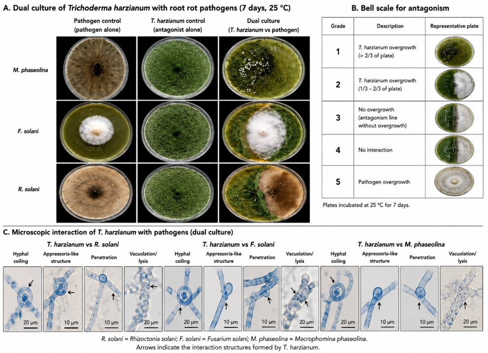
The antagonistic ability of T. harzianum was tested using the dual culture technique. Autoclave-sterilised PDA medium was prepared and dispensed into 9-cm-diameter Petri dishes, which were left until the medium solidified. Each dish was then inoculated by placing a 0.5-cm-diameter disc taken with a sterile cork borer from near the margin of the colony of each pathogenic isolate (R. solani at 3 days, F. solani at 7 days, and M. phaseolina at 4 days of age, each separately) cultured on PDA medium at the centre of one half of the dish. The centre of the other half of the dish was inoculated with a 0.5-cm-diameter disc taken with a sterile cork borer from near the margin of the biocontrol fungal colony cultured on PDA medium at 7 days of age. Four dishes were used per treatment, while the control treatment consisted of four dishes inoculated with the pathogenic fungi only. The dishes were incubated at 25 ± 2 ℃ for 7 days, and antagonism was assessed according to the five-grade scale of Bell et al. [51] as follows: (1) the antagonistic fungus overgrows the entire dish, including the pathogen; (2) the antagonistic fungus covers two-thirds of the dish; (3) the antagonistic fungus covers one-half of the dish; (4) the pathogenic fungus covers two-thirds of the dish; (5) the pathogenic fungus covers the entire dish. The antagonistic fungus was considered effective when the antagonism grade fell between grades 1 and 2.
2.3.1 Antagonistic activity assay and scoring system
The antagonistic activity of T. harzianum against the tested pathogens was evaluated using the five-grade scale proposed by Bell et al. [51] at 7 days post-inoculation. Grade 1 indicated that the antagonist overgrew the pathogen and colonised the entire surface of the dish; grade 2 indicated that the antagonist colonised approximately two-thirds of the dish; grade 3 indicated that neither organism dominated, each colonising approximately one-half of the dish; grade 4 indicated that the pathogen colonised approximately two-thirds of the dish; and grade 5 indicated that the pathogen overgrew the antagonist and colonised the entire dish. Isolates scoring grade 1 or 2 were considered effective antagonists, whereas grades 3–5 were considered ineffective. Because the Bell scale is a semi-quantitative ordinal criterion based on the extent of colony overgrowth rather than on radial-growth measurement, mycelial growth inhibition percentages and mean colony diameters were not calculated for this assay. Quantitative inhibition was determined only for the B. subtilis dual-culture assay (Section 2.4), in which colony diameters could be measured directly.
2.4 Propagation and antagonistic evaluation of Bacillus subtilis
Isolation of a bacterial strain tentatively named Bacillus subtilis (strain designation BS-1) was carried out from the rhizosphere of healthy broad beans grown in an agricultural field of Babylon, Iraq. Isolation of the bacterium was achieved on NA media and its preliminary identification was made by observing the morphological features and conventional phenotypic traits such as Gram-positive rods, spore formation, and catalase production. Since no molecular identification technique was applied, identification of the bacterium is only tentative. Source, criteria for identification, maintenance, method of counting cells, inoculum size, and use of the bacterial strain are given in Table 2.
For routine maintenance, the isolate was kept on nutrient agar slants at 4 ℃ and subcultured on fresh nutrient agar before experimental use. Working cultures were incubated at 28 ± 2 ℃ for 24–48 h. Fresh bacterial growth was suspended in sterile distilled water and thoroughly homogenized to obtain a uniform cell suspension [52]. Viable cell density was determined by serial dilution followed by plate counting on nutrient agar and expressed as colony-forming units per millilitre (CFU mL⁻¹). The suspension used for greenhouse application was adjusted to approximately 1 × 10⁸ CFU mL⁻¹.
The in vitro antagonistic activity of B. subtilis against Rhizoctonia solani, Fusarium solani, and Macrophomina phaseolina was evaluated using a dual-culture assay. A 5-mm agar plug taken from the actively growing margin of a 7-day-old culture of each fungal pathogen was placed on one side of a PDA plate. B. subtilis was streaked approximately 2 cm from the fungal plug. Control plates were inoculated with the corresponding fungal pathogen alone. Each pathogen–bacterium combination and its respective control were replicated four times. All plates were incubated at 25 ± 2 ℃ for 7 days.
Table 2. Source, identification, maintenance, and preparation of the Bacillus subtilis isolate used in the antagonism and greenhouse experiments
|
Parameter |
Description |
|
Microbial agent |
Bacillus subtilis |
|
Isolate code |
BS-1 |
|
Source |
Rhizosphere soil of healthy broad bean plants collected from an agricultural field in Babylon Province, Iraq |
|
Isolation medium |
Nutrient agar (NA) |
|
Preliminary identification |
Colony morphology, Gram-positive reaction, rod-shaped cells, endospore formation, catalase-positive reaction, and standard biochemical characteristics consistent with B. subtilis |
|
Molecular identification |
Not performed |
|
Short-term storage |
Nutrient agar slants at 4 ℃ |
|
Working culture |
Fresh culture prepared on nutrient agar and incubated at 28 ± 2 ℃ for 24–48 h |
|
Suspension preparation |
Bacterial growth was suspended in sterile distilled water and homogenized before use |
|
Cell-count method |
Viable plate-count method using serial dilution and colony counting on nutrient agar |
|
Target concentration |
Approximately 1 × 10⁸ CFU mL⁻¹ |
|
Greenhouse dose |
10 mL bacterial suspension per pot |
|
Application timing |
Applied 48 h before sowing |
|
In vitro application |
Bacterial streak placed approximately 2 cm from the fungal plug in dual-culture plates |
At the end of the incubation period, fungal colony growth was measured in the bacterial treatment and corresponding untreated control. Mycelial growth inhibition was calculated using Eq. (3) [53, 54]:
$I(\%)=\left(1-\frac{G_t}{G_c}\right) \times 100$ (3)
where,
I = Mycelial growth inhibition (%)
$G_t$ = Mean fungal colony diameter (or radial growth) in the bacterial treatment (mm)
$G_C$ = Mean fungal colony diameter (or radial growth) in the untreated control (mm)
For the greenhouse experiment, the standardized bacterial suspension (1 × 10⁸ CFU mL⁻¹) was applied at a rate of 10 mL per pot, 48 h before sowing, either individually or in combination with T. harzianum and/or garlic extract, according to the treatment structure described in Section 2.6.
2.5 Preparation and evaluation of plant aqueous extracts
Three plants were selected to study their effect against the pathogenic fungi: fresh cloves of garlic (Allium sativum, local cultivar), mature leaves of neem (Azadirachta indica), and dry fruits of fennel (Foeniculum vulgare). The method of Ahmed and Agnihotri [55] was followed in preparing the aqueous extracts by mixing 20 g of dried plant powder for each plant sample separately with 400 mL of distilled water in a 1000-mL glass flask. The suspension was placed in a water bath at 30 ℃ for half an hour, then filtered through several layers of gauze, and finally sterilised through a Millipore filter with a pore diameter of 0.22 µm. The filtrate was stored in tightly sealed containers in the refrigerator until use.
The method of the studies [56, 57] was followed by mixing the aqueous extract of each plant with PDA medium after it had been melted, sterilised, and cooled to 45 ℃. Volumes of 5, 10, and 15 mL of the extract were added to 95, 90, and 85 mL, respectively, of PDA medium, with three replicates per concentration. After the medium solidified, the dishes were inoculated at the centre with a 0.5-cm-diameter disc taken from the margin of an actively growing fungal colony on PDA medium for each of R. solani, F. solani, and M. phaseolina at 3, 7, and 4 days of age, respectively. The dishes were incubated at 25 ± 2 ℃. When the colony diameter in the control treatment (without extract) reached the edge of the dish, results were recorded by measuring the mean of two perpendicular diameters of each colony, and the percentage inhibition was calculated using the Eq. (3).
Although 15% garlic extract produced the highest inhibition of mycelial growth under in vitro conditions, the 10% concentration also provided consistently high antifungal activity against all three pathogens. Therefore, the intermediate concentration (10%) was selected for the greenhouse experiment to evaluate disease suppression under more practical application conditions while reducing extract input compared with the highest concentration tested.
2.6 Pot (greenhouse) experiment
The experiment was conducted using plastic pots 15 cm in diameter with a capacity of 3 kg of soil, sterilised in an autoclave at 121 ℃ and a pressure of 1.5 kg cm⁻² for one hour and left for 7 days for aeration before being distributed into the pots. T. harzianum inoculum, carried on local millet seeds, was added at a rate of 10 g per pot one week before adding the pathogen inoculum [58]. The B. subtilis suspension was added at a concentration of 1 × 10⁸ CFU mL⁻¹ one week before adding the pathogen inoculum [59]. Garlic extract was added at a concentration of 10% three days before adding the pathogen inoculum. A mixed inoculum of the three pathogenic fungi (R. solani, F. solani, and M. phaseolina), cultured on millet seeds in equal mass proportions, was added at 0.5% (w/w) of soil to provide the disease pressure for the pot experiment. The pots were sown with faba bean seeds three days after adding the pathogen inoculum and were irrigated as needed.
The experimental design was a randomised complete block design (RCBD) with three replicates per treatment, and the experiment comprised the following nine treatments: (T1) control, uninfected with the pathogen, to which sterilised millet seeds only were added; (T2) T. harzianum alone; (T3) B. subtilis alone; (T4) garlic extract alone; (T5) T. harzianum + B. subtilis; (T6) B. subtilis + garlic extract; (T7) T. harzianum + garlic extract; (T8) T. harzianum + B. subtilis + garlic extract; and (T9) pathogenic fungi alone. Germination percentage was recorded 10 days after sowing, and infection percentage and disease severity were recorded 30 days after sowing, using the same scoring system described in section 2.2.
2.7 Plant growth measurements
Thirty days after sowing, five plants were randomly selected from each replicate pot for growth assessment. Plant height was measured from the soil surface to the apical meristem using a graduated ruler. Root length was determined after carefully washing the root system under running water. Fresh weight was recorded immediately after harvesting using a digital balance, while dry weight was determined after oven-drying the plant material at 70 ℃ until constant weight was achieved.
2.8 Determination of chlorophyll content
The determination of the contents of chlorophyll was performed using a portable soil plant analysis development (SPAD) meter (SPAD-502 Plus, Konica Minolta, Japan). The SPAD meter was used on five randomly selected fully developed apical leaves of each plant. Three measurements were taken from each leaf, and the averages were calculated to have an exact SPAD value for each treatment.
2.9 Determination of defence-related enzyme activities
Fresh leaf samples were collected 30 days after sowing from plants subjected to the different greenhouse treatments. Fully expanded leaves of comparable physiological age were collected from each experimental unit. For each biological replicate, 0.5 g of fresh leaf tissue was homogenized under chilled conditions in 5 mL of 50 mM sodium phosphate buffer (pH 7.0). The homogenate was centrifuged at 12,000 × g for 15 min at 4 ℃, and the resulting clear supernatant was collected and used immediately as the crude enzyme extract for determination of peroxidase (POD), polyphenol oxidase (PPO), and phenylalanine ammonia-lyase (PAL) activities.
POD activity was determined using guaiacol as the electron-donor substrate. The 3.0-mL reaction mixture contained 2.5 mL of 50 mM sodium phosphate buffer (pH 6.0), 0.2 mL of 20 mM guaiacol, 0.1 mL of 40 mM H₂O₂, and 0.2 mL of crude enzyme extract. The reaction was initiated by adding H₂O₂, and the increase in absorbance resulting from guaiacol oxidation was monitored at 470 nm for 3 min using a UV–visible spectrophotometer. One unit (U) of POD activity was defined as the amount of enzyme producing an increase of 0.01 absorbance unit min⁻¹ under the assay conditions. POD activity was expressed as U g⁻¹ fresh weight (FW).
PPO activity was determined using catechol as the substrate. The 3.0-mL reaction mixture consisted of 2.5 mL of 50 mM sodium phosphate buffer (pH 6.5), 0.3 mL of 100 mM catechol, and 0.2 mL of crude enzyme extract. The reaction was initiated by adding the enzyme extract, and the increase in absorbance was recorded at 420 nm for 3 min. One unit of PPO activity was defined as the amount of enzyme causing an increase of 0.01 absorbance unit min⁻¹ under the assay conditions. PPO activity was expressed as U g⁻¹ FW.
PAL activity was determined based on the conversion of L-phenylalanine to trans-cinnamic acid. The reaction mixture contained 1.0 mL of 50 mM Tris-HCl buffer (pH 8.8), 0.5 mL of 20 mM L-phenylalanine, 0.5 mL of crude enzyme extract, and distilled water to a final volume of 3.0 mL. The mixture was incubated at 37 ℃ for 60 min, after which the reaction was terminated by adding 0.5 mL of 1 M HCl. The amount of trans-cinnamic acid formed was determined spectrophotometrically at 290 nm against an appropriate reaction blank. PAL activity was calculated using a trans-cinnamic acid calibration curve and expressed as µg trans-cinnamic acid g⁻¹ FW h⁻¹.
Enzyme assays were performed for each of the three biological replicates corresponding to the greenhouse experimental units. Two technical measurements were conducted for each biological replicate, and their mean was used as the replicate value for statistical analysis; technical replicates were not treated as independent observations. All extraction procedures were performed under chilled conditions, and enzyme activities were determined using freshly prepared extracts.
2.10 Field validation experiment
A field experiment was conducted during the 2024/2025 broad bean growing season in Babylon Province, Iraq, to validate the performance of the most promising treatments identified under greenhouse conditions. The experiment was established in an agricultural field with a documented history of broad bean root rot and naturally occurring soil-borne infection. No artificial fungal inoculum was introduced into the field; therefore, disease development was dependent on the naturally occurring pathogen population. The experiment was conducted using the same locally cultivated broad bean type used in the greenhouse evaluation.
The experiment was arranged in an RCBD with three replicates. Each experimental plot measured 3 × 4 m (12 m²). The treatments were assigned randomly within each block, with a proper buffer zone kept between the adjacent plots to avoid any interaction effect of the treatments. The seeds were planted at a distance of around 50 cm from each other, and the plants were kept at a spacing of 20 cm from each other within the row, producing an establishment density of six plants per square meter. The plots were uniformly managed with respect to irrigation, weed removal, and fertilizer application, without using any fungicides.
This field trial had four different treatments, which included an untreated naturally infected control plot, Trichoderma harzianum only, Trichoderma harzianum + Bacillus subtilis, and an integrated treatment of Trichoderma harzianum, Bacillus subtilis, and garlic extract. The microbial and botanical preparations were applied in concentrations similar to those used in the greenhouse trial. T. harzianum was applied as a conidial suspension adjusted to approximately 1 × 10⁷ conidia mL⁻¹, whereas B. subtilis was applied as a bacterial suspension adjusted to approximately 1 × 10⁸ CFU mL⁻¹. Garlic extract was applied at 10% (w/v). Treatments were applied in the planting zone immediately before or at sowing to maximize contact between the biological agents, seed/root zone, and naturally infested soil.
Disease incidence and disease severity were assessed during crop development using the same criteria and disease-rating scale described for the greenhouse experiment. Plants showing characteristic root-rot and damping-off symptoms were recorded within each plot, and disease incidence and severity were calculated using Eqs. (1) and (2), respectively. Assessments were made using plants sampled systematically from the central rows of each plot to minimize border effects.
At crop maturity, plants from the central 6 m² of each plot were harvested for yield determination, excluding border rows and plants at the ends of each row. Harvested seeds were cleaned, weighed, and plot yield was converted to t ha⁻¹. The experimental plot was considered the statistical unit. Field data were analysed according to the RCBD model described in Section 2.11, with treatment considered a fixed effect and block included as the blocking factor.
2.11 Statistical analysis
Statistical analyses were performed using SPSS software. The experimental unit and statistical model were defined separately for each experiment according to its design. Data from the pathogenicity assay and Bacillus subtilis antagonism experiment were analysed using one-way analysis of variance (ANOVA), with pathogen or treatment, as appropriate, considered the fixed factor. The antagonistic activity of Trichoderma harzianum evaluated using the Bell scale was treated as a semi-quantitative assessment and interpreted accordingly.
The plant-extract experiment was analysed using a three-way factorial ANOVA to evaluate the main effects of extract type, concentration, and fungal pathogen, as well as the extract × concentration, extract × pathogen, concentration × pathogen, and extract × concentration × pathogen interactions on mycelial growth inhibition.
Greenhouse data were analysed according to an RCBD, with treatment considered a fixed effect and block included as a blocking factor. The pot was considered the experimental unit. Measurements obtained from multiple plants or leaves within the same pot were treated as subsamples and averaged before statistical analysis. Field data were similarly analysed according to an RCBD, with treatment as the fixed effect and block as the blocking factor.
Percentage data were examined for normality and homogeneity of variance and were transformed using the arcsine square-root transformation when necessary to satisfy ANOVA assumptions. Statistical analyses were performed on transformed values where applicable, whereas untransformed means are presented in the tables for biological interpretation. When ANOVA indicated significant treatment effects, means were separated using Fisher's least significant difference (LSD) test at p ≤ 0.05. Results are presented as mean ± standard deviation (SD) where replicate-level observations were available. For each ANOVA, the corresponding F-statistics, degrees of freedom (df), and exact p-value were reported.
3.1 Fungal isolation and pathogenicity
During the survey of five broad bean fields in Babylon Province, Iraq, three fungi associated with symptomatic plants were consistently recovered and provisionally identified as Rhizoctonia solani, Fusarium solani, and Macrophomina phaseolina. Preliminary identification was based on colony morphology and cultural and microscopic characteristics, including hyphal features, conidial morphology where applicable, and the formation of characteristic sclerotial structures.
Subsequent pathogenicity assays confirmed the ability of all three isolates to induce root-rot symptoms in broad bean plants, and the respective fungi were re-isolated from symptomatic tissues, thereby fulfilling Koch's postulates.
Saprophytic Aspergillus and Penicillium species also appeared on the isolation plates but were not pursued. The pathogenicity test (Table 3) confirmed that all three target fungi were pathogenic on broad bean seedlings, with R. solani producing the most severe damage -germination dropped from 96.0% in the control to 40.0%, infection reached 76.0%, and disease severity was 71.3%. F. solani and M. phaseolina were both pathogenic but somewhat less aggressive than R. solani under the conditions of the test. Koch’s postulates were satisfied for each isolate by re-recovery from inoculated, symptomatic plants.
Table 3. Pathogenicity of three fungi isolated from symptomatic broad bean plants in Babylon Province, evaluated as germination percentage, infection percentage, and disease severity on broad bean seedlings in autoclaved soil with millet-seed inoculum at 0.5% (w/w)
|
Treatment |
Germination (%) |
Infection (%) |
Disease Severity (%) |
|
Control (sterile millet only) |
96.0 a |
0.0 d |
0.0 d |
|
Rhizoctonia solani |
40.0 d |
76.0 a |
71.3 a |
|
Fusarium solani |
60.0 c |
60.0 b |
54.7 b |
|
Macrophomina phaseolina |
68.0 b |
52.0 c |
47.5 c |
|
LSD (p < 0.05) |
4.8 |
5.1 |
4.6 |
3.2 Antagonistic ability of Trichoderma harzianum
The fungus Trichoderma harzianum displayed a potent antagonistic effect against all three root rot pathogens under the dual culture test conditions. Seven days after cultivation at 25 ℃, the antagonist was observed to be growing extensively on the culture medium, limiting the growth of the pathogen (Figure 1(A)). Based on the Bell system, T. harzianum exhibited potent antagonistic effects against R. solani and was considered to have grade 1 response, while the interactions with F. solani and M. phaseolina belonged to grade 2 response (Figure 1(B); Table 4). Microscopic examination of the interaction zone revealed typical mycoparasitic features, including hyphal coiling, appressorium-like structures, penetration pegs, and lysis of the pathogen hypha (Figure 1(C)) [13, 22].
|
Pathogen |
Bell Grade |
|
Rhizoctonia solani |
1 |
|
Fusarium solani |
2 |
|
Macrophomina phaseolina |
2 |
The antagonistic interaction between T. harzianum and the tested pathogens was evaluated using the Bell scale, which is a semi-quantitative method based on the degree of colony overgrowth rather than radial-growth inhibition. Consequently, inhibition percentages and mean colony diameters were not calculated for this assay because the Bell scale itself served as the predefined criterion for assessing antagonistic efficacy. Quantitative inhibition percentages were determined only for the B. subtilis antagonism assay, where fungal colony growth could be measured directly.
3.3 Antagonistic effect of Bacillus subtilis
B. subtilis produced inhibition of mycelial growth of all three pathogens (Table 5). Inhibition ranged from 51.4% (M. phaseolina) to 62.8% (R. solani), with F. solani at 57.6%. The ordering of sensitivity was R. solani > F. solani > M. phaseolina.
Table 5. Percentage inhibition of mycelial growth of three broad bean root rot pathogens by Bacillus subtilis on PDA medium
|
Pathogen |
Mean Colony Diameter (cm) -Control |
Mean Colony Diameter (cm) -B. subtilis |
Inhibition (%) |
|
Rhizoctonia solani |
8.6 |
3.2 |
62.8 a |
|
Fusarium solani |
8.5 |
3.6 |
57.6 b |
|
Macrophomina phaseolina |
8.7 |
4.2 |
51.4 c |
|
LSD (p < 0.05) |
— |
— |
3.4 |
3.4 Antifungal activity of plant aqueous extracts
The three plant extracts inhibited the pathogens in a dose-dependent manner (Table 6). Garlic extract was the most active across all three pathogens and all three concentrations: at 5% it gave 47.1–57.6% inhibition; at 10%, 73.5–85.9%; and at 15%, 90.2–100%, with complete inhibition of R. solani at the 15% concentration. Neem extract gave intermediate inhibition (12.4–56.5%), and fennel extract gave the weakest inhibition (5.9–40.0%). R. solani was the most sensitive pathogen to garlic and neem, while M. phaseolina required the highest extract concentrations to achieve equivalent inhibition.
Table 6. Percentage inhibition of mycelial growth of three broad bean root rot pathogens by aqueous extracts of garlic (Allium sativum), neem (Azadirachta indica), and fennel (Foeniculum vulgare) at three concentrations (5, 10, 15% v/v on PDA)
|
Extract |
Conc. (%) |
R. solani (%) |
F. solani (%) |
M. phaseolina (%) |
|
Garlic |
5 |
57.6 c |
52.3 c |
47.1 c |
|
Garlic |
10 |
85.9 b |
80.0 b |
73.5 b |
|
Garlic |
15 |
100.0 a |
95.3 a |
90.2 a |
|
Neem |
5 |
16.5 f |
14.1 e |
12.4 e |
|
Neem |
10 |
36.5 d |
30.6 d |
25.9 d |
|
Neem |
15 |
56.5 c |
48.2 c |
44.7 c |
|
Fennel |
5 |
8.2 g |
7.1 f |
5.9 f |
|
Fennel |
10 |
22.4 e |
18.8 e |
15.3 e |
|
Fennel |
15 |
40.0 d |
32.9 d |
28.2 d |
|
LSD (p < 0.05) |
— |
4.8 |
5.2 |
4.7 |
3.5 Greenhouse pot experiment
Inoculation of the pot substrate with the mixed three-pathogen complex drove germination of broad bean down from 96.0% in the healthy control to 38.0% in the pathogen-only treatment, infection up to 76.0%, and disease severity to 64.3% (Table 7). Under mixed-pathogen pressure, all individual biological or botanical treatments reduced disease development relative to the pathogen-only control. The pathogen + T. harzianum treatment reduced disease severity to 28.6%, while pathogen + B. subtilis and pathogen + garlic extract resulted in disease severities of 32.9% and 36.4%, respectively. Combined treatments provided greater disease suppression, with the three-component treatment (T8: pathogen + T. harzianum + B. subtilis + garlic extract) producing the highest germination (90.0%) and the lowest disease incidence (14.0%) and severity (10.8%) among the pathogen-inoculated treatments (Table 7).
Table 7. Effect of individual and combined biological and botanical treatments on germination, disease incidence, and root-rot severity of broad bean under mixed-pathogen pressure in the greenhouse (RCBD, three replicates)
|
Treatment |
Germination (%) |
Disease Incidence (%) |
Disease Severity (%) |
|
T1: Healthy control (non-inoculated) |
96.0 a |
0.0 h |
0.0 h |
|
T2: Pathogen + T. harzianum |
80.0 c |
33.3 d |
28.6 d |
|
T3: Pathogen + B. subtilis |
75.0 cd |
40.0 c |
32.9 c |
|
T4: Pathogen + garlic extract |
72.0 d |
44.0 c |
36.4 c |
|
T5: Pathogen + T. harzianum + B. subtilis |
85.0 b |
22.0 ef |
17.5 ef |
|
T6: Pathogen + B. subtilis + garlic extract |
82.0 bc |
26.0 e |
21.4 e |
|
T7: Pathogen + T. harzianum + garlic extract |
86.0 b |
20.0 f |
16.0 f |
|
T8: Pathogen + T. harzianum + B. subtilis + garlic extract |
90.0 b |
14.0 g |
10.8 g |
|
T9: Pathogen-only control |
38.0 e |
76.0 a |
64.3 a |
|
LSD (p < 0.05) |
5.1 |
4.8 |
4.4 |
Means within a column followed by different letters differ significantly (LSD, p < 0.05). Mixed inoculum of R. solani + F. solani + M. phaseolina at 0.5% (w/w) was applied to all pots except T1 (healthy control).
Beyond disease suppression, significant differences were also observed in plant growth performance among treatments. As shown in Table 8, the integrated application of T. harzianum, B. subtilis, and garlic extract (T8) produced the greatest improvements in plant height, root length, fresh biomass, and dry biomass compared with the pathogen-inoculated control.
Table 8. Effect of individual and combined biological and botanical treatments on growth characteristics of broad bean plants under mixed-pathogen pressure in the greenhouse
|
Treatment |
Plant Height (cm, mean ± SD) |
Root Length (cm, mean ± SD) |
Fresh Weight (g, mean ± SD) |
Dry Weight (g, mean ± SD) |
|
T1: Healthy control (non-inoculated) |
33.8 ± 1.2 bc |
15.4 ± 0.6 bcd |
21.8 ± 0.9 bc |
4.6 ± 0.2 bc |
|
T2: Pathogen + T. harzianum |
31.5 ± 1.0 de |
14.8 ± 0.5 de |
20.5 ± 0.8 cde |
4.3 ± 0.2 cde |
|
T3: Pathogen + B. subtilis |
30.8 ± 1.1 de |
14.2 ± 0.6 de |
19.7 ± 0.7 de |
4.1 ± 0.2 de |
|
T4: Pathogen + garlic extract |
29.6 ± 0.9 e |
13.7 ± 0.5 e |
18.9 ± 0.8 e |
3.9 ± 0.2 e |
|
T5: Pathogen + T. harzianum + B. subtilis |
34.2 ± 1.3 bc |
16.1 ± 0.7 bc |
22.7 ± 0.9 b |
4.8 ± 0.2 b |
|
T6: Pathogen + B. subtilis + garlic extract |
32.5 ± 1.1 cd |
15.2 ± 0.6 cd |
21.3 ± 0.8 bcd |
4.5 ± 0.2 bcd |
|
T7: Pathogen + T. harzianum + garlic extract |
34.8 ± 1.2 b |
16.5 ± 0.7 b |
23.1 ± 0.9 b |
4.9 ± 0.2 b |
|
T8: Pathogen + T. harzianum + B. subtilis + garlic extract |
37.6 ± 1.4 a |
18.3 ± 0.8 a |
25.4 ± 1.0 a |
5.5 ± 0.3 a |
|
T9: Pathogen-only control |
18.4 ± 0.8 f |
8.6 ± 0.4 f |
10.7 ± 0.6 f |
2.1 ± 0.1 f |
|
LSD (p < 0.05) |
2.1 |
1.2 |
1.8 |
0.4 |
Chlorophyll content followed a similar trend (Table 9). Plants receiving the combined biological and botanical treatment exhibited the highest SPAD values, indicating improved photosynthetic capacity and better physiological status compared with untreated infected plants.
Table 9. Effect of individual and combined biological and botanical treatments on chlorophyll content (SPAD values) of broad bean plants under mixed-pathogen pressure in the greenhouse
|
Treatment |
SPAD Value (mean ± SD) |
|
T1: Healthy control (non-inoculated) |
42.5 ± 1.0 ef |
|
T2: Pathogen + T. harzianum |
44.3 ± 1.1 cde |
|
T3: Pathogen + B. subtilis |
43.7 ± 0.9 def |
|
T4: Pathogen + garlic extract |
41.8 ± 1.0 f |
|
T5: Pathogen + T. harzianum + B. subtilis |
46.2 ± 1.2 bc |
|
T6: Pathogen + B. subtilis + garlic extract |
45.8 ± 1.1 bcd |
|
T7: Pathogen + T. harzianum + garlic extract |
46.8 ± 1.2 ab |
|
T8: Pathogen + T. harzianum + B. subtilis + garlic extract |
49.1 ± 1.3 a |
|
T9: Pathogen-only control |
28.6 ± 0.8 g |
|
LSD (p < 0.05) |
2.3 |
Significant increases in defense-related enzymes were recorded in treated plants (Table 10). The combined treatment (T8) resulted in the highest activities of POD, PPO, and PAL.
Table 10. Effect of individual and combined biological and botanical treatments on defense-related enzyme activities in broad bean plants under mixed-pathogen pressure in the greenhouse
|
Treatment |
POD (U g⁻¹ FW, mean ± SD) |
PPO (U g⁻¹ FW, mean ± SD) |
PAL (µg trans-cinnamic acid g⁻¹ FW h⁻¹, mean ± SD) |
|
T1: Healthy control (non-inoculated) |
12.8 ± 0.7 e |
8.4 ± 0.5 d |
21.6 ± 1.4 e |
|
T2: Pathogen + T. harzianum |
19.4 ± 0.9 c |
13.2 ± 0.7 c |
34.7 ± 1.8 c |
|
T3: Pathogen + B. subtilis |
18.1 ± 0.8 cd |
12.5 ± 0.6 c |
32.8 ± 1.7 cd |
|
T4: Pathogen + garlic extract |
16.5 ± 0.8 d |
11.2 ± 0.6 c |
28.6 ± 1.6 d |
|
T5: Pathogen + T. harzianum + B. subtilis |
24.3 ± 1.1 b |
17.1 ± 0.8 b |
42.8 ± 2.0 b |
|
T6: Pathogen + B. subtilis + garlic extract |
22.9 ± 1.0 b |
16.4 ± 0.8 b |
40.7 ± 1.9 b |
|
T7: Pathogen + T. harzianum + garlic extract |
25.6 ± 1.2 b |
17.9 ± 0.9 b |
44.1 ± 2.1 b |
|
T8: Pathogen + T. harzianum + B. subtilis + garlic extract |
31.4 ± 1.4 a |
22.6 ± 1.0 a |
56.3 ± 2.4 a |
|
T9: Pathogen-only control |
10.2 ± 0.6 e |
7.3 ± 0.4 d |
18.5 ± 1.2 e |
|
LSD (p < 0.05) |
2.8 |
2.1 |
4.5 |
3.6 Field validation trial
To validate the greenhouse findings under practical agricultural conditions, a field experiment was conducted during the subsequent growing season. The results demonstrated that the integrated treatment maintained superior disease suppression and yield performance under natural infection conditions (Table 11).
Table 11. Field performance of integrated disease management treatments against broad bean root rot under natural infection conditions
|
Treatment |
Disease Incidence (%) |
Disease Severity (%) |
Yield (t ha⁻¹) |
|
Untreated naturally infected control |
45.6 a |
37.8 a |
2.28 c |
|
T. harzianum |
28.4 b |
21.6 b |
2.74 b |
|
T. harzianum + B. subtilis |
19.6 c |
14.7 c |
2.89 ab |
|
T. harzianum + B. subtilis + garlic extract |
12.8 d |
9.7 d |
3.01 a |
|
LSD (p < 0.05) |
4.3 |
3.8 |
0.18 |
Three findings stand out from this work. First, the broad bean root rot complex in Babylon Province is dominated, in our isolations, by the same three fungi that have repeatedly emerged from comparable surveys elsewhere in the Mediterranean basin and North Africa: R. solani, F. solani, and M. phaseolina [7-9]. R. solani was the most aggressive of the three, both in the pathogenicity test and in dual culture, which is consistent with its well-documented capacity to cause rapid post-emergence collapse under warm, irrigated soil conditions [11, 12]. Second, the three biocontrol components evaluated -T. harzianum, B. subtilis, and garlic aqueous extract -all showed meaningful single-component activity in vitro, but none approached complete protection on its own under greenhouse conditions; the best single-component treatment (T. harzianum alone) still left disease severity at 28.6%. Third, combining a fungal biocontrol agent with a bacterial biocontrol agent and a botanical extract produced a markedly stronger effect: the three-way integrated treatment cut disease severity by approximately 83% relative to the pathogen-only control, and brought disease pressure down to a level only marginally above the healthy reference.
The Bell-scale grade 1 result for T. harzianum against R. solani is worth a brief comment. Grade 1 represents complete overgrowth of the pathogen by the antagonist, and microscopic examination of the contact zone confirmed the textbook mycoparasitic sequence: coiling, penetration peg formation, and cytoplasmic vacuolation of the host hyphae [22, 24, 60]. The slightly weaker (grade 2) response against F. solani and M. phaseolina reflects their thicker, more pigmented walls and -in the case of M. phaseolina -the formation of melanised microsclerotia that are inherently more resistant to enzymatic degradation [13, 61]. This pattern matches what has been reported for T. harzianum on other legume pathosystems [23, 25]. The fact that the T. harzianum isolate used here was a locally maintained strain rather than an introduced commercial product probably contributed to its performance under our soil and temperature conditions; the rhizosphere-competence advantage of indigenous Trichoderma isolates is now well documented [26, 62].
B. subtilis contributed a chemically distinct mode of action. The diffusible inhibition zones we observed are most plausibly attributable to surfactin, iturin, and fengycin lipopeptides, which destabilise fungal membranes and are now widely characterised as the main basis of Bacillus biocontrol activity [27-30]. The 51.4–62.8% inhibition range we obtained sits squarely in the range reported for other B. subtilis isolates against the same pathogens [31, 32, 63]. What matters most operationally is that lipopeptide and Trichoderma-mediated mechanisms are largely orthogonal: lipopeptides permeabilise membranes, whereas mycoparasitism degrades cell walls and competes for substrate. Pathogens that can evade one mechanism are not automatically able to evade the other, which is why T. harzianum + B. subtilis combinations have repeatedly outperformed either agent alone in published trials on legumes and Solanaceae [32, 33, 64].
Garlic aqueous extract was the most active of the three botanicals tested, and its near-complete inhibition of R. solani at 15% is consistent with the very extensive literature on allicin and related thiosulfinates [34-37]. The chemistry of garlic is somewhat unusual: allicin is formed almost instantly when the substrate alliin is exposed to the enzyme alliinase by tissue disruption, and it acts through several mechanisms simultaneously -reaction with cellular thiols, membrane permeabilisation, and inhibition of multiple fungal enzymes [36, 37, 65]. The dose response we recorded -strong activity at 10–15% but only modest activity at 5% -matches the pattern reported for aqueous garlic preparations on F. oxysporum, Botrytis cinerea, and other agriculturally important fungi [38, 39]. Neem extract’s intermediate performance and fennel’s weaker activity are likewise consistent with the published literature: azadirachtin and the broader triterpenoid pool in neem give moderate but reliable activity against several soil- borne fungi [39, 40, 65], while fennel’s phenolic and terpenoid mix is generally more active against airborne pathogens than against soil-borne complexes [42, 43, 66].
The strong in vitro performance of garlic at 15% did not translate directly into in vivo dominance -garlic alone in the greenhouse trial reduced disease severity to 36.4%, marginally weaker than T. harzianum alone (28.6%) and B. subtilis alone (32.9%). This in vitro–in vivo gap is one of the more important practical observations to come out of the work. Allicin is highly reactive and has a relatively short half-life once exposed to soil organic matter, microbial activity, and the buffering effects of soil pH [36, 65]. A pre-sowing soil application of garlic extract therefore delivers a useful but transient pulse of antifungal chemistry, which is precisely the kind of activity that complements the longer-term rhizosphere persistence of microbial biocontrol agents. The strong performance of the three-component treatment in our pot experiment is consistent with this complementarity -short-lived chemistry at the seed and seedling stage, sustained microbial activity through the longer crop cycle.
Our greenhouse result—a three-way combined treatment cutting disease severity by approximately 83%—sits within a broader pattern of improved effects of combined biocontrol strategies reported in previous studies. Essential-oil-plus-Trichoderma combinations gave better control of faba bean root rot than either alone in Egyptian work [45]; medicinal plant extracts combined with chemical inducers reduced Rhizoctonia damping-off on broad bean more effectively than single treatments [46]; vermicompost tea paired with Trichoderma and Serratia metabolites outperformed any single component on common bean [66, 67]; and Bacillus-plus-plant-extract packages have similarly outperformed single treatments on tomato and bean pathosystems [68, 69]. What our data add to this literature is a specific three-way combination tested under Iraqi soil and temperature conditions against the actual pathogen complex that dominates Babylon Province field surveys.
Apart from disease control, the treatment regime led to considerable improvements in the growth and physiology of the plants. The combination of Trichoderma harzianum, Bacillus subtilis, and garlic extract treatment resulted in higher plant height, root development, biomass production, and chlorophyll concentration compared to any other treatment. In addition, increased activities of defensive enzymes such as POD, PPO, and PAL were observed, which is an indication that the induced systemic resistance was triggered [29, 70]. Increased activity of these defensive enzymes has also been shown to occur after biological treatments in the tomato and legume pathosystems [70]. The favorable performance achieved in this research confirms previous observations where it was noted that the use of biological disease control measures is effective in real agricultural settings [71, 72].
Several limitations should be acknowledged. Although defence-related enzyme activities and field validation data were included in the present study, the field experiment was conducted at a single location and during one growing season only. Therefore, additional multi-location and multi-season evaluations are required before large-scale recommendations can be made [69]. In addition, the greenhouse design did not include non-inoculated plants treated separately with T. harzianum, B. subtilis, or garlic extract. Consequently, the observed improvements in plant growth, SPAD values, and defence-related enzyme activities cannot be unequivocally separated into direct plant-growth-promoting effects and indirect effects resulting from disease suppression. Future experiments should therefore include corresponding non- pathogen treatment controls to distinguish these mechanisms more clearly. In this study, the pathogens were identified mainly based on their morphology and culture properties without the use of any molecular techniques for their identification, such as internal transcribed spacer (ITS) region sequencing or other DNA-based methods. In future studies, molecular techniques for identification of the pathogens must be used, along with characterization of their rhizosphere microbial interactions and optimization of commercial products [72].
In general, the major causes of broad bean root rot in Babylon Province were associated with a pathogen complex of R. solani, F. solani, and M. phaseolina, with predominance of R. solani in greenhouse tests. The three biological and botanical components studied herein displayed an antagonistic activity towards the pathogens of interest. In particular, T. harzianum showed a highly antagonistic activity in dual culture tests with respect to R. solani, reaching Bell grade 1 and grade 2 for F. solani and M. phaseolina. Also, B. subtilis had an inhibitory effect on the mycelial growth of the pathogens by 51.4–62.8%. Garlic extract turned out to be the most efficient antifungal agent among the plant extracts studied, with complete inhibition of R. solani at the highest concentration.
In greenhouse experiments, the integrated treatment consisting of T. harzianum, B. subtilis, and garlic extract led to an increased seed germination rate and a decrease in disease incidence and severity compared to individual and dual- component treatments. In addition to reducing disease, the combination enhanced growth parameters of plants, increased chlorophyll content, and activated the activities of POD, PPO, and PAL enzymes, which means the induction of resistance.
Field validation trials showed the effectiveness of the proposed strategy under natural infection conditions. In particular, the integration significantly decreased the disease incidence and severity and increased grain yield from 2.28 t ha⁻¹ in the untreated naturally infected control to 3.01 t ha⁻¹, an increase of 32.0%, with disease incidence falling from 45.6% to 12.8% and disease severity from 37.8% to 9.7%.
Consistency of the results obtained in greenhouse and field experiments proves the practical value of integrating biological and botanical components as an alternative to regular fungicide usage in broad bean production systems.
Thus, this work has shown that the combination of T. harzianum, B. subtilis, and garlic extract is a good strategy for disease management. However, some additional work needs to be done to prove its efficacy in different locations and seasons, and to molecularly identify pathogen populations and formulations.
Overall, the combined application of T. harzianum, B. subtilis, and garlic extract represents a promising candidate for integrated management of broad bean root rot under the conditions evaluated in this study. However, the field evaluation was limited to one location and one growing season. Multi-location, multi-season, and cultivar-based trials, together with molecular confirmation of the pathogen and biocontrol isolates and optimization of application formulations, are required before large-scale field recommendations can be made.
The authors thank broad bean growers in Babylon Province for access to their fields during the disease survey, and the technical staff at the College of Agriculture laboratories for assistance with isolation, culture maintenance, and the greenhouse experiment.
|
DI |
Disease incidence, % |
|
DS |
Disease severity, % |
|
I |
Mycelial growth inhibition, % |
|
Gc |
Mean fungal colony diameter in the untreated control, mm |
|
Gt |
Mean fungal colony diameter in the treatment, mm |
|
N |
Total number of examined plants |
|
Ni |
Number of infected plants |
|
ni |
Number of plants in the ith disease severity class |
|
vi |
Disease severity rating assigned to the ith class (dimensionless) |
|
V |
Maximum disease severity rating (highest disease score) (dimensionless) |
|
POD |
Peroxidase activity, U g⁻¹ fresh weight |
|
PPO |
Polyphenol oxidase activity, U g⁻¹ fresh weight |
|
PAL |
Phenylalanine ammonia-lyase activity, µg cinnamic acid g⁻¹ h⁻¹ |
|
SPAD |
Soil plant analysis development chlorophyll index (dimensionless) |
|
LSD |
Least significant difference at p < 0.05 |
|
Subscripts |
|
|
c |
Control treatment |
|
i |
Disease severity class |
|
t |
Treatment |
[1] Etemadi, F., Hashemi, M., Barker, A.V., Zandvakili, O.R., Liu, X. (2019). Agronomy, nutritional value, and medicinal application of faba bean (Vicia faba L.). Horticultural Plant Journal, 5(4): 170-182. https://doi.org/10.1016/j.hpj.2019.04.004
[2] Karkanis, A., Ntatsi, G., Lepse, L., et al. (2018). Faba bean cultivation—Revealing novel managing practices for more sustainable and competitive European cropping systems. Frontiers in Plant Science, 9: 1115. https://doi.org/10.3389/fpls.2018.01115
[3] Jensen, E.S., Peoples, M.B., Hauggaard-Nielsen, H. (2010). Faba bean in cropping systems. Field Crops Research, 115(3): 203-216. https://doi.org/10.1016/j.fcr.2009.10.008
[4] Maalouf, F., Hu, J., O'Sullivan, D.M., et al. (2019). Breeding and genomics status in faba bean (Vicia faba). Plant Breeding, 138(4): 465-473. https://doi.org/10.1111/pbr.12644
[5] Lamichhane, J.R., Dürr, C., Schwanck, A.A., et al. (2017). Integrated management of damping-off diseases. A review: JR Lamichhane et al. Agronomy for Sustainable Development, 37(2): 10. https://doi.org/10.1007/s13593-017-0417-y
[6] Abdel-Monaim, M.F. (2013). Improvement of biocontrol of damping-off and root rot/wilt of faba bean by salicylic acid and hydrogen peroxide. Mycobiology, 41(1): 47-55. https://doi.org/10.5941/MYCO.2013.41.1.47
[7] Dugassa, A., Alemu, T., Woldehawariat, Y. (2021). In-vitro compatibility assay of indigenous Trichoderma and Pseudomonas species and their antagonistic activities against black root rot disease (Fusarium solani) of faba bean (Vicia faba L.). BMC Microbiology, 21(1): 115. https://doi.org/10.1186/s12866-021-02181-7
[8] Khaleil, M.M., Alnoman, M.M., Elrazik, E.S.A., Zagloul, H., Khalil, A.M.A. (2021). Essential oil of Foeniculum vulgare Mill. as a green fungicide and defense-inducing agent against Fusarium root rot disease in Vicia faba L. Biology, 10(8): 696. https://doi.org/10.3390/biology10080696
[9] Geiser, D.M., Aoki, T., Bacon, C.W., et al. (2013). One fungus, one name: Defining the genus Fusarium in a scientifically robust way that preserves longstanding use. Phytopathology, 103(5): 400-408. https://doi.org/10.1094/PHYTO-07-12-0150-LE
[10] Ajayi-Oyetunde, O.O., Bradley, C.A. (2018). Rhizoctonia solani: Taxonomy, population biology and management of rhizoctonia seedling disease of soybean. Plant Pathology, 67(1): 3-17. https://doi.org/10.1111/ppa.12733
[11] Sneh, B., Burpee, L., Ogoshi, A. (1991). Identification of Rhizoctonia Species. APS Press.
[12] Kaur, S., Dhillon, G.S., Brar, S.K., Vallad, G.E., Chand, R., Chauhan, V.B. (2012). Emerging phytopathogen Macrophomina phaseolina: Biology, economic importance and current diagnostic trends. Critical Reviews in Microbiology, 38(2): 136-151. https://doi.org/10.3109/1040841X.2011.640977
[13] Alwan, A.H. (2018). Effect of some ricinus communis secondary metabolites on phytophthora infestans and Fusarium solani. Al-Mustansiriyah Journal of Science, 29(1): 38-43. https://doi.org/10.23851/mjs.v29i1.288
[14] Ishii, H., Hollomon, D.W. (2015). Fungicide Resistance in Plant Pathogens: Principles and a Guide to Practical Management. Springer. https://doi.org/10.1007/978-4-431-55642-8
[15] Lucas, J.A., Hawkins, N.J., Fraaije, B.A. (2015). The evolution of fungicide resistance. Advances in Applied Microbiology, 90: 29-92. https://doi.org/10.1016/bs.aambs.2014.09.001
[16] Hollomon, D.W. (2015). Fungicide resistance: Facing the challenge. Plant Protection Science, 51(4): 170-176. https://doi.org/10.17221/42/2015-PPS
[17] Pal, K.K., Gardener, B.M. (2006). Biological control of plant pathogens. The Plant Health Instructor, 2(5): 1117-1142. https://doi.org/10.1094/PHI-A-2006-1117-02
[18] Saikia, B., Choudhary, P. (2021). Disease management and the role of antioxidants in combating plant pathogens upon PGPR inoculation with special reference to legumes. In Antioxidants in Plant-Microbe Interaction, pp. 383-394. https://doi.org/10.1007/978-981-16-1350-0_17
[19] Kareem, F.H. (2020). Control of damping-off and root rot diseases of Acacia tree using some biological control agents and plant extracts. Research on Crops, 21(1): 160-167. http://doi.org/10.31830/2348%E2%80%937542.2020.027
[20] Harman, G.E., Howell, C.R., Viterbo, A., Chet, I., Lorito, M. (2004). Trichoderma species—Opportunistic, avirulent plant symbionts. Nature Reviews Microbiology, 2(1): 43-56. https://doi.org/10.1038/nrmicro797
[21] Khaleel, A.I., Kareem, F.H., Alwan, K.F. (2020). The efficiency of plant extracts and biological control agents on some pathogens causing dampingoff and root rot disease of cucumber. Biopesticides International, 16(1): 71-76.
[22] Guzmán-Guzmán, P., Porras-Troncoso, M.D., Olmedo-Monfil, V., Herrera-Estrella, A. (2019). Trichoderma species: Versatile plant symbionts. Phytopathology, 109(1): 6-16. https://doi.org/10.1094/PHYTO-07-18-0218-RVW
[23] Tyśkiewicz, R., Nowak, A., Ozimek, E., Jaroszuk-Ściseł, J. (2022). Trichoderma: The current status of its application in agriculture for the biocontrol of fungal phytopathogens and stimulation of plant growth. International Journal of Molecular Sciences, 23(4): 2329. https://doi.org/10.3390/ijms23042329
[24] Zeilinger, S., Omann, M. (2007). Trichoderma biocontrol: Signal transduction pathways involved in host sensing and mycoparasitism. Gene Regulation and Systems Biology, 1: 227-234. https://doi.org/10.4137/GRSB.S397
[25] El-Sharkawy, H.H., Rashad, Y.M., El-Blasy, S.S., et al. (2025). Mycorrhizal symbiosis and application of vitamin B3-treated Trichoderma harzianum HE24 additively trigger immunity responses in faba bean plants against Rhizoctonia root rot and promote the plant growth and yield. BMC Plant Biology, 25(1): 926. https://doi.org/10.1186/s12870-025-06950-8
[26] Hermosa, R., Viterbo, A., Chet, I., Monte, E. (2012). Plant-beneficial effects of trichoderma and of its genes. Microbiology, 158(1): 17-25. https://doi.org/10.1099/mic.0.052274-0
[27] Ongena, M., Jacques, P. (2008). Bacillus lipopeptides: Versatile weapons for plant disease biocontrol. Trends in Microbiology, 16(3): 115-125. https://doi.org/10.1016/j.tim.2007.12.009
[28] Fira, D., Dimkić, I., Berić, T., Lozo, J., Stanković, S. (2018). Biological control of plant pathogens by Bacillus species. Journal of Biotechnology, 285: 44-55. https://doi.org/10.1016/j.jbiotec.2018.07.044
[29] Raaijmakers, J.M., De Bruijn, I., Nybroe, O., Ongena, M. (2010). Natural functions of lipopeptides from Bacillus and pseudomonas: More than surfactants and antibiotics. FEMS Microbiology Reviews, 34(6): 1037-1062. https://doi.org/10.1111/j.1574-6976.2010.00221.x
[30] Pieterse, C.M.J., Zamioudis, C., Berendsen, R.L., Weller, D.M., Van Wees, S.C.M., Bakker, P.A.H.M. (2014). Induced systemic resistance by beneficial microbes. Annual Review of Phytopathology, 52(1): 347-375. https://doi.org/10.1146/annurev-phyto-082712-102340
[31] Akladious, S.A., Gomaa, E.Z., El-Mahdy, O.M. (2019). Efficiency of bacterial biosurfactant for biocontrol of Rhizoctonia solani (AG-4) causing root rot in faba bean (Vicia faba) plants. European Journal of Plant Pathology, 153: 1237-1257. https://doi.org/10.1007/s10658-018-01639-1
[32] Kareem, F.H., Matloob, A.A. (2020). Efficiency of some of bio-formulas against fungi caused sunflower root rot disease. International Journal of Agricultural and Statistical Sciences, 16(1): 1485-1493. https://connectjournals.com/03899.2020.16.1485.
[33] Shaban, W.I., El-Bramawy, M.A. (2011). Impact of dual inoculation with rhizobium and trichoderma on damping off, root rot diseases and plant growth parameters of some legumes field crop under greenhouse conditions. International Research Journal of Agricultural Science and Soil Science 1(3): 98-108.
[34] Slusarenko, A.J., Patel, A., Portz, D. (2008). Control of plant diseases by natural products: Allicin from garlic as a case study. European Journal of Plant Pathology, 121(3): 313-322. https://doi.org/10.1007/s10658-007-9232-7
[35] Matloob, A.A.A.H., Kareem, F.H., Al-Baldawy, M.S.M. (2021). Efficiency of biological control agents and plant extracts against Rhizoctonia solani kuhn causing of damping off in cotton. Indian Journal of Ecology, 48(13): 203-207.
[36] Borlinghaus, J., Albrecht, F., Gruhlke, M.C.H., Nwachukwu, I.D., Slusarenko, A.J. (2014). Allicin: Chemistry and biological properties. Molecules, 19(8): 12591-12618. https://doi.org/10.3390/molecules190812591
[37] Miron, T., Rabinkov, A., Mirelman, D., Wilchek, M., Weiner, L. (2000). The mode of action of allicin: Its ready permeability through phospholipid membranes may contribute to its biological activity. Biochimica et Biophysica Acta (BBA)-Biomembranes, 1463(1): 20-30. https://doi.org/10.1016/S0005-2736(99)00174-1
[38] Hayat, S., Cheng, Z., Ahmad, H., Ali, M., Chen, X., Wang, M. (2016). Garlic, from remedy to stimulant: Evaluation of antifungal potential reveals diversity in phytoalexin allicin content among garlic cultivars; allicin containing aqueous garlic extracts trigger antioxidants in cucumber. Frontiers in Plant Science, 7: 1235. https://doi.org/10.3389/fpls.2016.01235
[39] Jamel, D.S., Malik, S.T.A., Hassan, A.K., Ayesh, A.A. (2025). Biological functions of garlic (Allium sativum L.) and its active compounds against pathogens. Plant Protection, 9(1): 193-201. https://doi.org/10.33804/pp.009.01.5551, 2617-1287
[40] Singh, U.P., Singh, H.B., Singh, R.B. (1980). The fungicidal effect of neem (Azadirachta indica) extracts on some soil-borne pathogens of gram (Cicer arietinum). Mycologia, 72(6): 1077-1093. https://doi.org/10.1080/00275514.1980.12021288
[41] Mondall, N.K., Mojumdar, A., Chatterje, S.K., Banerjee, A., Datta, J.K., Gupta, S. (2009). Antifungal activities and chemical characterization of neem leaf extracts on the growth of some selected fungal species in vitro culture medium. Journal of Applied Sciences and Environmental Management, 13(1): 49-53.
[42] Diao, W.R., Hu, Q.P., Zhang, H., Xu, J.G. (2014). Chemical composition, antibacterial activity and mechanism of action of essential oil from seeds of fennel (Foeniculum vulgare Mill.). Food Control, 35(1): 109-116. https://doi.org/10.1016/j.foodcont.2013.06.056
[43] Kadhim, I.J., Kareem, F.H., Segar, S.H. (2021). Effect of eggplant root extracts on the growth of storage fungi of wheat grains and their ability for aflatoxin production in Babylon silos in Iraq. Arab Journal of Plant Protection, 39(1): 29-38.
[44] El-Mougy, N.S., El-Gamal, N.G., Abdel-Kader, M.M. (2007). Control of wilt and root rot incidence in Phaseolus vulgaris L. by some plant volatile compounds. Journal of Plant Protection Research, 47(3): 255-265.
[45] Al-Askar, A.A., Rashad, Y.M. (2010). Efficacy of some plant extracts against Rhizoctonia solani on pea. Journal of Plant Protection Research, 50(3): 239-243. https://doi.org/10.2478/v10045-010-0042-0
[46] Heflish, A.A., Behiry, S.I., Al-Askar, A.A., Su, Y., Abdelkhalek, A., Gaber, M.K. (2023). Rhaphiolepis indica fruit extracts for control Fusarium solani and Rhizoctonia solani, the causal agents of bean root rot. Separations, 10(7): 369. https://doi.org/10.3390/separations10070369
[47] Rojo, F.G., Reynoso, M.M., Ferez, M., Chulze, S.N., Torres, A.M. (2007). Biological control by Trichoderma species of Fusarium solani causing peanut brown root rot under field conditions. Crop Protection, 26(4): 549–555. https://doi.org/10.1016/j.cropro.2006.05.006
[48] Al khafaji, H., Al-Ghanimi, G.M. (2025). Evaluation of the effectiveness of chitosan and threonine in protecting tomato plants from root and stem rot disease caused by the fungus Fusarium solani. International Innovations Journal of Applied Science, 2(2). https://doi.org/10.61856/rvbgtq43
[49] Dewan, M.M. (1988). Identity and frequency of occurrence of fungi in roots of wheat and rye grass and their effect on take-all and host growth. Doctoral dissertation, University of Western Australia.
[50] McKinney, H.H. (1923). Influence of soil temperature and moisture on infection of wheat seedlings by Helminthosporium sativum. Journal of Agricultural Research, 26(5): 195-217.
[51] Bell, D.K., Wells, H.D., Markham, C.R. (1982). In vitro antagonism of trichoderma species against six fungal plant pathogens. Phytopathology, 72(4): 379-382. https://doi.org/10.1094/Phyto-72-379
[52] Chowdhury, S.P., Hartmann, A., Gao, X., Borriss, R. (2015). Biocontrol mechanism by root-associated Bacillus amyloliquefaciens FZB42—A review. Frontiers in Microbiology, 6: 780. https://doi.org/10.3389/fmicb.2015.00780
[53] Montealegre, J.R., Reyes, R., Pérez, L.M., Herrera, R., Silva, P., Besoain, X. (2003). Selection of bioantagonistic bacteria to be used in biological control of Rhizoctonia solani in tomato. Electronic Journal of Biotechnology, 6(2): 115-127. https://www.ejbiotechnology.info/index.php/ejbiotechnology/article/view/v6n2-8.
[54] Fira, D., Dimkić, I., Berić, T., Lozo, J., Stanković, S. (2018). Biological control of plant pathogens by Bacillus species. Journal of Biotechnology, 285: 44-55. https://doi.org/10.1016/j.jbiotec.2018.07.044
[55] Ahmed, S.R., Agnihotri, J.P. (1977). Antifungal activity of some plant extracts. Indian Journal of Mycology and Plant Pathology, 7(2): 180-181.
[56] Bajpai, V.K., Kang, S.C. (2012). In vitro and in vivo inhibition of plant pathogenic fungi by essential oil and extracts of Magnolia liliflora Desr. Journal of Agricultural Science and Technology, 14(4): 845-856. https://dor.isc.ac/dor/20.1001.1.16807073.2012.14.4.2.8
[57] Soylu, E.M., Soylu, S., Kurt, Ş. (2006). Antimicrobial activities of the essential oils of various plants against tomato late blight disease agent Phytophthora infestans. Mycopathologia, 161(2): 119-128. https://doi.org/10.1007/s11046-005-0206-z
[58] Dubey, S.C., Suresh, M., Singh, B. (2007). Evaluation of Trichoderma species against Fusarium oxysporum f. sp. ciceris for integrated management of chickpea wilt. Biological Control, 40(1): 118-127. https://doi.org/10.1016/j.biocontrol.2006.06.006
[59] Choudhary, D.K., Johri, B.N. (2009). Interactions of Bacillus spp. and plants—with special reference to induced systemic resistance (ISR). Microbiological Research, 164(5): 493-513. https://doi.org/10.1016/j.micres.2008.08.007
[60] Benhamou, N., Chet, I. (1996). Parasitism of sclerotia of Sclerotium rolfsii by Trichoderma harzianum: Ultrastructural and cytochemical aspects of the interaction. Phytopathology, 86(4): 405-416. https://doi.org/10.1094/Phyto-86-405
[61] Khan, M.R., Mohiddin, F.A. (2018). Trichoderma: Its multifarious utility in crop improvement. In Crop Improvement Through Microbial Biotechnology, pp. 263-291. https://doi.org/10.1016/B978-0-444-63987-5.00013-X
[62] Hermosa, R., Rubio, M.B., Cardoza, R.E., Nicolás, C., Monte, E., Gutiérrez, S. (2013). The contribution of Trichoderma to balancing the costs of plant growth and defense. International Microbiology, 16(2): 69-80. https://doi.org/10.2436/20.1501.01.181
[63] Chen, Y., Yan, F., Chai, Y., et al. (2013). Biocontrol of tomato wilt disease by Bacillus subtilis isolates from natural environments depends on conserved genes mediating biofilm formation. Environmental Microbiology, 15(3): 848-864. https://doi.org/10.1111/j.1462-2920.2012.02860.x
[64] Karuppiah, V., Sun, J., Li, T., Vallikkannu, M., Chen, J. (2019). Co-cultivation of Trichoderma asperellum GDFS1009 and Bacillus amyloliquefaciens 1841 causes differential gene expression and improvement in the wheat growth and biocontrol activity. Frontiers in Microbiology, 10: 1068. https://doi.org/10.3389/fmicb.2019.01068
[65] Kareem, F.H., Matloob, A.A. (2019). Efficiency of some biological control agents and plant extracts against Fusarium solani causing agent of damping off disease on tomato. Plant Archives, 19(2): 937-942. https://cms.atu.edu.iq/wp-content/uploads/2020/02/1-5-5.pdf.
[66] Badgujar, S.B., Patel, V.V., Bandivdekar, A.H. (2014). Foeniculum vulgare Mill: A review of its botany, phytochemistry, pharmacology, contemporary application, and toxicology. BioMed Research International, 2014: 842674. https://doi.org/10.1155/2014/842674
[67] Helmy, K.G., Abu-Hussien, S.H. (2024). Root rot management in common bean (Phaseolus vulgaris L.) through integrated biocontrol strategies using metabolites from Trichoderma harzianum, serratia marcescens, and vermicompost tea: KG Helmy et al. Microbial Ecology, 87(1): 94. https://doi.org/10.1007/s00248-024-02400-4
[68] Latha, P., Anand, T., Ragupathi, N., Prakasam, V., Samiyappan, R. (2009). Antimicrobial activity of plant extracts and induction of systemic resistance in tomato plants by mixtures of PGPR strains and Zimmu leaf extract against Alternaria solani. Biological Control, 50(2): 85-93. https://doi.org/10.1016/j.biocontrol.2009.03.002
[69] Hussein, S.N., Safaie, N., Shamsbakhsh, M., Al-Juboory, H.H. (2025). Rhizobacteria from Iraq: A novel biocontrol approach for tomato root rot disease. Agricultural Science Digest, 45(4): 672-678. http://doi.org/10.18805/ag.DF-707
[70] Larkin, R.P. (2015). Soil health paradigms and implications for disease management. Annual Review of Phytopathology, 53(1): 199-221. https://doi.org/10.1146/annurev-phyto-080614-120357
[71] Mostafa, Y.S., Alamri, S.A., Alrumman, S.A., Hashem, M., Taher, M.A., Baka, Z.A. (2022). In vitro and in vivo biocontrol of tomato Fusarium wilt by extracts from brown, red, and green macroalgae. Agriculture, 12(3): 345. https://doi.org/10.3390/agriculture12030345
[72] Woo, S.L., Ruocco, M., Vinale, F., et al. (2014). Trichoderma-based products and their widespread use in agriculture. The Open Mycology Journal, 8(1): 71-126. http://doi.org/10.2174/1874437001408010071