Aqila Ammaraghani | Delianis Pringgenies* | Ali Ridlo | Sunelsya Surya
© 2026 The authors. This article is published by IIETA and is licensed under the CC BY 4.0 license (http://creativecommons.org/licenses/by/4.0/).
OPEN ACCESS
Symbiotic marine bacteria represent a promising yet underexplored source of bioactive enzymes and metabolites with significant biotechnological potential. Gastropod-associated microbiota, in particular, may contribute to host adaptation through enzymatic activity and secondary metabolite production. This study aimed to evaluate extracellular proteolytic activity, identify high-potential bacterial isolates using molecular approaches, and characterise their secondary metabolites through Gas Chromatography–Mass Spectrometry (GC–MS) analysis. Symbiotic bacteria were isolated from the marine gastropods Mauritia arabica (syn. Monetaria arabica), Cypraea tigris, and Cypraea vitellus. Proteolytic activity was assessed using skim milk agar followed by a consortium interaction test to evaluate the compatibility of selected isolates. Selected isolates were identified through 16S rRNA gene sequencing, and their metabolite profiles were analysed using GC–MS. Among the isolates screened, isolates MA147 and MA1414 exhibited the highest extracellular proteolytic activity, producing hydrolysis zone diameters of 18.20 ± 0.84 mm and 31.83 ± 0.70 mm, respectively, after 48 hours of incubation. The two isolates were identified as Vibrio sp. MA147 and Vibrio sp. MA1414, showing the closest gene sequence similarity to Vibrio tubiashii and Vibrio owensii, respectively. Both isolates demonstrated stable enzymatic activity and an absence of antagonistic interactions in the consortia. GC–MS profiling revealed that the metabolite profiles of both isolates were dominated by compounds identified as long-chain fatty acid methyl esters (FAMEs), including 10-octadecenoic acid methyl ester, 9-hexadecenoic acid methyl ester, hexadecanoic acid methyl ester, and trans-13-octadecenoic acid, suggesting active lipid biosynthesis pathways. These findings highlight the potential of gastropod-associated Vibrio spp. as preliminary candidates for enzyme production and lipid-associated bioactive compound development in marine biotechnology for further research.
marine symbiotic bacteria, gastropod-associated microbiota, proteolytic activity, Gas Chromatography–Mass Spectrometry profiling, marine biotechnology
Mollusca is the second largest animal phylum (≈90,000 species), mentions its ubiquity across habitats (deep sea to mountains), and highlights its diverse morphological and ecological roles [1]. Among the classes, gastropods dominate coastal and tropical ecosystems. This group is characterised by its soft body, which relies on muscular feet for movement. Molluscs are also known for their ability to adapt to fluctuations in temperature, salinity, and nutrient availability. Dynamic environments compel these organisms to form symbiotic interactions with microorganisms, particularly bacteria [2].
In this study, two gastropods from the cowrie group, namely Mauritia arabica and Cypraea sp., collected from the waters of Sopapei Beach, Ambon, Indonesia, were selected as hosts for the exploration of symbiotic bacteria. The coral reef and intertidal habitats occupied by these species experience high ecological pressure and intense microbial competition. In this context, symbiotic bacteria, whether living as endosymbionts or epibionts, support the host’s physiology, enhance metabolic efficiency and assist in environmental adaptation [2].
Symbiotic bacteria in marine invertebrates are known to produce extracellular enzymes that degrade organic substrates and support natural biogeochemical cycles [3]. From a biotechnology perspective, utilising symbiotic microorganisms is more sustainable than directly exploiting host organisms, as functional compounds can be obtained through bacterial culture [4]. The characteristics of enzymes are significantly influenced by the environmental origin of the microorganism [5]. Thus, marine bacteria can produce enzymes with the potential to maintain high stability against variations in temperature, pH, and salinity.
Protease is an enzyme of considerable economic value that hydrolyses peptide bonds in proteins into amino acid derivatives [6]. This enzyme is found in nearly all living organisms, and its commercial production is predominantly driven by microorganisms due to their fermentative efficiency and simplicity of production [7]. Protease enzymes derived from marine environments often exhibit high tolerance to extreme conditions, making them suitable for industrial applications.
Aside from enzymatic activities, symbiotic bacteria from marine molluscs are also known to produce secondary metabolite compounds with varying biological activities, including antibacterial, antifungal, antitumor, antifouling, antiparasitic and antiviral properties [8]. The presence of these compounds reflects the adaptive strategies of microorganisms in defending themselves and competing in their natural habitat. Therefore, characterising the metabolite profile is a critical step in evaluating the functional capacity of an isolate.
Previous research demonstrated that bacterial symbionts from gastropods and seagrass in Indonesian waters possess proteolytic, amylolytic, cellulolytic, and lipolytic activities, as well as antibacterial activity against Staphylococcus aureus and Escherichia coli [8]. These findings confirm that symbiotic microorganisms from Indonesian marine organisms are a potential source of enzymes and bioactive metabolites. However, approaches that integrate the screening of extracellular enzyme activity and metabolite profiling based on Gas Chromatography–Mass Spectrometry (GC–MS) in bacteria associated with cowrie gastropods, particularly M. arabica and Cypraea sp. from Ambon waters, remain limited.
Despite increasing numbers of reports on marine symbiotic bacteria as sources of bioactive compounds, limited studies have integrated extracellular screening involving GC–MS-based metabolite profiling for bacteria associated with cowrie gastropods from the Ambon region of Indonesia. Furthermore, the comparative selection of bacterial isolates based on proteolytic performance, followed by molecular identification and metabolite characterisation, remains insufficiently explored. Based on the findings above, this study aims to screen extracellular enzyme activity with a focus on proteases, identify isolates with the highest enzymatic potential through a molecular approach, and characterise the secondary metabolite profiles of selected isolates from Mauritia arabica (Linnaeus, 1758), Cypraea tigris (Linnaeus, 1758), and Cypraea vitellus (Linnaeus, 1758) using GC–MS analysis. Additionally, this study is hoped to provide a structured selection framework for identifying bacterial candidates with potential biotechnological relevance.
2.1 Sample collection
Marine gastropods were collected from the intertidal zone of Sopapei Beach, Ambon, Indonesia, in September 2024. One specimen was collected from each host species, namely Mauritia arabica (Linnaeus, 1758), Cypraea tigris (Linnaeus, 1758), and Cypraea vitellus (Linnaeus, 1758). Sampling procedures, location selection, and handling methods are based on the protocol described by the study [9]. The samples were placed in sterile containers and transported to the laboratory under cooled conditions, specifically at 4 ℃, until further processing. Identification of the gastropod samples was done to the species level [9]. The whole soft tissue of each gastropod specimen was used as the source material for bacterial isolation.
2.2 Isolation and purification of symbiotic bacteria
Symbiotic bacteria were isolated from the whole soft tissue of each gastropod using the spread plate method on nutrient agar (NA) prepared with 100% seawater. One gram of a gastropod tissue sample was suspended in 1 mL of sterile seawater and serially diluted up to 10⁻⁴ to obtain the appropriate microbial density [10]. The suspension from each dilution was inoculated onto the surface of the medium and spread using a sterile glass spreader. The inoculated Petri dishes were then incubated at 28 ℃ for 48 h under aerobic conditions [11]. A total of 21 isolates were obtained.
The colonies that grew were observed visually and purified using the 4-quadrant streak method to obtain a single culture [12]. Sterilisation of the loop was performed at each stage of the streak to ensure the purity of the isolate [13]. The pure isolate was then stored and used for further characterisation and testing.
2.3 Morphological characterisation and culture preparation
Initial characterisation was conducted based on colony morphology, which included shape, colour, elevation, margin, and surface texture [14]. To obtain active cultures, each isolate was inoculated into Nutrient Broth (NB) and incubated on a shaker for 24–48 hours to increase cell density and homogeneity [15].
2.4 Protease activity screening
The protease production capabilities of the bacterial symbionts were tested using the disk diffusion method on NA medium enriched with 1% skimmed milk [10]. Active culture suspensions were inoculated onto sterile paper discs, which were then placed at equal distances on the surface of the medium. Each isolate was tested in three replicates. The incubation process was carried out at 28 ℃ for 48 hours with periodic observations [16]. The formation of a hydrolysis zone around the disc indicates protein hydrolysis. The diameter of the zone was measured and expressed as a proteolytic index in millimetres (mm) [17]. The proteolytic activity values represented the average hydrolysis zone diameters obtained from three independent replicates of each isolate, expressed as mean ± standard deviation (SD).
2.5 Consortium interaction test
Interactions between selected isolates were evaluated using the streak method on NA medium [18]. The cultures were incubated for 24 hours at 28 ℃, and the colony growth patterns were then observed. Interactions were categorised as antagonistic, synergistic, or neutral based on the presence of inhibition zones or growth integration [19].
2.6 Molecular identification
Genomic DNA from selected isolates was extracted using the Quick-DNA Fungal/Bacterial Miniprep Kit (Zymo Research) [20]. The 16S rRNA gene was amplified using universal primers 27F and 1429R with MyTaq HS Red Mix 2×, following the protocol described by research [21]. The Polymerase Chain Reaction (PCR) programme included a pre-denaturation step, followed by 33 cycles of denaturation, annealing at 55 ℃, elongation at 72 ℃, and a final extension. The amplification products were visualised by agarose gel electrophoresis in 1% TBE buffer at 100 V for 40 minutes [22]. The resulting DNA fragments were then sequenced and analysed using MEGA software version 7. The obtained 16S rRNA gene sequences were compared against the GenBank database using Basic Local Alignment Search Tool (BLAST) from NCBI (http://www.ncbi.nlm.nih.gov) to determine their closest phylogenetic relatives [23]. Close phylogenetic relationships were inferred when query coverage exceeded 98%, and sequence similarity was ≥99%. The obtained 16S rRNA gene sequences were deposited in the GenBank database under accession numbers PZ368869 (MA147) and PZ368914 (MA1414).
2.7 Secondary metabolite profile analysis
Selected isolates were cultured in 250 mL of NB for five days with continuous agitation [7]. The cultures were then extracted using 96% ethanol at a 1:1 (v/v) ratio between the culture and solvent, followed by incubation at 4 ℃ for 72 hours [24]. The mixture was centrifuged, and the supernatant was collected as a crude extract. The solvent blank used in this study was 96% ethanol (v/v). The ingredients of the culture medium were evaluated and did not correspond to any of the compounds identified in the GC–MS analysis. The major constituents of the medium are generally non-volatile and therefore are not expected to be detected under the GC–MS operating conditions.
The extract was analysed using GC–MS in splitless injection mode. Operating conditions included an inlet temperature of 230 ℃, a carrier gas flow rate of 1 mL/min, and an HP-5MS UI column (30 m × 0.25 mm × 0.25 µm). The temperature programme started at 60 ℃ and increased to 280 ℃, with a total analysis time of 32 minutes. Detection was performed using electron ionisation (EI) with a scan range of m/z 40–500. Compound identification was based on matching mass spectra with the National Institute of Standards and Technology (NIST) library according to fragmentation patterns and retention time (RT) matching [25]. Relative abundance was determined using peak-area normalisation and expressed as percentage peak area relative to the total chromatographic area. The similarity index (SI) was used as a measure of agreement between the experimental electron ionisation mass spectrum and the corresponding reference spectrum in the database, with higher values indicating stronger spectral correspondence and greater confidence in compound assignment [26]. Only compounds with SI ≥ 800 were retained for interpretation and are presented only in the supplementary dataset.
Bacterial metabolites were extracted using 96% ethanol, and no derivatisation, transesterification, or methylation procedures were applied during sample preparation. Therefore, compounds detected as fatty acid methyl ester (FAME) compounds in the chromatograms were assigned based on NIST library spectral matching and retention characteristics during sample processing [27]. Consequently, because no derivatisation was performed, these compound identifications should be considered tentative and interpreted with appropriate caution. Compounds commonly reported as analytical contaminants, particularly siloxane derivatives, were interpreted cautiously and excluded from biological interpretation.
The selected bacteria were characterised by observing the morphological properties of the colonies, including size, colour, shape, elevation, margin, and surface characteristics. Each isolate exhibited distinct colony morphologies, ranging from small to large colonies, with colouration varying from white to yellowish. Some isolates formed punctiform colonies, and these traits varied with the host species from which the isolates were obtained. Variations in colony shape were also evident, with some exhibiting round, irregular, and swarm-like characteristics. Despite the diversity in size, colour, and elevation, all isolates shared a common feature in their colony margins, which consistently displayed a uniform type.
3.1 Isolation and morphological characterisation of symbiotic bacteria
The isolation of symbiotic bacteria from several species of marine gastropods yielded several colonies with diverse morphological characteristics. Based on morphological observations of the colonies on NA medium, characterisation was performed on colony size, colour, elevation, shape (form), margin, and surface as observed in Table 1.
Table 1. Morphological characterisation of symbiotic bacteria isolates
|
Host |
Isolate Codes |
Size |
Color |
Elevation |
Form |
Margin |
Surface |
|
Cypraea tigris (Linnaeus, 1758) |
CP041 |
Medium |
White |
Convex |
Circular |
Entire |
Smooth |
|
CP042 |
Medium |
White |
Convex |
Circular |
Lobate |
Smooth |
|
|
CP043 |
Small |
White |
Convex |
Circular |
Entire |
Smooth |
|
|
CP044 |
Small |
White |
Convex |
Circular |
Entire |
Smooth |
|
|
CP047 |
Large |
White Yellowish |
Flat |
Irregular |
Entire |
Smooth |
|
|
Mauritia arabica (Linnaeus, 1758) |
MA161 |
Medium |
White |
Flat |
Circular |
Entire |
Dry |
|
MA162 |
Medium |
White |
Flat |
Circular |
Entire |
Dry |
|
|
MA163 |
Small |
White |
Crateriform |
Circular |
Entire |
Smooth |
|
|
MA164 |
Medium |
White |
Flat |
Irregular |
Lobate |
Smooth |
|
|
MA165 |
Small |
White Yellowish |
Flat |
Circular |
Entire |
Smooth |
|
|
MA141 |
Medium |
White |
Convex |
Circular |
Entire |
Smooth |
|
|
MA142 |
Small |
Yellowish |
Flat |
Circular |
Entire |
Dry |
|
|
MA143 |
Medium |
White Yellowish |
Convex |
Circular |
Entire |
Smooth |
|
|
MA144 |
Small |
White Yellowish |
Flat |
Circular |
Entire |
Smooth |
|
|
MA145 |
Large |
White |
Flat |
Irregular |
Lobate |
Dry |
|
|
MA146 |
Large |
White Yellowish |
Draughtsman |
Swarm |
Entire |
Smooth |
|
|
MA147 |
Medium |
Yellowish |
Raised |
Circular |
Entire |
Smooth |
|
|
MA1414 |
Small |
Clear Yellowish |
Flat |
Circular |
Entire |
Dry |
|
|
Cypraea vitellus (Linnaeus, 1758) |
CV2243 |
Medium |
White Yellowish |
Convex |
Circular |
Entire |
Smooth |
|
CV241 |
Punctiform |
White |
Convex |
Circular |
Entire |
Smooth |
|
|
CV242 |
Small |
White |
Flat |
Circular |
Entire |
Smooth |
From the host C. tigris, five isolates were obtained with the codes CP041, CP042, CP043, CP044, and CP047. Most colonies were white, with convex elevation and a round shape. The colony margins were generally entire, with a smooth surface. Morphological variations were observed in isolate CP042, which had lobate margins, and CP047, which exhibited a large colony size with a yellowish-white colour, flat elevation, and irregular shape.
From the host M. arabica, several isolates were obtained with codes MA161–MA165, MA141–MA147, and MA1414. The morphological characteristics of the colonies showed greater variation compared to other hosts. Some isolates were medium to large in size, with a white or yellowish-white colour and convex or flat elevation. Isolates MA145 and MA146 displayed large colony sizes with irregular shapes and lobate or entire margins. Additionally, isolates MA142 and MA1414 had dry surface characteristics with flat elevations, while most other isolates exhibited smooth surfaces.
Colour variations were also observed, ranging from white and yellowish-white to clear yellow in some isolates. Three characterised isolates, CV2243, CV241 and CV242, were obtained from the host C. vitellus. Isolate CV241 displayed a punctiform colony size with convex elevation, a round shape, entire margins, and a smooth surface. Meanwhile, CV242 exhibited a small colony size with a white colour, flat elevation, a round shape, an entire margin, and a smooth surface.
Based on the enzymatic activity screening results in Table 2, not all symbiotic bacterial isolates demonstrated the ability to produce extracellular enzymes. Several isolates, namely MA161, MA163, CV2243, CV241, and CV242, did not form an activity zone during the entire incubation period (6–48 hours). The absence of a hydrolysis zone is indicated by a value of 0 ± 0. This indicates that, under the test conditions used, these isolates did not exhibit detectable enzymatic activity.
Table 2. Proteolytic screening results
|
Isolate |
6 h |
12 h |
18 h |
24 h |
30 h |
36 h |
42 h |
48 h |
|
(-) Control |
0 ± 0 |
0 ± 0 |
0 ± 0 |
0 ± 0 |
0 ± 0 |
0 ± 0 |
0 ± 0 |
0 ± 0 |
|
CP042 |
0 ± 0 |
0 ± 0 |
2.15 ± 0.21 |
2.55 ± 0.35 |
2.15 ± 0.49 |
1.95 ± 0.21 |
2.05 ± 0.07 |
2.05 ± 0.07 |
|
CP047 |
1.30 ± 1.70 |
3.60 ± 1.2 |
2.95 ± 1.76 |
3.65 ± 2.75 |
3.95 ± 2.89 |
4.15 ± 2.61 |
4.20 ± 2.54 |
4.25 ± 2.19 |
|
MA161 |
0 ± 0 |
0 ± 0 |
0 ± 0 |
0 ± 0 |
0 ± 0 |
0 ± 0 |
0 ± 0 |
0 ± 0 |
|
MA163 |
0 ± 0 |
0 ± 0 |
0 ± 0 |
0 ± 0 |
0 ± 0 |
0 ± 0 |
0 ± 0 |
0 ± 0 |
|
MA164 |
1.00 ± 0.90 |
1.60 ± 0.84 |
1.75 ± 0.49 |
1.65 ± 0.07 |
1.75 ± 0.21 |
2.20 ± 0.42 |
3.00 ± 0 |
3.00 ± 0 |
|
MA165 |
1.90 ± 0 |
0 ± 0 |
0 ± 0 |
0 ± 0 |
0 ± 0 |
0 ± 0 |
0 ± 0 |
0 ± 0 |
|
MA147 |
1.60 ± 0.42 |
3.83 ± 0.46 |
7.60 ± 0.98 |
10.48 ± 0.96 |
14.13 ± 0.60 |
14.28 ± 0.67 |
14.55 ± 0.35 |
18.20 ± 0.84 |
|
MA1414 |
0.95 ± 0.07 |
5.50 ± 0.98 |
17.40 ± 0.84 |
25.05 ± 0.91 |
21.98 ± 0.96 |
21.70 ± 0.56 |
28.10 ± 0.84 |
31.83 ± 0.70 |
|
CV2243 |
0 ± 0 |
0 ± 0 |
0 ± 0 |
0 ± 0 |
0 ± 0 |
0 ± 0 |
0 ± 0 |
0 ± 0 |
|
CV241 |
0 ± 0 |
0 ± 0 |
0 ± 0 |
0 ± 0 |
0 ± 0 |
0 ± 0 |
0 ± 0 |
0 ± 0 |
|
CV242 |
0 ± 0 |
0 ± 0 |
0 ± 0 |
0 ± 0 |
0 ± 0 |
0 ± 0 |
0 ± 0 |
0 ± 0 |
In contrast, several isolates displayed enzymatic activity with varying patterns. Isolate CP042 began to show activity at 18 hours of incubation (2.15 ± 0.21 mm) and remained relatively stable until 48 hours, with a zone diameter range of 1.95–2.55 mm. Isolate CP047 exhibited activity from the early incubation phase, showing a gradual increase and reaching 4.25 ± 2.19 mm at 48 hours. Isolate MA164 demonstrated an increase in activity up to 36 hours of incubation; however, at 42 and 48 hours, swarming occurred, which could affect the clarity of the observation zone.
Bacterial isolates obtained from M. arabica, C. tigris, and C. vitellus were selected following proteolytic activity screening; isolates MA147 and MA1414, both originating from M. arabica, exhibited the highest proteolytic activity and were selected for molecular identification and GC–MS analysis. Although isolates from C. tigris and C. vitellus were included in the screening stage, none met the selection criteria for further characterisation. Isolate MA147 showed an increase in zone diameter from 1.6 ± 0.42 mm (6 hours) to 18.20 ± 0.84 mm at 48 hours of incubation and throughout all replicates. Similarly, isolate MA1414 demonstrated an overall increasing trend with fluctuations, from 0.95 ± 0.07 mm at 6 hours to 31.83 ± 0.70 mm at 48 hours.
Differences in zone diameter among isolates indicate variations in extracellular enzyme production capacity among symbiotic bacteria isolated from gastropods. Based on these results, isolates MA1414 and MA147 possess the highest enzymatic potential and are suitable for further analysis.
3.2 Enzymatic results and consortium interaction
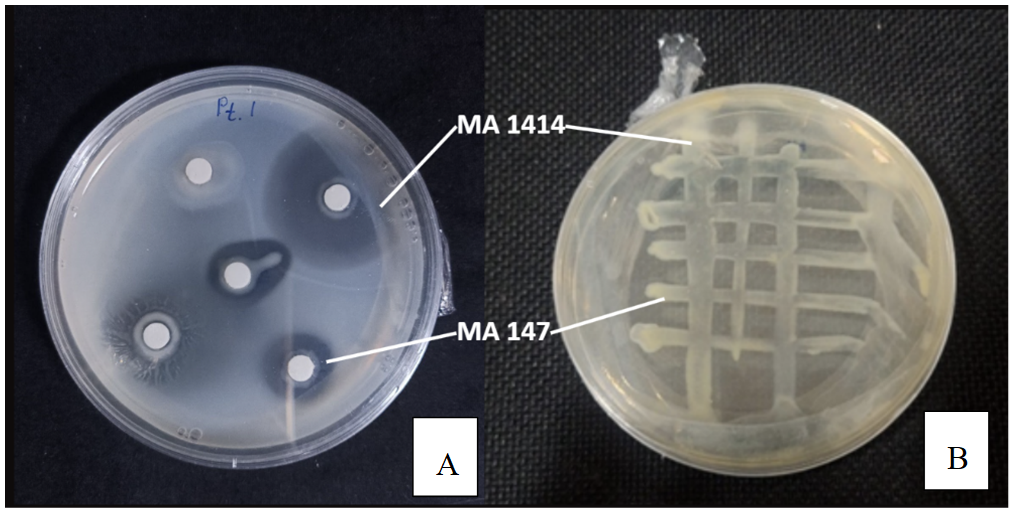
Observation of extracellular enzyme activity after 36 hours of incubation revealed the formation of hydrolysis zones around the colonies in both selected isolates. The observation was carried out using disposable Petri dishes with a 90 mm diameter and a 6 mm paper disc inoculated with bacterial culture. Proteolytic activity was evaluated by measuring the width of the clear hydrolysis zone extending beyond the edge of the paper disc. The inhibition or hydrolysis zones were clear and well defined, indicating the secretion of extracellular enzymes capable of degrading the skimmed milk in the medium. Visually, the diameter of the zones formed differed between isolates, consistent with the quantitative measurement of the enzyme index presented earlier.
Evaluation of interactions with the consortia test showed that both isolates could grow on the same medium, with no visible inhibition zone observed between the two isolates under the tested conditions, as shown in Figure 1. The colony growth pattern appeared close together, showing no visual indication of antagonism. No significant changes in growth morphology were observed compared with single cultures. These results suggest the absence of antagonistic interactions.
3.3 Molecular identification of selected bacterial isolates
The bacterial isolates with the highest enzymatic activity, namely MA147 and MA1414, were further analysed molecularly through amplification of the 16S rRNA gene using the PCR method. Genomic DNA from selected isolates was extracted using the Quick-DNA Fungal/Bacterial Miniprep Kit, and the 16S rRNA gene was amplified using universal primers 27F (5′-AGAGTTTGATCMTGGCTCAG-3′) and 1429R (5′-GGTTACCTTGTTACGACTT-3′) with MyTaq HS Red Mix 2×. The PCR electrophoresis results shown in Figure 2 indicate the formation of a single, clear DNA band in the range of approximately 1500 bp in both isolates.
The two isolates with the highest enzymatic activity were further analysed through amplification of the 16S rRNA gene using the PCR method. The electrophoresis results showed the formation of a single DNA band in the range of approximately 1500 bp in each isolate. The size of these fragments corresponds to the length of the 16S rRNA gene in bacteria, indicating the success and specificity of the amplification process.

Figure 2. Electrophoresis results of selected bacterial isolates
The PCR products were then sequenced and analysed using BLAST in the GenBank database. A summary of the identification results is presented in Table 3. One isolate showed the highest sequence similarity to Vibrio tubiashii, a query cover of 100%, and a percent identity of 100%. The second isolate showed the highest sequence similarity to Vibrio owensii, a query cover of 100%, and a percent identity of 99.9%. A query cover value of 100% in both isolates indicates that the entire length of the amplified sequence is aligned with the reference sequence in the database. An identity percentage above 99% indicates a very high genetic similarity to the corresponding reference sequences in the GenBank database. However, because closely related Vibrio species may share highly similar 16S rRNA gene sequences, these results are interpreted to indicate that the isolates are closely related to V. tubiashii and V. owensii.
Table 3. Molecular identification of selected isolates
|
Isolate |
Host Species |
Closest GenBank Match |
Query Cover (%) |
P. Ident (%) |
GenBank Accession Number |
|
MA147 |
Mauritia arabica (Linnaeus, 1758) |
Vibrio tubiashii |
100 |
100% |
PZ368869 |
|
MA1414 |
Vibrio owensii |
100 |
99.9% |
PZ368914 |
Based on these results, the two isolates exhibiting the highest enzymatic activity were assigned to the genus Vibrio and designated as Vibrio sp. MA147 and Vibrio sp. MA1414. BLAST analysis of the 16S rRNA gene sequences showed the highest similarity to V. tubiashii and V. owensii, respectively. The corresponding sequences have been deposited in GenBank under accession numbers PZ368869 (MA147) and PZ368914 (MA1414). These isolates were subsequently selected for secondary metabolite profile analysis.
3.4 Gas Chromatography–Mass Spectrometry results
GC–MS analysis of the MA147 isolate extract produced 19 compound peaks with a total area of 3,619,062.134 counts·min, as shown in Figure 3. The chromatogram profile contained multiple detected peaks; however, only compounds with an SI ≥ 800 were retained for interpretation. Based on this criterion, the metabolite profile was dominated by 10-octadecenoic acid, methyl ester.

Figure 3. Gas Chromatography–Mass Spectrometry (GC–MS) profile of metabolites detected in isolate MA147
The peak with the highest relative area percentage was detected at an RT of 19.15 minutes and was identified as 10-octadecenoic acid, methyl ester (C₁₉H₃₆O₂), with an SI value of 835 and a relative area of 46.61%. This compound is a derivative of long-chain unsaturated fatty acids.
The predominance of the identified FAMEs suggests that lipid-associated metabolic pathways contribute substantially to the metabolite profile of isolate MA147. These compounds are known to play a role in cell membrane structure and may exhibit certain biological activities, including antibacterial and anti-inflammatory properties [28].
Based on the retained high-confidence compound identification, the metabolite profile of isolate MA147 is dominated by 10-octadecanoic acid methyl ester, indicating the predominance of a lipid-associated metabolite. This suggests strong metabolic potential in the lipid pathway, which may correlate with certain biological activities and possible contributions to the adaptation mechanisms of symbiotic bacteria in the marine environment [27].
GC–MS analysis of the MA1414 isolate extract produced 49 compound peaks with a total area of 1,889,882.720 counts·min, as shown in Figure 4. The chromatogram profile showed a dominance of fatty acids and their derivatives, particularly in the form of methyl esters, similar to the metabolic pattern observed in the previous isolate.

Figure 4. Gas Chromatography–Mass Spectrometry (GC–MS) profile of metabolites detected in isolate MA1414
The peak with the highest relative area percentage was detected at an RT of 17.20 minutes and was identified as 9-hexadecenoic acid, methyl ester (C₁₇H₃₂O₂), with an SI of 935 and a relative area of 27.60%, as shown in Table 4. This compound is a derivative of palmitic acid, commonly found as a component of microbial lipids.
Table 4. Comparative Gas Chromatography–Mass Spectrometry (GC–MS) profiles of Vibrio sp. MA147 and Vibrio sp. MA1414 isolates
|
Isolate |
Retention Time (RT) (min) |
Relative Area (%) |
SI |
Compound Name |
Molecular Formula |
|
Vibrio sp. MA147 |
19.15 |
46.61 |
835 |
10-Octadecenoic acid methyl ester |
C₁₉H₃₆O₂ |
|
Vibrio sp. MA1414 |
17.20 |
27.60 |
935 |
9-Hexadecenoic acid methyl ester |
C₁₇H₃₂O₂ |
|
17.39 |
13.34 |
849 |
Hexadecanoic acid methyl ester |
C₁₇H₃₄O₂ |
|
|
19.05 |
24.86 |
912 |
Trans-13-Octadecenoic acid methyl ester |
C₁₉H₃₆O₂ |
The next dominant peak appeared at an RT of 19.05 minutes as trans-13-octadecenoic acid, methyl ester (C₁₉H₃₆O₂), with an SI of 912 and a relative area of 24.86%. Additionally, hexadecanoic acid, methyl ester (C₁₇H₃₄O₂) was detected at an RT of 17.39 minutes with a relative area of 13.34% and an SI of 849. The identified compounds indicate that the metabolite profile of isolate MA1414 was dominated by long-chain saturated as well as unsaturated FAMEs.
Additional compounds with SI values below 800 were excluded from further analysis because they did not meet the confidence threshold established for compound identification.
Overall, the metabolite profile of isolate MA1414 was dominated by saturated and unsaturated FAMEs. Consistent with the profile observed in MA147, major compounds detected in both isolates include FAMEs, particularly 10-octadecenoic acid, hexadecanoic acid methyl ester, and trans-13-octadecenoic acid methyl ester in MA1414, which all exhibited the highest relative peak areas. These compounds represented the major metabolite class detected in the bacterial extracts.
The morphological data presented in Table 1 indicate a high level of phenotypic diversity among gastropod-associated symbiotic bacteria, which may reflect differences in metabolic and physiological capacities. Such variability provides a critical foundation for downstream selection, as phenotypic traits are frequently associated with functional capabilities, including the production of extracellular enzymes and secondary metabolites in marine bacteria [10]. Thus, the observed morphological diversity is not merely descriptive but serves as an early indicator of the potential biotechnological value of individual isolates.
This functional relevance is further supported by the proteolytic activity screening results in Table 2, which reveal clear differences in enzymatic performance among isolates. The data show that not all isolates exhibited detectable activity, indicating heterogeneity in metabolic expression within the symbiotic bacterial community. The absence of hydrolysis zones in certain isolates suggests either limited protease production under the tested conditions or the requirement for specific environmental cues to induce enzyme expression [29].
In contrast, isolates such as MA147 and MA1414 demonstrated a significant increase in hydrolysis zone diameter over the incubation period, reflecting high and sustained proteolytic activity. The progressive enlargement of these zones indicates continuous enzyme secretion and efficient extracellular degradation of protein substrates. This pattern suggests stable extracellular enzyme production and efficient protein substrate degradation [7]. Furthermore, the marked increase in activity after 18 hours of incubation may indicate a transition from the adaptation phase to the exponential phase of enzyme production, which is typically associated with enhanced metabolic activity under favourable nutrient conditions in marine bacteria [10].
The swarming behaviour observed in certain isolates, such as MA164, also suggests high motility, which is often associated with bacteria from the genus Vibrio. This motility can influence the spatial distribution of colonies and the diffusion dynamics of extracellular enzymes within the medium, potentially affecting the visibility and uniformity of hydrolysis zones [30]. Therefore, the interpretation of enzymatic activity should consider not only zone diameter but also biological factors such as motility and growth patterns.
Taken together, the results presented in Table 2 demonstrate that only a subset of isolates exhibits strong enzymatic potential, emphasising the importance of selective screening in identifying candidates for further analysis, including GC–MS-based metabolite profiling. The formation of hydrolysis zones on the assay medium indicates that both isolates are capable of secreting extracellular protease enzymes that actively degrade protein substrates. As observed after 36 hours of incubation, the presence of clear and well-defined zones surrounding the colonies reflects effective enzyme diffusion into the medium and efficient extracellular protein degradation.
In marine bacteria, the production of extracellular enzymes represents an adaptive strategy to acquire nitrogen and carbon sources from environments that are relatively low in dissolved nutrients [18]. Such nutrient-limited conditions are characteristic of tropical marine ecosystems, where fluctuating resource availability necessitates the utilisation of hydrolytic enzymes to access complex organic substrates [28]. The enzymatic activity detected from the mid to late incubation phase suggests that enzyme production likely occurs during the late exponential or early stationary phase, when nutrient competition becomes more pronounced [10]. This temporal pattern reflects typical metabolic regulation in marine bacteria, where extracellular enzyme expression is triggered by nutrient limitation and coordinated through quorum-sensing mechanisms [31].
Variations in hydrolysis zone diameter among isolates indicate differences in secretion capacity and catalytic efficiency. The visual differences, which are consistent with the measured enzyme index values, indicate that the size of the clear zone can serve as a relative quantitative indicator of protease activity. Such variability may arise from differences in genetic regulation, growth kinetics, and protein secretion systems across bacterial taxa [19]. In the context of marine symbiotic bacteria, the ability to produce proteolytic enzymes is often associated with ecological roles in degrading complex organic matter surrounding the host, thereby contributing to localized nutrient cycling on host surfaces [10]. Additionally, enhanced proteolytic capability may confer a competitive advantage by enabling more efficient nutrient acquisition compared to coexisting microorganisms within the same microhabitat [5].
The consortium interaction assay revealed no formation of inhibition zones or clear growth boundaries, indicating the absence of strong antagonistic interactions between the two isolates. The closely associated colony growth pattern, without the appearance of an inhibition line, further suggests their compatibility under the consortium conditions. This pattern suggests a neutral or compatible relationship in artificial culture conditions. In marine microbial systems, non-antagonistic interactions are commonly observed among communities originating from the same microhabitat, reflecting long-term co-existence and ecological adaptation [18]. Such interactions are consistent with the concept of stable microbial assemblages, in which interspecies relationships tend to be cooperative or non-inhibitory [32].
This observation suggests that the consortium conditions do not suppress enzymatic expression and may instead contribute to maintaining metabolic stability. In some studies, bacterial consortia have been shown to enhance substrate degradation efficiency through metabolic complementation or cross-regulation mechanisms among species [18]. Furthermore, synergistic interactions within microbial communities may stimulate enzyme-related gene expression through cell-to-cell communication processes such as quorum sensing [16].
These findings demonstrate that both isolates possess not only strong individual proteolytic capabilities but also physiological compatibility in co-culture systems. The combination of high enzymatic activity and non-antagonistic interaction represents a key characteristic for the development of microbial consortia in biotechnological applications. Such stability is particularly advantageous, as microbial consortia often outperform single cultures in biomolecule degradation and in the production of industrial enzymes and bioactive compounds [4].
Molecular identification based on the 16S rRNA gene confirmed that the two isolates with the highest proteolytic activity belong to the genus Vibrio. Electrophoretic analysis revealed a single DNA band of approximately 1500 bp, indicating successful and specific amplification of the target gene, consistent with the typical length of bacterial 16S rRNA sequences. With a query cover of 100% and sequence identity above 99%, the taxonomic assignment can be considered highly reliable [23]. Although 16S rRNA gene sequence similarity above 98.7–99% is commonly used as an operational criterion for species‑level assignment, ecological and genomic studies have revealed cryptic species complexes that remain unresolved at this threshold, thereby providing a robust molecular foundation for downstream biological interpretation [33].
The isolates were identified as closely related to Vibrio sp. MA147 (V. tubiashii) and Vibrio sp. MA1414 (V. owensii) indicates that the selected gastropod-associated symbiotic bacteria belong to a group known for high metabolic plasticity. The genus Vibrio comprises facultative marine bacteria capable of adapting to a wide range of salinity and nutrient conditions and is characterised by efficient protein secretion systems for extracellular enzyme production [19]. In addition, members of this genus possess dynamic and flexible genomes, enabling rapid responses to environmental fluctuations and facilitating the expression of genes involved in metabolism and ecological adaptation [34]. These findings reinforce the correlation between genetic identity and enzymatic performance observed during the screening stage.
Such a relationship is particularly relevant in the context of biotechnological exploration, where enzymatic traits often reflect broader metabolic capabilities. Protease production by Vibrio spp. not only serves as a mechanism for nutrient acquisition but also reflects broader metabolic capabilities. In marine environments, hydrolytic enzymes play a key role in the decomposition of complex organic matter and contribute to nitrogen and carbon biogeochemical cycles [10]. Accordingly, the high proteolytic activity observed in the selected isolates in Table 2 can be interpreted as an indicator of integrated metabolic potential rather than solely an artefact of in vitro conditions. This suggests that the enzymatic activity is closely linked to ecological adaptation and may be associated with the biosynthesis of secondary metabolites.
Furthermore, several studies have reported that Vibrio species are capable of producing a wide range of bioactive compounds, including fatty acid derivatives, cyclic peptides, and small molecules with antibacterial and antifouling activities [13]. Recent genomic analyses have also identified diverse biosynthetic gene clusters (BGCs) within Vibrio, supporting their capacity to synthesise structurally diverse secondary metabolites with potential pharmacological relevance [16]. The identification of the most enzymatically active isolates within this genus provides a strong biological rationale for proceeding with GC–MS-based metabolite profiling. In this context, molecular identification extends beyond taxonomic classification and functions as a predictive framework for assessing biosynthetic potential.
This finding supports the hypothesis that gastropod-associated symbiotic bacteria represent a potential source of microbial candidates for biotechnological applications, particularly in enzyme production and bioactive compound discovery. By integrating molecular identification Table 3 with enzymatic activity and metabolic profiling, this approach demonstrates the effectiveness of function-driven selection in marine microbial exploration. Consequently, the molecular identification stage serves as a conceptual bridge between enzymatic screening and subsequent chemical analysis of bioactive compounds.
GC–MS analysis of the two selected isolates, V. tubiashii (MA147) and V. owensii (MA1414), revealed metabolite profiles dominated by composed identified as of long-chain FAMEs, as clearly illustrated in Figures 3 and 4. Only compounds with an SI ≥ 800 were retained. Based on this criterion, the consistent chromatographic patterns observed across both figures indicate a conserved metabolic orientation toward lipid biosynthesis.
The presence of octadecenoic acid and hexadecanoic acid methyl ester as major components suggests that fatty acid biosynthesis represents the dominant metabolic pathway in both isolates. As shown in Figure 3, isolate MA147 exhibited a pronounced dominance of 10-octadecenoic acid methyl ester, whereas MA1414 contained both saturated and unsaturated FAMEs, including hexadecanoic acid methyl ester and trans-13-octadecanoic acid methyl ester.
In marine bacteria, unsaturated fatty acids play a crucial role in maintaining membrane fluidity in response to osmotic pressure and temperature fluctuations in coastal environments. Such adaptive mechanisms are essential for preserving membrane integrity and cellular function under dynamic marine conditions [33, 34]. Consequently, the high proportion of oleic and palmitic acid derivatives likely reflects physiological adaptation strategies to tropical marine habitats.
From a biotechnological perspective, the predominance of compounds identified as FAMEs suggests active lipid-associated metabolic pathways in the selected isolates. Long-chain FAMEs are not only structural components of cellular membranes but have also been reported to exhibit biological activities, including antibacterial, anti-inflammatory, and surface-active properties. Compounds such as hexadecanoic acid methyl ester have been associated with antibacterial activity in various marine microbial extracts [10]. In addition, fatty acid derivatives are increasingly recognised for their ability to disrupt microbial membranes and modulate inflammatory responses, highlighting their potential in pharmaceutical and cosmeceutical applications [4].
Although both isolates exhibited lipid-dominated metabolite profiles, distinct differences in relative composition were observed. Both chromatograms were dominated by compounds identified as FAMEs. Among the retained high-confidence identifications (SI ≥ 800), isolate MA147 was characterised predominantly by 10-octadecenoic acid methyl esters. Meanwhile, isolate MA1414 contained 9-hexadecenoic acid methyl ester, hexadecanoic acid methyl ester, and trans-13-octadecenoic acid methyl ester.
This contrast suggests differences in metabolic regulation, enzyme expression, or adaptive strategies between species within the same genus [32]. Such variation may also indicate the potential for differential biological activity, which warrants further investigation in downstream bioassays.
Compounds identified with lower confidence and commonly reported analytical contaminants, particularly siloxane derivatives, were excluded from biological interpretation to ensure a conservative interpretation of the GC–MS data.
Taken together, the dominance of FAMEs in both isolates reinforces the central theme and literature background of this study: enzymatic activity-based selection effectively identifies bacteria with high metabolic potential, as reflected in their lipid-associated metabolite profiles. However, this study has several limitations that should be considered. Each gastropod host species was represented by a single specimen, which limits the generalizability of the observed bacterial diversity and enzymatic characteristics. The bacterial community associated with individual hosts may vary according to environmental conditions, host physiology, and geographic location. Therefore, the present findings should be regarded as preliminary and require further validation using larger sample sizes, multiple biological replicates, and broader geographic sampling.
Furthermore, GC–MS analysis was performed without derivatization and therefore provides only a partial metabolite profile that does not represent the full metabolic capacity of the isolates. Polar, thermally unstable, and high-molecular-weight metabolites are likely underrepresented under the analytical conditions employed. Accordingly, the detected metabolites should be interpreted as a preliminary characterization of the volatile and semi-volatile metabolite composition rather than a comprehensive metabolomic profile.
The integration of enzymatic screening (Table 2), molecular identification (Table 3), and metabolite profiling (Figures 3 and 4) highlights the effectiveness of a function-driven selection strategy. In the context of gastropod symbiosis, lipid-associated metabolites may contribute to chemical defence, microbial competition, and the stability of host–microbe interactions. These compounds may also play roles in chemical signalling and antimicrobial defence within the host microenvironment [2].
This study demonstrates that the preliminary screening approach combining enzymatic screening, molecular identification, and GC–MS analysis can be used to determine the enzymatic and metabolic potential of gastropod-associated symbiotic bacteria. A total of 21 bacterial isolates were obtained from three marine gastropod species: M. arabica, C. tigris, and C. vitellus, with one individual host specimen each. The isolates exhibiting the highest proteolytic activity were identified as V. tubiashii (MA147) and V. owensii (MA1414), which originated from M. arabica, showed higher proteolytic activity compared to other isolates and was further characterised for GC–MS analysis.
Metabolite profiling further showed that both isolates are dominated by long-chain FAMEs, particularly hexadecenoic acid methyl ester and octadecanoic acid derivatives, indicating active lipid biosynthesis pathways that may contribute to ecological adaptation and bioactivity. These findings indicate gastropod-associated Vibrio spp. may serve as preliminary candidates for further investigation for enzyme production and metabolite-based applications. Additional quantitative enzyme assays, purification studies, and bioactivity validation may be required to expand these observations.
The authors extend their sincere appreciation to the leadership of Diponegoro University for their continued support throughout the completion of this work.
[1] Sousa, R. (2024). A brief global agenda for advancing the study of molluscs. Frontiers in Ecology and Evolution, 12: 1176380. https://doi.org/10.3389/fevo.2024.1176380
[2] Zhukova, N.V., Eliseikina, M.G., Balakirev, E.S., Ayala, F.J. (2022). Multiple bacterial partners in symbiosis with the nudibranch mollusk Rostanga alisae. Scientific Reports, 12(1): 169. https://doi.org/10.1038/s41598-021-03973-7
[3] Zaman, W., Ayaz, A., Puppe, D. (2025). Biogeochemical cycles in plant–soil systems: Significance for agriculture, interconnections, and anthropogenic disruptions. Biology, 14(4): 433. https://doi.org/10.3390/biology14040433
[4] Rojas-Villalta, D., Núñez-Montero, K., Pizarro-Cerdá, J., Chavarría-Pizarro, L. (2023). Draft genome sequences of Tsukamurella sp. 8F and 8J strains isolated from social wasps (Vespidae; Polistinae: Epiponini). Microbiology Resource Announcements, 12(6): 9-10. https://doi.org/10.1128/mra.00237-23
[5] Maghraby, Y.R., El-shabasy, R.M., Ibrahim, A.H., Azzazy, H.M.E. (2023). Enzyme immobilization technologies and industrial applications. ACS Omega, 8: 5184-5196. https://doi.org/10.1021/acsomega.2c07560
[6] David Troncoso, F., Alberto Sánchez, D., Luján Ferreira, M. (2022). Production of plant proteases and new biotechnological applications: An updated review. ChemistryOpen, 11(3): e202200017. https://doi.org/10.1002/open.202200017
[7] Song, P., Zhang, X., Wang, S., Xu, W., Wang, F., Fu, R., Wei, F. (2023). Microbial proteases and their applications. Frontiers in Microbiology, 14: 1236368. https://doi.org/10.3389/fmicb.2023.1236368
[8] Pringgenies, D., Soenardjo, N., Setyati, W.A., Pramesti, R. (2024). Secondary metabolites from symbiotic bacteria of seagrass and molluscs as a reference for natural food preservatives. Trends in Sciences, 21(5): 7404. https://doi.org/10.48048/tis.2024.7404
[9] Surya, S., Pringgenies, D., Pathak, Y., Sedjati, S., Brotosudarmo, T.H.P., Ariyanto, D., Mailoa, M.N. (2026). Enzymes from mollusk-associated bacteria in Ambon waters of Wallacea: Prospective cosmetic applications. Open Veterinary Journal, 16(3): 1704-1715. https://doi.org/10.5455/OVJ.2026.v16.i3.28
[10] Surya, S., Pringgenies, D., Sedjati, S. (2025). Antibacterial and enzymatic activities of symbiotic bacteria from gastropods and bivalves in marine skincare applications. International Journal of Design and Nature and Ecodynamics, 20(1): 83-90. https://doi.org/10.18280/ijdne.200109
[11] Su, H., Jiang, W., Verweij, P.E., Li, L., Zhu, J., Han, J., Deng, S. (2023). The in vitro activity of echinocandins against clinical Trichophyton rubrum isolates and review of the susceptibility of T. rubrum to echinocandins worldwide. Infection and Drug Resistance, 2023: 5395-5403. https://doi.org/10.2147/IDR.S423735
[12] Mahmud, M.K., Rahman, M.M., Chy, M.A.H., Kabir, M.R., Nazmul, K.M., Akhter, M.K., Chowdhury, M.H. (2025). Isolation and characterization of soil bacteria: A review. International Journal of Modern Science and Research Technology, 3(10): 22-33.
[13] Pringgenies, D., Wilis, A.S., Feliatra, F., Ariyanto, D. (2024). The antibacterial and antifungal potential of marine natural ingredients from the symbiont bacteria of mangrove. Global Journal of Environmental Science and Management, 9(4): 819-832. https://doi.org/10.22035/gjesm.2023.04.11
[14] Tarigan, D.M., Barus, W.A., Munar, A., Lestami, A. (2023). Exploration and morphological characteristics of phosphate solubilizing and nitrogen-fixing bacteria in saline soil. SABRAO Journal of Breeding and Genetics, 55: 550-563.
[15] Dutta, D. (2024). Bioprocess strategies for enhanced performance in single-use bioreactors for biomolecule synthesis: A biokinetic approach. Food and Bioengineering. https://doi.org/10.1002/fbe2.12104
[16] Simpson, C.A., Celentano, Z.R., Haas, N.W., et al. (2024). Quorum sensing in Vibrio controls carbon metabolism to optimize growth in changing environmental conditions. PLoS Biology, 22(11): e3002891. https://doi.org/10.1371/journal.pbio.3002891
[17] Zafrida, S., Ethica, S.N., Ernanto, A.R., Wijanarka, W. (2022). Optimization of crude protease production from Bacillus thuringiensis HSFI-12 and thrombolytic activity its enzyme dialysate. Trends in Sciences, 19(23): 1952. https://doi.org/10.48048/tis.2022.1952
[18] Dahdah, K., Charchar, N., Bouchaala, L., Nourine, H., Belkabla, N., Melo, J., Nabti, E.H. (2022). Isolation, in vitro and construction of versatile microbial consortia. Cellular and Molecular Biology, 68(8): 173-181. https://doi.org/10.14715/cmb/2022.68.8.31
[19] Akram, M.A., Savitha, R., Kinsella, G.K., Nolan, K., Ryan, B.J., Henehan, G.T. (2024). Microbial and enzymatic biodegradation of plastic waste for a circular economy. Applied Sciences, 14(24): 11942. https://doi.org/10.3390/app142411942
[20] Obaretin, N.N., Daokoru-Olukole, C.G., Pondei, J.O., et al. (2024). Molecular assessment of dominant genus in heavy metal contaminated soil from selected dumpsites in Port Harcourt, Nigeria. Asian Journal of Environment and Ecology, 23(8): 128-139. https://doi.org/10.9734/ajee/2024/v23i8589
[21] Regueira-Iglesias, A., Balsa-Castro, C., Blanco-Pintos, T. (2023). Critical review of 16S rRNA gene sequencing workflow in microbiome studies: From primer selection to advanced data. Molecular Oral Microbiology, 38(5): 347-399. https://doi.org/10.1111/omi.12434
[22] Nwe, M.K., Jangpromma, N., Taemaitree, L. (2024). Evaluation of molecular inhibitors of loop-mediated isothermal amplification (LAMP). Scientific Reports, 14(1): 5916. https://doi.org/10.1038/s41598-024-55241-z
[23] Yang, S., Chen, S., Mo, J., Liu, J., Pan, Q., Song, C. (2024). Application of DNA barcoding to identify medicinal plants. JoVE Journal of Visualized Experiments, 213: e66925. https://doi.org/10.3791/66925
[24] Mohd Kamal, K., Mahamad Maifiah, M.H., Abdul Rahim, N., Hashim, Y.Z. H.Y., Abdullah Sani, M.S., Azizan, K.A. (2022). Bacterial metabolomics: Sample preparation methods. Biochemistry Research International, 2022(1): 9186536. https://doi.org/10.1155/2022/9186536
[25] Russo, F.F., Benner, P., Parr, M.K., Lisec, J. (2024). Machine learning methods for compound annotation in non-targeted mass spectrometry — A brief overview of fingerprinting, in silico fragmentation and de novo methods. Rapid Communications in Mass Spectrometry, 38(20): e9876. https://doi.org/10.1002/rcm.9876
[26] Banerjee, P., Emanuel, K., Dunkel, M., Preissner, R. (2024). Reductions in cod and acute ProTox 3.0: A webserver for the prediction of toxicity of chemicals. Nucleic Acids Research, 52(W1): W513-W520. https://doi.org/10.1093/nar/gkae303
[27] Verpoorte, R., Kim, H.K., Choi, Y.H. (2022). Trivialities in metabolomics: Artifacts in extraction and analysis. Frontiers in Molecular Biosciences, 9: 972190. https://doi.org/10.3389/fmolb.2022.972190
[28] Xu, B., Kong, L., Sun, J., Zhang, J., et al. (2025). Molluscan systematics: Historical perspectives and the way ahead. Biological Reviews, 100(2): 672-697. https://doi.org/10.1111/brv.13157
[29] Mangena, P. (2022). Pleiotropic effects of recombinant protease inhibitors in plants. Frontiers in Plant Science, 13: 994710. https://doi.org/10.3389/fpls.2022.994710
[30] Baquero, F., Coque, T.M., Bastón-Paz, N., Pérez-Cobas, A.E. (2025). Biophysics of Bacterial Colonial Structures and the Occupancy of Microecological Spaces. Biology, 15(1): 56. https://doi.org/10.3390/biology15010056
[31] Rajpurohit, H., Eiteman, M.A. (2022). Nutrient-limited operational strategies for the microbial production of biochemicals. Microorganisms, 10(11): 2226. https://doi.org/10.3390/microorganisms10112226
[32] Pappalardo, P., Hemmi, J.M., Machida, R.J., Leray, M., Collins, A.G., Osborn, K.J. (2025). Taxon-specific BLAST percent identity thresholds for identification of unknown sequences using metabarcoding. Methods in Ecology and Evolution, 16(10): 2380-2394. https://doi.org/10.1111/2041-210X.70147
[33] Wani, A.K., Akhtar, N., Sher, F., Navarrete, A.A., Américo-Pinheiro, J.H.P. (2022). Microbial adaptation to different environmental conditions: Molecular perspective of evolved genetic and cellular systems. Archives of Microbiology, 204(2): 144. https://doi.org/10.1007/s00203-022-02757-5
[34] Gerhards, R., Seston, R.M., Kozerski, G.E., McNett, D.A., Boehmer, T., Durham, J.A., Xu, S. (2022). Basic considerations to minimize bias in collection and analysis of volatile methyl siloxanes in environmental samples. Science of the Total Environment, 851: 158275. https://doi.org/10.1016/j.scitotenv.2022.158275