Eco-Friendly Synthesis of Cobalt Oxide Nanoparticles Using Capparis spinosa Fruit Extract and Their Antibacterial Application

Marwa Waleed Mahmood | Nada K. Abbas*![]()
© 2025 The authors. This article is published by IIETA and is licensed under the CC BY 4.0 license (http://creativecommons.org/licenses/by/4.0/).
OPEN ACCESS
In this work, we report the synthesis of cobalt oxide nanoparticles prepared by green synthesis using Caparison spinosa. The extract and chemical method using cobalt chlorides. The product was described by means of energy-dispersive X-ray spectroscopy (EDX), field emission scanning electron microscopy (FESEM), X-ray diffraction (XRD), UV-visible (UV-Vis) spectroscopy, and Fourier-transform infrared spectroscopy (FTIR). The sample made using the green approach had a crystalline size of 14.6 nm, but the average crystal size determined by XRD using the chemical process was 47 nm. The energy band gap value for the chemically prepared sample was 2.47 eV, while the energy band gap value for the green-synthesized sample was 2.68 eV. The shape was observed by FESEM to be the same as spherical in both methods. EDX analysis indicated the existence of peaks that represent Co and O. As shown by the FTIR spectrum, peaks in the 569 cm region were due to Co and cobalt oxide. The results showed that the cobalt oxide particles prepared by the green method are better, less hazardous, and environmentally friendly than the chemical method, which allows their use in medical applications. The material also showed bactericidal activity against Staphylococcus aureus bacteria and recorded the highest inhibition diameter at a high concentration of 100% Mg/ml (27 mm), which was higher than that of Escherichia coli (E. coli), which recorded the highest inhibition diameter of (17 mm) at a concentration of 100% Mg/ml. The Co3O4 NPS prepared by green technology also showed an effective effect against the A549 cell line compared to the normal HdFn cells by measuring MTT at different concentrations, as it showed a gradual decrease in the cell survival rates the great concentration where the vital rates reached 61.99±1.90%, 75.27±3.96%, 87.26±1.68%, 93.32±0.24%, 95.25±0.91%, and 97.14±0.57% for A549 compared to the normal HdFn cells where the live cells reached (82.60±0.26%, 89.73±1.49%, 94.56±0.20%, 95.17±1.14%, 96.06±0.91%, and 98.07±0.46%) respectively. These results confirm the high biological effectiveness of the Co3O4 NPs manufactured from the extract of the Caparison spinosa plant.
Capparis fruit plant, Co3O4 NPs, green synthesis, chemical synthesis
Nanomaterials are matter in tiny forms. Metals, semiconductors, organics, and metal oxides are the "building blocks" of nanostructures. Clusters, thin films, multiple layers, and nanocrystalline materials are examples of nanomaterials exhibiting structure [1]. Since nanoscience relies on the study and use of materials, it has significantly changed several fields in the modern period at the nanoscale level (1 to 100 nanometers). Thanks to this progress, advanced technologies have been developed in the fields of industry, energy, electronics, and medicine [2]. Nanomaterials are characterized by their small sizes compared to large materials, which gives them distinct chemical and physical attributes, including greater surface area in relation to volume and unique optical, biological, and magnetic properties [3, 4]. Given this, nanomaterials are utilized in many different domains, including electronics, medicine, the environment, solar cells, and pharmaceuticals [5]. One-dimensional, two-dimensional, and three-dimensional Nanomaterials can be distinguished based on their structure, chemical makeup, and methods of synthesis. [6]. The methods of manufacturing nanomaterials vary between chemical methods, physical methods, and biological methods that are environmentally friendly [7]. These techniques seek to achieve the necessary characteristics by employing the size, shape, and distribution of nanoparticles [8]. There are many approaches to fabricating nanostructures, which are broadly divided into two main classes, namely bottom-up methods and top-down methods [9]. Physical techniques include spraying, physical grinding, and physical pulverization. Chemical vapor is one of the chemical processes. Chemical precipitation, sol-gel, hydrothermal, template, and deposition techniques [10]. Recently, nanotechnology has witnessed increasing interest and development, which has led to its usage in a number of industries, including energy, environmental, and medical domains [11]. The green method, which uses plant extracts from fruits, leaves, roots, and fungi, has developed as an environmentally acceptable way for creating nanomaterials [12]. The green method has several advantages, including low cost, harmlessness, environmental friendliness, low toxicity, and biocompatibility [13, 14]. Flavonoids, polyphenols, and terpenoids—active substances found in plant extracts that function as reducing agents and catalysts—are essential to the green approach [15, 16]. Many researchers have recently become interested in cobalt oxide because of its thermal and chemical stability and unique electronic, magnetic, and catalytic properties, which make it very versatile [17]. It is also used in biological applications such as cancer treatment and drug delivery [18]. One frequent transition metal oxide and significant p-type semiconductor is cobalt oxide nanoparticles [19]. Co3O4 is very effective and can be easily prepared in several ways. However, safer and more readily available methods must be used. Therefore, green chemistry methods were used, which are safer and more environmentally friendly. A safe and easy-to-prepare method for creating green nanomaterials is considered, and one of the most widely used methods for producing nanomaterials is the use of living organisms [20]. Among these organisms, plants are the ideal choice. Plant extracts are easy to use, readily available, inexpensive, and safe because they contain effective organic reducing agents [21, 22]. Plant extracts usually contain flavonoids, terpenoids, and phenols [23]. In previous studies, Capparis spinosa leaves were used in previous studies, Capparis spinosa plant was used to produce nanoparticles such as ZnO, fruits, and leaves [24]. Cobalt oxide nanoparticles were created in this study using two distinct methods: the chemical approach and the green method, which uses the fruits of the Capparis spinosa plant to reduce and coat the nanoparticles. This study's objective is to create cobalt oxide nanoparticles via a biological process and assess their biological and medicinal properties.
2.1 Preparation of extracts from Capparis spinosa fruits
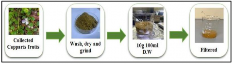
Capparis spinosa fruits were gathered from the northern Diyala region, repeatedly cleaned with tap water, deionized water, let to dry at room temperature, and then pulverized using a pestle. A magnetic stirrer was used to dissolve 10 grams of the plant in 100 milliliters of deionized water (Model: VS-130Sh, Made in Korea) at a speed of 4000 rpm and a temperature of 60℃ for 3 hours. After that, it was filtered by a centrifuge (Model 1710 made in Germany) for 20 minutes, followed by filter paper, and the extract was kept at 4℃ for storage (Figure 1).
Figure 1. The steps of extract preparation
2.2 Synthesis of nano cobalt oxide by the biological method
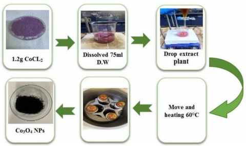
First, nano cobalt oxide was prepared by taking 25 ml of the previously prepared plant extract and distilling it in 0.1 M of 75 ml cobalt chloride and stirring it with a magnetic stirrer at a speed of 400 and a temperature of 60℃ for 3 hours until a color change was observed. After that, it was centrifuged for 20 minutes and washed twice with deionized water and once with alcohol. Figure 2 illustrates preparing pure Co3O4 NPs using Capparis spinosa fruits by the green method.
Figure 2. The steps to prepare the pure Co3O4 NPs using Capparis spinosa fruits using a green approach
2.3 Manufacturing of nano-cobalt oxide by chemical method
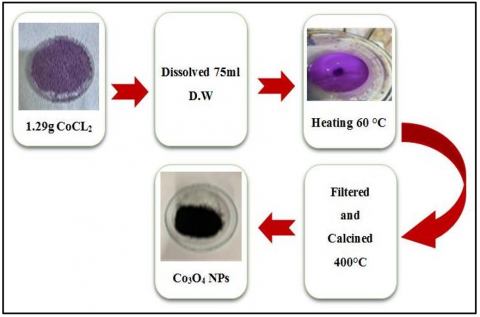
Cobalt chloride and sodium hydroxide were used as raw materials to prepare cobalt oxide nanoparticles. After dissolving 1.29 g in 100 milliliters of 0.1 M deionized water, 1 M NaOH was added. Dropwise and continuously stirred using a magnetic stirrer at 60℃ and observed for 3 h. A color change and the formation of a suspension. After that, the sample was filtered using filter paper and then cleaned with a magnetic stirrer for 20 minutes twice with deionized water and once with alcohol. After that, the dough was dried at 60℃, and then it was ground with a manual pestle. Figure 3 illustrates preparing pure Co3O4 NPs by the chemical precipitation method.
Figure 3. The preparation of the pure Co3O4 NPs using the chemical precipitation method
2.4 Bacterial preparation
The bacteria (Staphylococcus aureus and Escherichia coli (E. coli)) had been obtained from the Iraqi Ministry of Health Bacteriological Laboratory, and the concentration of bacteria was examined by use of the McFarland standard at a concentration of 1.5 × 10⁸. The organisms' pure cultures were subcultured on Müller-Hinton broth at 37℃ while spinning at 200 rpm on a rotary shaker. A sterile well borer was used to create a 6-mm-diameter well on Müller-Hinton agar plates. Every strain was evenly swabbed onto the two separate plates or each type of bacteria using sterile cotton swabs each well in each plate for the same bacterial test were receive three different concentrations of prepared cobalt oxide nanoparticles as 100%, 75% and 50% of the original cobalt oxide nanoparticles solution at same volume into each well (20 μL), distilled water had been used as control. After incubation for 24 hours at 37℃. The inhibition zone of each different cobalt oxide nanoparticle concentration (100%, 75% and 50%) was measured for the two types of bacteria mentioned. The minimum and maximum concentration of nanoparticles that prevent microbial growth was designated as the MIC.
2.5 MTT protocol
Cancer cells, Lung cancer (A549), and normal human dermal fibroblasts (HdFn). were transplanted into 96-well plates with a concentration (1×10⁴ - 1×10⁶ cells/ml) and a final size 200 microliters after full transplantation, then incubated at 37℃ with 5% CO2 for 24 hours. The sequential concentrations of compounds (400-25 mg/ml) were then added three bis with controls, and the cells were exposed for the specified period. After the treatment, 10 microliters of MTT solution were added and the dishes were incubated for 4 hours to form formazan crystals. Remove the medium and add 100 microliters of solubilizing solution to decompose the crystals within 5 minutes. Absorbance was measured using an ELISA reader at 575 nm. Finally, the values were statistically analyzed to calculate the half inhibitory concentration (IC50) according to the logistic [25].
3.1 X-ray diffraction (XRD)
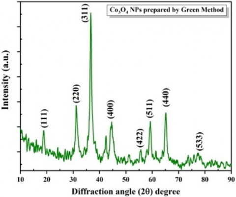
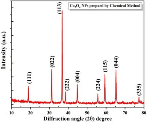
XRD analysis was performed using an XRD device from Shimadzu – Japan, with a scan speed of 5.000 (deg/min) was carried out from 2θ = 20° to 80° to study the structure of cobalt oxide nanoparticles prepared by two different techniques: the green method and the chemical method. For the green method, we see the perfect matching of the peaks at (2θ = 18.96°, 31.26°, 36.70°, 44.61°, 55.54°, 59.22°, 65.21°, 77,43°) with the standard card JCPDS (01-078-1969) for Co3O4, in agreement with Sultan et al. [26], as shown in Figure 4.
Figure 4. X-ray diffraction pattern of pure Co3O4 NPs recorded by the green and chemical methods
For the chemical method, we can see the matching of peaks at (2θ = 19.01°, 31.26°, 36.84°, 38.53°, 44.80°, 55.36°, 59.36°, 65.23°, 77.36°) with the standard card JCPDS (98-002-8158) for Co3O4, in agreement with Sabir et al. [27].
The crystally size was calculated from the Debye-Scherrer equation below [28, 29]:
$D=\frac{\mathrm{k} \lambda}{\beta \cos \theta}$ (1)
where, D is the average crystallite size, k is Scherrer's constant, β is the full-width at half-maximum (FWHM), and θ is the Bragg angle calculated based on particle size with the Debye-Scherrer equation, as shown in Tables 1 and 2. It became clear to us that the size of the cobalt oxide nanoparticles eco-friendly synthesized (14.6 nm), while the particle size was (47 nm) for those fabricated by the chemical method. This is due to the presence of organic molecules in plant extracts that serve as reductants, which limit the growth of crystals and reduce their agglomeration. The peaks of the green-prepared sample in the XRD pattern show a larger width at FWHM, indicating the presence of small-sized crystals according to the inverse Debye-Scherrer equation between FWHM and crystal size [30, 31] and Akram et al. [32].
Table 1. Crystalline size and structural parameters of bio-prepared cobalt oxide nanoparticles
|
2θ (Deg) |
FWHM (rad.) |
Crystallin Size (nm) |
(hkl) |
|
18.8913 |
0.750 |
10.73 |
(111) |
|
31.3229 |
0.6375 |
12.94 |
(220) |
|
36.755 |
0.6501 |
12.87 |
(311) |
|
37.5357 |
0.275 |
30.50 |
(222) |
|
44.7309 |
0.60 |
14.31 |
(400) |
|
59.2621 |
0.775 |
11.79 |
(511) |
|
65.1268 |
0.7417 |
11.79 |
(440) |
Table 2. Grain size and lattice parameters of Co3O4 NPs fabricated by the chemical method
|
2θ (Deg) |
FWHM (rad.) |
Crystallin Size (nm) |
(hkl) |
|
19.0174 |
0.1666 |
48.33 |
(111) |
|
31.2641 |
0.1775 |
46.45 |
(022) |
|
36.8401 |
0.1781 |
47.00 |
(113) |
|
38.533 |
0.1485 |
56.64 |
(222) |
|
44.803 |
0.2009 |
42.75 |
(004) |
|
55.667 |
0.1605 |
55.95 |
(224) |
|
59.3645 |
0.1902 |
48.05 |
(115) |
|
65.2358 |
0.2172 |
43.40 |
(044) |
3.2 Field emission scanning electron microscopy (FE-SEM)
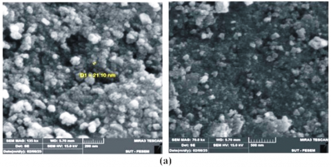
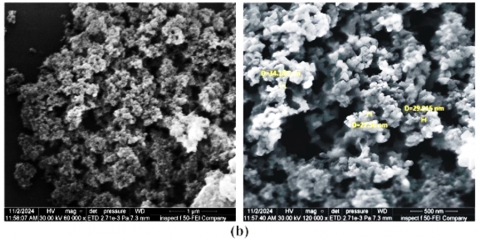
To detect and examine the morphology of the surfaces of Co3O4 NPs prepared via two separate methods, the precipitation method and the biological method, a scanning electron microscope with field emission was used, as shown in Figure 5. It was revealed through image analysis that the Co3O4 NPs manufactured by both methods were spherical in shape [30]. Also, Figure 5 shows that the Co3O4 NPs prepared by the green method are smaller in size than the particles prepared by the chemical method due to the effect of the plant extract of the Capparis fruit, which is a substance that reduces. The greater the reducing power, the greater the number of nuclei formed in a short time, which leads to the formation of a large number of nanoparticles. This is consistent with Sultan et al. [26] and Sanjeev et al. [33]. Less agglomeration of particles due to the presence of the extract, which is consistent with Barzinjy and Haji [34].
Figure 5. FE-SEM images of Co3O4 NPs made by (a) biological techniques and (b) chemical method
3.3 Energy dispersive X-ray spectroscopy (EDX)
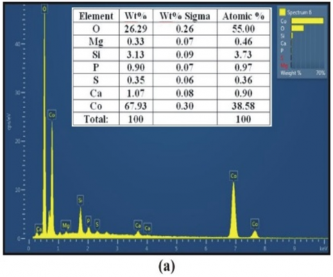
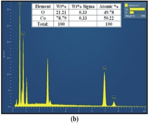
As shown in Figure 6, EDX analysis to determine the elements present in the sample confirmed the presence of characteristic peaks of nano-cobalt oxide (Co) and (O), confirming the successful formation of nano-cobalt oxide in both methods. In the green method, some spectral peaks of other elements, such as silicon, phosphorus, sulfur, and magnesium, were observed. The reason for the presence of these elements is due to the utilization of plant extracts in the process of preparation. In the chemical method, the elements (Co) and (O) are only present [30, 31].
Figure 6. EDX patterns of Co3O4 NPs synthesis by (a) biological approach and (b) chemical method
3.4 Fourier transform infrared spectroscopy
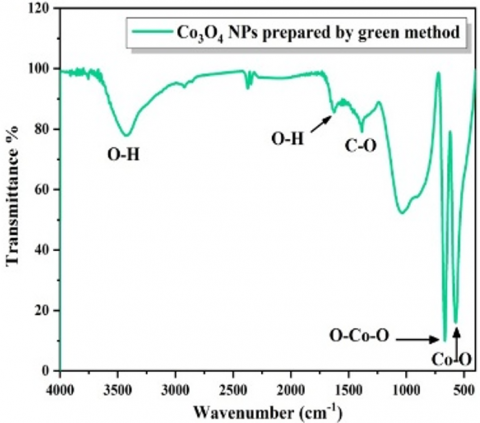
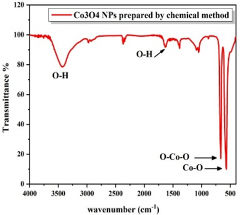
Figure 7 displays the infrared spectrum of the Fourier transform of cobalt oxide prepared from the green extract. It shows the appearance of Co-O and O-Co-O stretching bonds produced by either water or the plant extract for the synthesis process at wavelengths of 569 and 661 cm-1, respectively. These bonds result from cobalt oxide bonds, confirming the formation of nanoparticles. The peak at 1627 cm-1 is due to the symmetric O-H bond. The peak at 3435 cm-1 is attributed to the O-H bond [35]. The Fourier transform infrared (FTIR) spectrum of the chemically prepared cobalt oxide shows a Co-O symmetric Cobalt oxide is responsible for bond at 573 cm-1 and the O-Co-O symmetric bond at 675 cm-1. Additionally, a weak symmetric bond at 1638 cm-1 is seen, which is thought to be an O-H bond. The O-H bond caused by alcohol or water residues is responsible for the peak at 3429 cm-1 [36]. The FTIR with the 100 scan/sec from Perkin Elmer, TWO, USA, was used.
Figure 7. FTIR spectrum for Co3O4 nanoparticle prepared by the green and chemical methods
3.5 UV-Vis analysis
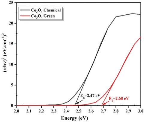
The UV-visible (UV-Vis) absorption spectra of Co3O4 NPs made using Capparis fruit extract, utilizing chemical and environmentally friendly procedures, were examined. The Tuac equation was used to determine the optical energy band gap, Eg, of Co3O4 nanoparticles [37].
$\alpha h v=B(h v-E g) r$ (2)
Figure 8. Allowed direct optical band gap using Tauc plots for Co3O4 NPs
As indicated in Figure 8, the allowed direct transitions are dominant between conduction and valence bands. In the chemical method, the results showed that. The gap energy, the value was 2.47 eV compared to the green method, which had a value of 2.68 eV. The difference in value is because of the fact that the size of the Co3O4 NPs prepared by the green method is smaller than the size of the particles prepared by the chemical method. This leads to the phenomenon that the smaller the particle size, the larger the energy gap, according to quantum confinement, and this is consistent with studies [38, 39].
3.6 Antibacterial activity of Co3O4 NPs
Many studies have shown that nanoparticles have antibacterial properties mainly due to their small size, which facilitates their penetration into bacterial cells [37, 40]. Co3O4 NPs are known to be safe, stable, and effective against many types of bacteria [41]. In particular, these Co3O4 NPs have shown promising potential as antimicrobials that combat a variety of microbes [42]. Bacteria consist of three distinct components: cytoplasm, cell wall, and plasma membrane. The cell wall is mostly made up of a consistent peptidoglycan layer, which is made up of sugars and amino acids. The osmotic pressure of the cytoplasm is maintained by the cell wall, which gives it a distinctive shape. One of the properties of spore-forming bacteria is that they consist of a thick cell wall (20-80 nm) and a single plasma membrane covered with a layer of peptidoglycan. There are two cell membranes in Gram-negative bacteria: an inner plasma membrane and an outer membrane. Between these two membranes is a thin layer of peptidoglycan, 7-8 nm thick [43].
3.7 The disk-diffusion technique of Agar
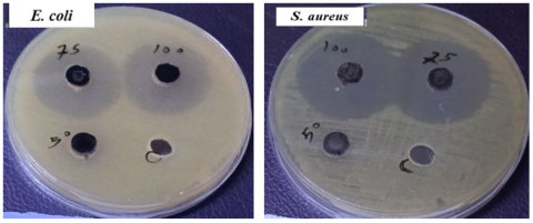
The agar disk diffusion was employed to manage Gram-negative and Gram-positive Escherichia coli and Staphylococcus aureus [44]. When using cobalt oxide nanoparticles, the concentrations used vary to affect the growth of these bacteria [41]. It was observed that the nanomaterial showed greater antibacterial activity against Gram-positive bacteria than against Gram-negative bacteria [45]. This is because the permeability of the cell wall is determined by its structure. Gram-negative bacteria have a smaller layer encircled by an outer membrane, whereas Gram-positive bacteria have a thicker peptidoglycan layer that makes it easier to access many drugs containing many lipopolysaccharides that act as an entry barrier for antibiotics [46, 47]. Table 3 shows the diameter of the inhibitory zone for bacteria using cobalt oxide nanoparticles at three different concentrations (A = 100%, B = 75%, C = 50%) M against these two types of bacteria. The results showed a comparison of the inhibition zone between the two types of bacteria for cobalt oxide nanoparticles prepared by the green method using caper plant extract. As shown in Figure 9, Co3O4 NPs prepared by the green method showed remarkable antibacterial activity. The antibacterial activity is influenced by several factors, most importantly the size, surface area, and shape of the nanomaterial. This activity occurs through mechanisms such as damaging the bacterial cell wall, inhibiting protein or nucleic acid synthesis, or disrupting enzyme activity. The prepared nanomaterial is characterized by its capacity to produce reactive oxygen species (ROS), including hydroxyl and free radicals. Bacterial cell death is the ultimate outcome of these ROS's disruption of biological processes, which damages the cell membrane, mitochondria, and DNA [48].
Figure 9. Antibacterial activity of Co3O4 NPs using disk diffusion with concentration (100%, 75%, 50%)
Table 3. Antibacterial activity of Co3O4 NPs using disk diffusion
|
Antibacterial |
Type Bacteria |
Zone of Inhibition(mm) |
||
|
Conc. (100%) Mg/ml |
Conc. (75%) Mg/ml |
Conc. (50%) Mg/ml |
||
|
Escherichia Coli |
Gram-negative |
16 |
15 |
0 |
|
Staphylococcus aureus |
Gram Positive |
27 |
26 |
0 |
The diminutive size of the nanoparticles and the increased surface area enhance their efficiency in interacting and penetrating the cell wall.
3.8 Cytotoxicity effect of Co3O4 NPs
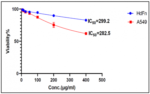
According to the findings, the cytotoxicity of the toxin with HdFn cells line (normal cell line) and A549 cell line (Lung cancer cell line)increased with increasing of the concentration of Co3O4, the killing ratio of Co3O4 on HdFn cells that 26.4%, 9.3%, 4.5%, 4.1%, 4%, 2% and 1.2 occurred with concentration: 400, 200, 100, 50, 25, 12.5 and 6.2 μg/mL respectively shown in Figure 10, the results are presented in Table 4. While the result of the killing ratio of Co3O4 ON A549 cells was 38.1%, 24.8%, 12.8%, 6.7%, 4.8 %, 2.9% and 1.4% occurred with concentration: 400, 200, 100, 50, 25, 12.5 μg/mL [49]. Cancer cells are char 400, 200, 100, 50, 25, and 12.5 μg/mL concentration acterized by their ability to absorb nanoparticles compared to normal cells. This is due to the increased permeability of their cell membranes and a disruption in the regulation of metabolic balance, which makes them more susceptible to interaction with nanomaterials that are characterized by their ability to produce ROS. Therefore, nanoparticles selectively target cancer cells, which increases the effectiveness of treatment and reduces the impact on normal cells. The results indicate that the A549 lung cancer cell line exhibits a significantly lethal effect at high concentrations compared to normal HdFn cells. This indicates that Co3O4 NPs selectively targeted cancer cells with less effect on normal cells.
Figure 10. Cytotoxicity effect of Co3O4 NPs
Table 4. The cytotoxic effect of Co3O4 NPs on HdFn and A549 cell lines
|
Concentration (µg/ml) |
Mean Viability (%) ± SD |
|
|
HdFn |
A549 |
|
|
400 |
82.60±0.26 |
61.99±1.90 |
|
200 |
89.73±1.4 |
75.27±3.96 |
|
100 |
94.56±2.0 |
87.26±1.68 |
|
50 |
95.17±1.14 |
93.32±0.24 |
|
25 |
96.06±0.91 |
95.25±0.91 |
|
12.5 |
98.07±0.46 |
97.14±0.57 |
In this study, cobalt oxide nanoparticles were synthesized by chemical and green methods. The Co3O4 NPs were characterized by XRD, confirming that the formed particles were polycrystalline for both cobalt oxide nanoparticles that have crystal diameters between 46 and 14.6 nm. Both types of compounds were found to contain Co3O4 NPs according to EDX analysis. FESEM image analysis showed that the spherical particles fluctuated in size according to the preparation methods. There was a slight difference in the energy gap based on UV examination, and finally, it appears that there is a higher effect on the was observed that nanomaterials demonstrate more antibacterial activity against Gram-positive bacteria than Gram-negative bacteria, as compared to the control group. (distilled water). The material also showed a significant effect on the A549 cell line compared to normal HdFn cells. So, the cobalt oxide, because of its low toxicity, affordability, and environmental friendliness, green method preparation makes it safer and better for use in medicinal applications.
[1] Ali, R.M., Abbas, N.K., Abbas, A.K., Abbas, L.K. (2020). Biochemical analyses of male rat’s serum administered dexamethasone and green synthesis cerium oxide nanoparticles treatment. Indian Journal of Forensic Medicine & Toxicology, 14(1): 1402-1407.
[2] Khan, F., Shariq, M., Asif, M., Siddiqui, M.A., Malan, P., Ahmad, F. (2022). Green nanotechnology: Plant-mediated nanoparticle synthesis and application. Nanomaterials, 12(4): 673-694. https://doi.org/10.3390/nano12040673
[3] Fehim, F. (2021). Nanomaterials and their applications. Periodicals of Engineering and Natural Sciences, 9(3): 62-75.
[4] Muhammed, S.A., Abbas, N.K. (2023). Synthesis and investigation of structural and optical properties of CdO: Ag nanoparticles of various concentrations. Baghdad Science Journal, 20(5): 32. https://doi.org/10.21123/bsj.2023.7292
[5] Slepička, P., Kasálková, N.S., Siegel, J., Kolská, Z., Švorčík, V. (2020). Methods of gold and silver nanoparticles preparation. Materials, 13(1): 1. https://doi.org/10.3390/ma13010001
[6] Sannino, D. (2021). Types and classification of nanomaterials. In Nanotechnology, pp. 15-38. https://doi.org/10.1007/978-981-15-9437-3_2
[7] Xie, Y., Kocaefe, D., Chen, C., Kocaefe, Y. (2016). Review of research on template methods in preparation of nanomaterials. Journal of Nanomaterials, 2016(1): 2302595. https://doi.org/10.1155/2016/2302595
[8] Sindhwani, S., Chan, W.C.W. (2021). Nanotechnology for modern medicine: Next step towards clinical translation. Journal of Internal Medicine, 290(3): 486-498. https://doi.org/10.1111/joim.13254
[9] Selmani, A., Kovačević, D., and Bohinc, K. (2022). Nanoparticles: From synthesis to applications and beyond. Advances in Colloid and Interface Science 303: 102640. https://doi.org/10.1016/j.cis.2022.102640
[10] Yousaf, H., Azhar, M., Bashir, M., Riaz, S., Kayani, Z.N., Naseem, S. (2020). Effect of capping agent on microwave assisted sol-gel synthesized zirconia coatings for optical applications. Optik, 222: 165297. https://doi.org/10.1016/j.ijleo.2020.165297
[11] Obead, A.H., Abbas, N.K. (2025). Green Synthesis of MgO nanoparticles via two different methods and studying their structural, morphological, and optical properties. Baghdad Science Journal, 22(8): 2638-2646. https://doi.org/10.21123/2411-7986.5027
[12] Ali, R.M., Abbas, N.K., Abbas, A.K., Abbas, L.K. (2020). Histological sections of pancreas and serum biochemical changes in rats after dexamethasone and zinc oxide nanoparticles injection. Medico-legal Update, 20(1): 694-699. https://doi.org/10.37506/v20/i1/2020/mlu/194405
[13] Hafeez, M., Shaheen, R., Akram, B., Zain ul, A., Haq, S., Mahsud, S., Ali, S., Khan, R.T. (2020). Green synthesis of cobalt oxide nanoparticles for potential biological applications. Materials Research Express, 7: 025019. https://doi.org/10.1088/2053-1591/ab70dd
[14] Ulwali, R.A., Abass, N.K., Majed, M.D., Alwally, H.A. (2021). Green synthesis of MgO NPs by Olea Europeae leaves extract from bulk MgO and study physical properties. NeuroQuantology, 19(5): 114-119. https://doi.org/10.14704/nq.2021.19.5.NQ21055
[15] Stozhko, N.Y., Bukharinova, M.A., Khamzina, E.I., Tarasov, A.V., Vidrevich, M.B., Brainina, K.Z. (2019). The effect of the antioxidant activity of plant extracts on the properties of gold nanoparticles. Nanomaterials, 9(12): 1655. https://doi.org/10.3390/nano9121655
[16] Taha, J.H., Abbas, N.K., Al-Attraqchi, A.A.F. (2023). Synthesis and evaluation of platinum nanoparticles using F. Carica fruit extract and their antimicrobial activities. Baghdad Science Journal. 20(3): 26. https://doi.org/10.21123/bsj.2023.7294
[17] Govindasamy, R., Raja, V., Singh, S., Govindarasu, M., Sabura, S., Rekha, K., Rajeswari, V.D., Alharthi, S.S., Vaiyapuri, M., Sudarmani, R., Jesurani, S., Venkidasamy, B., Thiruvengadam, M. (2022). Green synthesis and characterization of cobalt oxide nanoparticles using Psidium guajava leaves extracts and their photocatalytic and biological activities. Molecules, 27(17): 5646. https://doi.org/10.3390/molecules27175646
[18] Samuel, M.S., Selvarajan, E., Mathimani, T., Santhanam, N., Phuong, T.N., Brindhadevi, K., Pugazhendhi, A. (2020). Green synthesis of cobalt-oxide nanoparticle using jumbo Muscadine (Vitis rotundifolia): Characterization and photo-catalytic activity of acid Blue-74. Journal of Photochemistry and Photobiology B: Biology, 211: 112011. https://doi.org/10.1016/j.jphotobiol.2020.112011
[19] Al Nafiey, A., Addad, A., Sieber, B., Chastanet, G., Barras, A., Szunerits, S., Boukherroub, R. (2017). Reduced graphene oxide decorated with Co3O4 nanoparticles (rGO-Co3O4) nanocomposite: A reusable catalyst for highly efficient reduction of 4-nitrophenol, and Cr(VI) and dye removal from aqueous solutions. Chemical Engineering Journal, 322: 375-384. https://doi.org/10.1016/j.cej.2017.04.039
[20] Dewi, N.O.M., Yulizar, Y., Apriandanu, D.O.B. (2019). Green synthesis of Co3O4 nanoparticles using Euphorbia heterophylla L. leaves extract: characterization and photocatalytic activity. IOP Conference Series: Materials Science and Engineering, 509: 012105. https://doi.org/10.1088/1757-899X/509/1/012105
[21] Saeed, M., Akram, N., Atta-ul-Haq, Naqvi, S.A.R., Usman, M., Abbas, M.A., Adeel, M., Nisar, A. (2019). Green and eco-friendly synthesis of Co3O4 and Ag-Co3O4: Characterization and photo-catalytic activity. Green Processing and Synthesis, 8(1): 382-390. https://doi.org/10.1515/gps-2019-0005
[22] Nagar, N., Devra, V. (2018). Green synthesis and characterization of copper nanoparticles using Azadirachta indica leaves. Materials Chemistry and Physics, 213: 44-51. https://doi.org/10.1016/j.matchemphys.2018.04.007
[23] Hano, C., Abbasi, B.H. (2022). Plant-based green synthesis of nanoparticles: Production, characterization and applications. Biomolecules, 12(1): 31. https://doi.org/10.3390/biom12010031
[24] Nilavukkarasi, M., Vijayakumar, S., Prathipkumar, S. (2020). Capparis zeylanica mediated bio-synthesized ZnO nanoparticles as antimicrobial, photocatalytic and anti-cancer applications. Materials Science for Energy Technologies, 3: 335-343. https://doi.org/10.1016/j.mset.2019.12.004
[25] Najim, S.A., Hussain, S.S., Abdulrazaq, R.A. (2024). Synergistic and cytotoxic effects of the combination of inerolysin and vaginolysin toxins on VK2 and HeLa cells isolated from an Iraqi woman who had an abortion. Journal of Contemporary Medical Sciences, 10(4). https://doi.org/10.22317/jcms.v10i4.1595
[26] Sultan, F.A., El-Salamony, R.A., Said, S.A., Abdu, K.S. (2025). Preparation and characterization of cobalt and zinc oxide nanoparticles using green synthesis method for technological development. Al-Azhar Bulletin of Science, 2025(1).
[27] Sabir, F.K., Bekele, E.T., Gonfa, B.A., Edossa, G.D., Adino, A.T. (2021). Synthesis of cobalt oxide nanoparticles through chemical and biological pathways for antibacterial activity. Journal of Nanostructures. 11(3): 577-587.
[28] Al Kiey, S.A., Ramadan, R., El-Masry, M.M. (2022). Synthesis and characterization of mixed ternary transition metal ferrite nanoparticles comprising cobalt, copper and binary cobalt–copper for high-performance supercapacitor applications. Applied Physics A, 128: 473. https://doi.org/10.1007/s00339-022-05590-1
[29] Alterkaoui, A., Gonca, S., Dogan, S., Isik, Z., Ozdemir, S., Filiz, V., Dizge, N. (2025). Preparation of cobalt oxide powders synthesized by green chemistry method and improving the antibacterial performance of PES membrane. Water, Air, & Soil Pollution, 236(4): 220. https://doi.org/10.1007/s11270-025-07796-4
[30] Hussein, A., Al-Abodi, E.E. (2023). A review article: Green synthesis by using different plants to preparation oxide nanoparticles. Ibn AL-Haitham Journal for Pure and Applied Sciences, 36(1): 246-259. https://doi.org/10.30526/36.1.2933
[31] Shanan, Z.J., Ali, H.M.J., Abdalameer, N.K. (2024). Evaluation of the properties of magnetite (Fe3O4) nanoparticles prepared by the green method using Phoenix dactylifera extract. Iraqi Journal of Science, 65(12): 6996-7004. https://doi.org/10.24996/ijs.2024.65.12.16
[32] Akram, S., Hussain, S., Arif, M., Ali, M.H., Tariq, M., Rauf, A., Munawar, K.S., Alkahtani, H.M., Zen, A.A., Shah, S.A.A. (2025). Psidium guajava mediated green synthesized cobalt oxide nanoparticles dispersed on reduced graphene oxide for electrocatalytic water splitting. RSC Advances, 15(18): 13786-13798. https://doi.org/10.1039/D5RA00040H
[33] Sanjeev, N.O., Valsan, A.E., Zachariah, S., Vasu, S.T. (2023). Synthesis of zinc oxide nanoparticles from Azadirachta indica extract: A sustainable and cost-effective material for wastewater treatment. Journal of Hazardous, Toxic, and Radioactive Waste, 27(4): 04023027. https://doi.org/10.1061/JHTRBP.HZENG-1217
[34] Barzinjy, A.A., Haji, B.S. (2024). Green synthesis and characterization of Ag nanoparticles using fresh and dry Portulaca Oleracea leaf extracts: Enhancing light reflectivity properties of ITO glass. Micro & Nano Letters, 19(3): e12198. https://doi.org/10.1049/mna2.12198
[35] Safdar, A., Mohamed, H.E.A., Hkiri, K., Muhaymin, A., Maaza, M. (2023). Green synthesis of cobalt oxide nanoparticles using Hyphaene thebaica fruit extract and their photocatalytic application. Applied Sciences, 13(16): 9082. https://doi.org/10.3390/app13169082
[36] Ratchagar, V., Muralidharan, M., Silambarasan, M., Jagannathan, K., Kamaraj, P., Subbiah, S.K., Vivekanand, P.A., Periyasami, G., Rahaman, M., Karthikeyan, P., Gonfa, G. (2023). Coprecipitation methodology synthesis of cobalt-oxide nanomaterials influenced by pH conditions: Opportunities in optoelectronic applications. International Journal of Photoenergy, 2023(1): 2493231. https://doi.org/10.1155/2023/2493231
[37] Alzaid, M., Abu-Dief, A.M., Hadia, N.M.A., Ezzeldien, M., Mohamed, W.S. (2024). Synthesis and characterization study of spinel Co3O4 nanoparticles synthesized via the facial co-precipitation route for optoelectronic application. Optical and Quantum Electronics, 56(7): 1156. https://doi.org/10.1007/s11082-024-07095-y
[38] Chelliah, P., Wabaidur, S.M., Sharma, H.P., Jweeg, M.J., Majdi, H.S., AL. Kubaisy, M.M.R., Iqbal, A., Lai, W.-C. (2023). Green Synthesis and characterizations of cobalt oxide nanoparticles and their coherent photocatalytic and antibacterial investigations. Water, 15(5): 910. https://doi.org/10.3390/w15050910
[39] Salman, W.K., Jubier, N.J. (2025). Structural and optical properties study of cobalt oxide and copper oxide composite: Energy band gap evaluation. Journal of Physics: Conference Series, 2974(1): 012031. https://doi.org/10.1088/1742-6596/2974/1/012031
[40] Menichetti, A., Mavridi-Printezi, A., Mordini, D., Montalti, M. (2023). Effect of size, shape and surface functionalization on the antibacterial activity of silver nanoparticles. Journal of Functional Biomaterials. 14(5): 244. https://doi.org/10.3390/jfb14050244
[41] Waris, A., Din, M., Ali, A., Afridi, S., Baset, A., Khan, A.U., Ali, M. (2021). Green fabrication of Co and Co3O4 nanoparticles and their biomedical applications: A review. Open Life Sciences, 16(1): 14-30. https://doi.org/10.1515/biol-2021-0003
[42] Ifijen, I.H., Faderin, E., Okafor, C.E., Onyinyechi, O.L., et al. (2025). Dual functionality of cobalt oxide nanoparticles: Exploring their potential as antimicrobial and anticancer agents. Biomedical Materials & Devices, 4: 143-188. https://doi.org/10.1007/s44174-025-00276-7
[43] Vaiwala, R., Sharma, P., Ganapathy Ayappa, K. (2022). Differentiating interactions of antimicrobials with Gram-negative and Gram-positive bacterial cell walls using molecular dynamics simulations. Biointerphases, 17(6): 061008. https://doi.org/10.1116/6.0002087
[44] Al-Hada, N.M., Kamari, H.M., Baqer, A.A., Shaari, A.H., Saion, E. (2018). Thermal calcination-based production of SnO2 nanopowder: An analysis of SnO2 nanoparticle characteristics and antibacterial activities. Nanomaterials, 8(4): 250. https://doi.org/10.3390/nano8040250
[45] Kolahalam, L.A., Prasad, K.R.S., Krishna, P.M., Supraja, N. (2024). Lawsonia inermis plant-based cobalt oxide nanoparticles: Synthesis, characterization and their biological studies. Results in Chemistry, 7: 101367. https://doi.org/10.1016/j.rechem.2024.101367
[46] Li, C., Li, Z., Zeng, Y., Cao, X., Zhao, H., Yang, Y.Y., Yuan, P., Lu, X., Ding, X. (2022). Co3O4 nanowires capable of discharging low voltage electricity showing potent antibacterial activity for treatment of bacterial skin infection. Advanced Science Letters, 11(3): 2102044. https://doi.org/10.1002/adhm.202102044
[47] Hajri, A.K., Albalawi, M.A., Alsharif, I., Jamoussi, B. (2022). Marine Algae extract (Grateloupia Sparsa) for the green synthesis of Co3O4NPs: Antioxidant, antibacterial, anticancer, and hemolytic activities. Bioinorganic Chemistry and Applications, 2022(1): 3977935. https://doi.org/10.1155/2022/3977935
[48] Ajarem, J.S., Maodaa, S.N., Allam, A.A., Taher, M.M., Khalaf, M. (2022). Benign synthesis of cobalt oxide nanoparticles containing red algae extract: Antioxidant, antimicrobial, anticancer, and anticoagulant activity. Journal of Cluster Science, 33(2): 717-728. https://doi.org/10.1007/s10876-021-02004-9
[49] Abudayyak, M., Gurkaynak, T.A., Özhan, G. (2017). In vitro evaluation of cobalt oxide nanoparticle-induced toxicity. Toxicology and Industrial Health, 33(8): 646-654. https://doi.org/10.1177/0748233717706633