Effect of Intake of Lidah Mertua (Sansevieria trifasciata Laurentii) Leaf Extract on Free Fatty Acid, Glucose and Triglyceride Levels in Obese Wistar Rats and Compound Identification by LC-MS/MS

Ni Wayan Bogoriani*![]() | I Gusti Ayu Putu Eka Pratiwi
| I Gusti Ayu Putu Eka Pratiwi![]() | I Gusti Agung Gede Bawa
| I Gusti Agung Gede Bawa![]() | Komang Tria Noviana Dewi
| Komang Tria Noviana Dewi![]() | Ahmad Fudholi
| Ahmad Fudholi![]()
© 2025 The authors. This article is published by IIETA and is licensed under the CC BY 4.0 license (http://creativecommons.org/licenses/by/4.0/).
OPEN ACCESS
Lidah mertua (Sansevieria trifasciata Laurentii) is one of the plants that contains secondary metabolites, namely saponins, flavonoids, sapogenins, tannins, and steroids. The aim of this study was to determine the effect of Lidah mertua leaf extract on triglycerides, glucose, and free fatty acids in obese rats, as well as to identify the compounds. This study used Wistar rats as test animals. There were 4 treatment groups, namely normal control, obesity group, obesity plus LM 100 mg/kg, and obesity plus LM 200 mg/kg. The treatment duration was four weeks, and the parameters measured were body weight, Lee's obesity index, and levels of glucose, free fatty acids, and triglycerides in the serum of Wistar rats, as well as identification of the compounds using LC-MS/MS. The results of the study showed that Lidah mertua leaf extract was able to reduce body weight, Lee's obesity index, and levels of glucose, free fatty acids, and triglycerides with significant differences (p<0.05) from the obese group. The results of LC-MS/MS of Lidah mertua leaf extract identified 15 compounds, namely 3 phenolic acids, 6 flavonoids, a steroid, sapogenin, flavonoid glycosides, tannin, and 2 saponins. This study can be concluded that all identified compounds have a potential role as hypoglycemic and hypolipidemic agents.
Lidah mertua leaves, free fatty acids, triglycerides, glucose
Obesity is a metabolic disorder that occurs because the incoming calorie intake is higher than the outgoing calories, resulting in fat accumulating in the body. Factors that cause obesity are improper diet, low physical activity, and stress. Excess fat can accumulate in the abdominal area, which can hinder the work of insulin, thereby triggering insulin resistance [1, 2].
Fat metabolism in obese sufferers experiences an increase in the lipid fraction in plasma, which is called hyperlipidemia. Excess fat is stored in the form of triglycerides, which are esters of glycerol that are formed from 3 fatty acids and glycerol [3].
Obesity can cause various degenerative diseases; one of the diseases caused by obesity is type 2 diabetes mellitus, which occurs due to disorders in insulin secretion or insulin function. In conditions of insulin deficiency, there is a decrease in the entry of fatty acids from the blood into adipose tissue cells, so that triglyceride levels increase. While in conditions of insulin resistance, the sensitive lipase hormone in adipose tissue will become active, so that triglyceride lipolysis increases and the free fatty acids produced are also excessive. This condition will cause insulin resistance, which is a trigger for type 2 diabetes mellitus [4, 5].
Based on data from the International Diabetes Federation (IDF), Indonesia is currently on a diabetes alert status because it is ranked seventh out of ten countries with the largest number of diabetes sufferers. However, the dangerous condition is that 50.1% of diabetes is undiagnosed, making diabetes a silent killer that haunts the world. The number of diabetes sufferers is estimated to increase by 45% or equivalent to 629 million sufferers per year, by 2045 [6].
Treatment for diabetes sufferers usually involves taking synthetic drugs, but most synthetic drugs can cause side effects, which can trigger most diabetes sufferers to choose natural drugs that have fewer side effects. Herbal medicines have secondary metabolites that can play a role in lowering blood glucose levels [5].
According to Mutiara and Wildan [7], flavonoids can lower blood glucose levels by inhibiting damage to pancreatic beta cells. Flavonoids are a group of antioxidant compounds that can neutralize free radicals, and they can increase insulin sensitivity.
Lidah mertua is a plant that contains flavonoids, saponins, tannins, polyphenols, alkaloids, and steroids. These compounds are thought to have antidiabetic activity in obese mice. Ardalani et al. [8] reported that alkaloids of isquinoline of Captis japonica have strong hypoglycemic activity against inhibitors of aldose reductase [8]. Andong plant, which is in the same family as Lidah mertua, contains saponins, flavonoids, tannins, and steroids, which have been proven to be able to reduce blood glucose levels in obese rats [5, 9]. Phenol, flavonoids, saponins, tannins, and terpenoids in Lidah mertua are thought to have antibacterial, antiulcer, antifungal, antidiabetic, anti-inflammatory, anti-quorum, and antioxidant activity [10-21]. This study was conducted with the aim of identifying compounds that play an active role as hypoglycemic and hypolipidemic agents in obese Wistar rats.
2.1 Plant collection and determination
The materials used in this research were Lidah mertua (Sansevieria trifasciata Lauretii) leaves taken from the Gianyar area, 96% methanol, distilled water, female Wistar rats, high-fat feed, CP 550 standard feed, and pp indicator.
Lidah mertua leaves were taken from Sanding of Tampaksiring, May-June. Determined by the Head of the Plant Conservation Center, LIPI of Bali. The leaves that were taken were then cleaned and cut into small pieces and dried at room temperature (25℃). The dried leaves were blended and sieved up to 100 mesh, and then the water content was determined up to 8% and the powder obtained was extracted.
2.2 Extraction of Lidah mertua leaves
One kg of fine powder was placed in a 2.5-liter beaker, then methanol solvent (2 L) was added for the extraction of all secondary metabolites and left to soak for 24 hours with maceration. The mixture is filtered, and the filtrate is collected. Maceration was carried out for 3 × 24 hours and 1 × 24 hours at room temperature (25℃). All the macerate obtained was concentrated with a rotary vacuum evaporator until a thick methanol extract was obtained. The filtrate was combined, concentrated, and continued with LC-MS/MS identification and activity testing using Wistar rats, according to the method of Bogoriani et al. [22].
2.3 Experimental animals
The minimum number of rats used in this research was 24, which were adapted for one week so they could adapt to the new environment. During the adaptation stage, body weight was measured, and uniform standard feed was given and drinking water ad libitum. A total of 24 female Wistar rats were divided into four groups and placed in cages at room temperature according to the treatment group. A total of six mice as a normal control group were given standard food and drinking water ad libitum, while the other 18 rats were given high-fat food and given drinking water ad libitum. Rats were declared obese if the Lee obesity index value was ˃ 0.3 or showed an increase in body weight of more than 50% compared to normal control rats with an induction mass for 4 weeks, using the method of Bogoriani et al. [23].
$Indeks\ Lee=\frac{\sqrt{ { Body\ weight }\ ( { gram }) \times 10}}{ { Nasoanal\ length }\ ({mm})}$ (1)
The division of groups based on treatment is as follows:
Group I (normal control)
Female Wistar rats in this group were given standard food and drinking water ad libitum. Every week, the weight and nasoanal length of the rats were measured for four weeks.
Group II (control obese rats on a high-fat diet)
Female Wistar rats in this group were given high-fat feed and drinking water ad libitum. Every week, the weight and nasoanal length of the rats were measured for four weeks.
Group III (obese rat group with 100 mg/kg bw extract)
Obese female Wistar rats in this group were given high-fat feed and drinking water ad libitum and given methanol extract of Lidah mertua leaves at a dose of 100 mg/kg bw by sonde. Every week, the weight and nasoanal length of the rats were measured for 4 weeks.
Group IV (obese rat group with 200 mg/kk bw extract)
Obese female Wistar rats in this group were given high-fat feed and drinking water ad libitum and given methanol extract of Lidah mertua leaves at a dose of 200 mg/kg bw by sonde. Every week, the weight and nasoanal length of the rat were measured for 4 weeks.
After that, the mice were fasted for approximately 10 hours before blood samples were taken. The mouse blood was taken from the eye orbital using a syringe, then the blood that had been collected in the blood tube was centrifuged at a speed of 1000 rpm for 6 minutes. The serum was separated and put into a closed bottle. This blood serum was used to determine the levels of glucose, free fatty acids, and triglycerides. In accordance with the method of Sukadana et al. [4].
2.4 Analysis of Lidah mertua leaf extract using the LC-MS/MS method
The methanol extract of Lidah mertua leaves was prepared using the SPE (Solid Phase Extraction) method, namely solid phase separation by chromatography using Sep-Pak C18 Cartridge (1 cc, 100 mg), which had been conditioned with 1 ml 80:20 (acetonitrile: air), a water brand HLB (Hydrophilic-Lipophilic-Balanced) stationary phase made from N-vinylpyrrolidine for hydrophilic and divinyl benzene for lipophilic. Samples that have been dissolved with methanol are placed in a syringe container containing the stationary phase, then eluted with methanol and then collected (Shimadzu Corporation, Japan, 2014) [24]. The elution results were analyzed by LC-MS/MS, using Bogoriani et al.'s method [25]. LC-MS/MS analysis was performed using a Shimadzu LCMS-8040 LC/MS system equipped with a Shimadzu Shim Pack FC-ODS column (22 mm × 150 mm, 3 µm).
2.5 Data analysis
The research data were analyzed statistically using the ANOVA method with SPSS 23.0 with a confidence level of 95%. Analysis of research data was carried out in the following stages:
The normality test was carried out using the Shapiro-Wilk test (sample ˂30. The data obtained is normally distributed if p ˃ 0.05. Homogeneity test of variance between groups with Levene's test. Data is homogeneous if p ˃ 0.05.
3.1 Results
3.1.1 Effect of Lidah mertua (Sansevieria trifasciata Lauretii) leaf extract on the body weight of Wistar rats
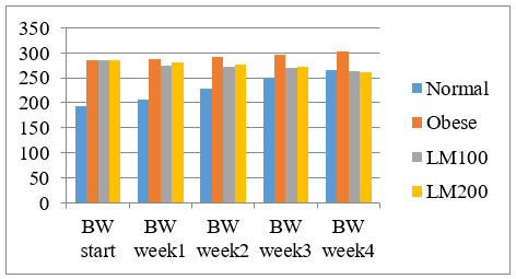
The development of the rats' body weight over 4 weeks is presented in Table 1 and Figure 1.
Table 1. The average weight development of Wistar rats is 4 weeks
|
Group |
Normal |
Obese |
LM100 |
LM200 |
|
BW. Start |
192.38±2.36b,c,d |
286.20±2.18a |
285.32±2.15a |
286.11±1.84a |
|
BW.Week1 |
207.01±4.37b,c,d |
288.47±2.59a,c,d |
275.12±6.81a,b |
279.92±190a,b, |
|
BW.Week2 |
227.37±4.73b,c,d |
292.81±2.67a,c,d |
272.62±2.23a,b |
275.56±2.79a,b |
|
BW.Week3 |
249.59±2.82b,c,d |
296. 45±3.07a,c,d |
270.26±1.82b,d |
272.43±0.84a,b |
|
BW.Week4 |
265.71±2.06b |
302.02±3.51a,c,d |
262.41±1.65b,d |
260.20±0.60b, |
Note: a indicates Normal; b shows Obese; c shows LM100; d shows LM200
Figure 1. Average weight development of Wistar rats after 4 weeks of research
Mean ± SD followed by letters in the same row indicates a significant difference = p<0.05; group 1 (normal = standard feed); group 2 (Obese = high fat feed), group 3 (LM100 = Lidah mertua leaf extract with 100 mg/kg), and group 4 (LM200 = Lidah mertua leaf extract 200 mg/kg).
Figure 1 and Table 1 show that the average body weight of the normal group differed significantly at the beginning of the study (p<0.05) from the other three groups. The average weight of the normal group was 192.38 ± 2.36 g, the obese group was 286.20 ± 2.18 g, the obese group with LM 100 extract was 285.32 ± 2.15 g, and the obese group with LM200 extract was 286.11 ± 1.84 g. Weight gain every week occurred in the normal group and the obese group, with a significant difference (p<0.05). In contrast, in the LM 100 and LM 200 groups, there was a significant decrease in weight every week (p<0.05).
3.1.2 The effect of Lidah mertua Leaf extract on glucose, triglycerides, free fatty acids in rat blood serum, and Lee's obesity index
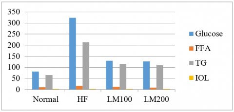
The effect of Lidah mertua leaf extract at doses of 100 mg/kg and 200 mg/kg on glucose, triglyceride, free fatty acid, and Lee's obesity index levels in obese Wistar rats can be indicated in Table 2 and Figure 2.
Mean ± SD followed by letters in the same row indicates a significant difference = p<0.05; group 1(normal = standard feed); group 2 (Obese = high fat feed), group 3 (LM 100 = Lidah mertua 100 mg/kg), and treatment group 4 (LM 200 = Lidah mertua leaves with 100 mg/kg), were compared with the normal. TG (triglycerides); IOL (Index Obesity Lee).
Table 2. Average levels of glucose, free fatty acids, and triglycerides in rat blood serum, and Lee obesity index
|
|
Normal |
Obese |
LM100 |
LM200 |
|
TG mg/dl |
65.83± 2.93b,c,d |
213.17±4.25a,c,d |
115.83±6.32a,b |
109.17±4.43a,b,c |
|
Blood glucose (mg/dL) |
81.17± 2.79b,c,d |
232.33±8.36a,c,d |
129.83±3.97a,b |
126.50±0.59a,b |
|
Free Fatty acids (mmol/L) |
11.42± 1.10b,d |
27.51± 0.81a,c,d |
12.83± 1.13b,d |
9.87± 0.59a,b,c |
|
IOL |
0.28± 0.009b |
0.33± 0.007a,c,d |
0.23± 0.005b |
0.22± 0.004b |
Note: a indicates normal; b shows Obese; c shows LM 100; d indicates LM 200
Figure 2. Effect of Lidah mertua leaf extract on levels of glucose, free fatty acids (FFA), triglycerides (TG) in rat blood serum, and the index obesity Lee (IOL)
3.1.3 Analysis of Lidah mertua leaf extract using LC-MS/MS
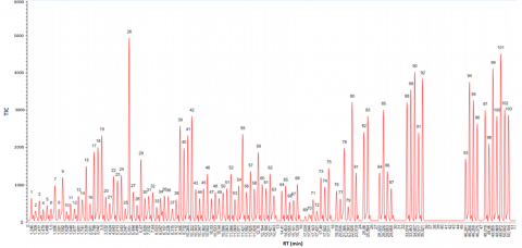
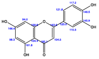
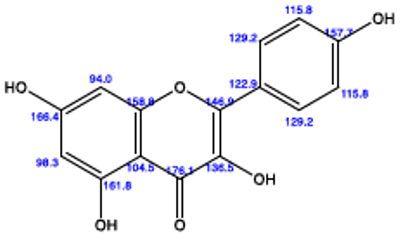
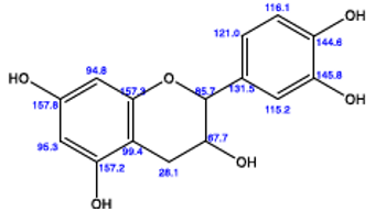
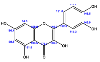
The results of compound identification from Lidah mertua leaf extract using LC-MS/MS produced several chromatography spectrum peaks with different retention times. Before conducting LC-MS/MS testing, separation was carried out using SPE, which aims to reduce impurities in the sample. The chromatograms obtained can be seen in Figure 3 and Table 3.
Figure 3. LCMS chromagram result of Lidah mertua leaves extract
Table 3. LC-MS/MS chromatogram results
|
Peak Number |
RT(min) |
Curva Area |
Composition (%) |
Analysis |
Structure |
|
15 |
1.839 |
1503.66362 |
1.02810 |
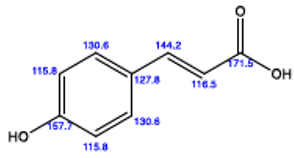
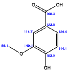
p-coumaric acid Chemical Formula: C6H8O3 Exact Mass: 164.0473 Molecular Weight: 164.1600 m/z: 164.0473 (100.0%), 169.0456 (9.7%) |
|
|
917 |
2.799 |
1884.55263 |
1.28852 |
Vanillic acid Formula: C8H8O4 Mass: 168.0423 Weight: 168.1480 m/z: 168.0423 (100.0%), 169.0456 (8.7%) |
|
|
18 |
3.042 |
2013.66521 |
1.37680 |
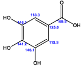
Gallic acid Chemical Formula: C7H6O5 Exact Mass: 170.0215 Molecular Weight: 170.1200 m/z: 170.0215 (100.0%), 171.0249 (7.6%), 172.0258 (1.0%) |
|
|
39 |
9.365 |
2584.66352 |
1.76720 |
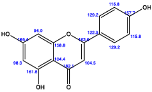
Apigenin Formula: C15H10O5 Mass: 270.0528 Weight: 270.2400 m/z: 270.0528 (100.0%), 271.0562 (16.2%) |
|
|
40 |
9.732 |
1986.55236 |
1.35826 |
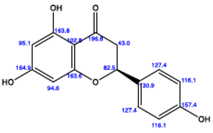
Naringenin Chemical Formula: C15H12O5 Exact Mass: 272.0685 Molecular Weight: 272.2560 m/z: 272.0685 (100.0%), 273.0718 (16.2%), 274.0752 (1.2%), 274.0727 (1.0%) |
|
|
41 |
10.265 |
2325.51214 |
1.59002 |
Luteolin Formula: C15H10O6 Mass: 286.0477 Weight: 286.2390 m/z: 286.0477 (100.0%), 287.0511 (16.2%), 288.0520 (1.2%), 288.0544 (1.2%) |
|
|
42 |
10.322 |
2845.52260 |
1.94556 |
Kaempferol Formula: C15H10O6 Mass: 286.0477 Weight: 286.2390 m/z: 286.0477 (100.0%), 287.0511 (16.2%), 288.0520 (1.2%), 288.0544 (1.2%) |
|
|
46 |
10.502 |
1302.25144 |
0.89038 |
Catechin Chemical Formula: C15H14O6 Exact Mass: 290.0790 Molecular Weight: 290.2710 m/z: 290.0790 (100.0%), 291.0824 (16.2%), 292.0833 (1.2%), 292.0857 (1.2%) |
|
|
55 |
11.427 |
2362.20211 |
1.61510 |
Quercetin Molecular Formula: C15H10O7 Mass: 302.0427 Weight: 302.2380 m/z: 302.0427 (100.0%), 303.0460 (16.2%), 304.0469 (1.4%), 304.0494 (1.2%) |
|
|
67 |
15.101 |
594.32026 |
0.40635 |
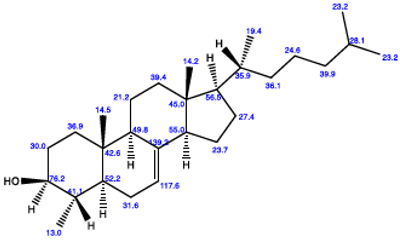
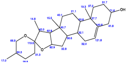
Lophenol Formula: C28H48O Mass: 400.3705 Weight: 400.6910 m/z: 400.3705 (100.0%), 401.3739 (30.3%), 402.3772 (2.7%), 402.3772 (1.7%) |
|
|
70 |
17.046 |
216.5535 |
0.14806 |
Diosgenin Chemical Formula: C27H42O3 Exact Mass: 414.3134 Molecular Weight: 414.6300 m/z: 414.3134 (100.0%), 415.3168 (29.2%), 416.3201 (2.7%), 416.3201 (1.4%) |
|
|
90 |
34.007 |
4021.33652 |
2.7495 |
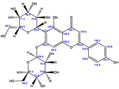
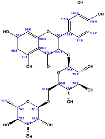
Saponarin Chemical Formula: C27H30O15 Exact Mass: 594.1585 Molecular Weight: 594.5220 m/z: 594.1585 (100.0%), 595.1618 (29.2%), 596.1627 (3.1%), 596.1652 (2.7%), 596.1652 (1.4%) |
|
|
91 |
35.517 |
2398.55236 |
1.63996 |
Rutin Chemical Formula: C27H30O16 Exact Mass: 610.1534 Molecular Weight: 610.5210 m/z: 610.1534 (100.0%), 611.1567 (29.2%), 612.1576 (3.3%), 612.1601 (2.7%), 612.1601 (1.4%) |
|
|
94 |
46.256 |
3756.61222 |
2.56850 |
Sansevierin A Chemical Formula: C39H62O13 Exact Mass: 738.1490 Molecular Weight: 738.9120 m/z: 740.4190 (100.0%), 739.4224 (42.2%), 740.4258 (8.7%), 740.4233 (2.7%), 741.4291(1.2%), 741.4266(1.1%) |
|
|
101 |
49.972 |
4513.65523 |
3.08611 |
Sansevistatin 1 Chemical Formula: C45H70O16 Exact Mass: 866.4664 Molecular Weight: 867.0390 m/z: 866.4664(100.0%), 867.4697(48.7%), 868.4731(11.6%), 868.4706(3.3%), 869.4740(1.6%) |
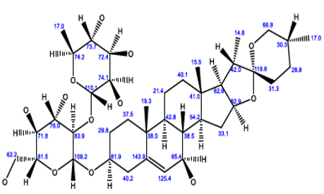
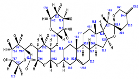
Based on the chromatogram in Figure 3 and Table 3, it shows that Lidah mertua leaf extract identified 15 peaks with different retention times and 15 different compounds, namely 3 phenols (p-coumaric acid, vanillic acid, gallic acid), 6 flavonoids (apigenin, naringenin, luteolin, kaemferol, quercetin, cathecin), flavonoid glycosides (saponarin), steroids (lophenol), Sapogenin (diosgenin), tannins(rutin), saponin (Sansevirin A and Sansevistatin 1).
3.2 Discussion
Data from statistical analysis in Table 1 shows that the body weight of rats in the normal and high fat groups increased every week, while those in the group treated with 100 mg/kg and 200 mg/kg of Lidah mertua extract experienced a decrease in body weight every week.
This indicates that administration of Lidah mertua leaf extract at doses of 100 mg/kg and 200 mg/kg was able to suppress the weight gain of rats, or reduce the weight of rats, and also Lee's obesity index decreased with a significant difference compared to the high-fat/obese rats group.
The weight loss of rats was caused by the presence of bioactive compounds in Lidah mertua leaf extract, which involves several complex biochemical and physiological pathways. The results of analysis of Lidah mertua leaf extract using LC-MS/MS showed that there were 15 compounds identified, namely 3 phenolic acids (p-coumaric acid, vanillic acid, gallic acid), 6 flavonoids (apigenin, naringenin, luteolin, kaemferol, quercetin, catechin), flavonoid glycosides (saponarin), steroids (lophenol), Sapogenin (diosgenin), tannins (rutin), saponins (Sansevierin A and Sansevistatin 1). Sansevierin A and Sansevistatin 1 are steroid saponins found in the Lidah mertua plant leaf extract that play an important role in reducing the weight of obese rats. The mechanism of action of saponins in reducing weight is to reduce fat accumulation by increasing fat excretion through feces and inhibiting fat absorption from the intestines. Bogoriani et al. [9] also reported that steroid saponins from Andong leaves had anti-obesity activity in Wistar rats with significant differences.
The flavonoids contained are thought to have anti-obesity potential. The role of flavonoids in losing weight is by increasing the activity of lipolytic enzymes involved in breaking down fat. In addition, flavonoids are also known to increase thermogenesis and fat metabolism through activation of the AMPK and PPAR-α pathways. Dzomba and Musekiwa [26] reported that flavonoids in the saphoricoside compound, which is an anti-obesity and antioxidant compound, are thought to act as pancreatic lipase inhibitors and reduce appetite. The results of this research are also supported by research by Suastuti et al. [27] reported that dragon fruit extract (Hylocereus costarioensis), which contains flavonoids, has anti-obesity activity.
Based on the results of statistical analysis, the data in Table 2 and Figure 2 show that the normal group has lower levels of glucose, triglycerides, and free fatty acids than the obese group, with a significant difference (p<0.05). This is caused by the impact of obesity. Food fat in the intestine will be broken down by the lipase enzyme released by the pancreas into a simpler form, namely monoglycerides, where the monoglycerides are broken down again by bile salts and the lipase enzyme into free fatty acids. When monoglycerides and free fatty acids pass through the intestinal membrane, triglycerides are formed again. These triglycerides will combine with apolipoprotein and cholesterol to form chylomicrons. Chylomicrons will carry triglycerides into the blood circulation. In muscle, triglycerides will be degraded into acetyl coenzyme A, while excess triglycerides will be stored in adipose tissue, which can be used again through the lipolysis process. In adipose tissue, triglycerides are broken down into fatty acids and glycerol. The fatty acids produced will be converted into acetyl coenzyme A for use in muscles; the excess is stored in ketone bodies to be used again through the gluconeogenesis process to be converted into glucose [3].
In obesity, there will be an increase in free fatty acids in the blood, which will be followed by an increase in the uptake of free fatty acids by muscle tissue. Muscles, under normal circumstances, will use glucose to produce energy. In this way, fatty acid oxidation in the muscles increases, which will inhibit glucose uptake by the muscles, resulting in hyperglycemia. The same situation occurs in the liver, where the liver will accommodate most of the free fatty acids and become material for gluconeogenesis. With increased gluconeogenesis, fasting blood glucose will increase, resulting in hyperglycemia. This hyperglycemic state will result in insulin resistance in the liver [3].
The data in Table 2 and Figure 2 show that the group treated with extracts of 100 mg/kg and 200 mg/kg of Lidah mertua leaves had lower levels of blood glucose, triglycerides and free fatty acids on average than the obese group with a significant difference (p < 0.05) and was in the range of normal mouse glucose levels is 50-145 mg/dL. Rats in the obese group had blood glucose levels that exceeded the glucose levels of normal rats, namely >145 mg/dL, so the rats were declared to have hyperglycemia [28]. The levels of blood glucose, TG, and free fatty acids in the group treated with 100 mg/kg of Lidah mertua leaf extract experienced a percentage decrease of respectively of 44.12%; 45.66%; 53.36% and the 200 mg/kg treatment group experienced a decrease respectively of 45.55%; 48.79% and 63.82% of the obese group experienced an increase in glucose, triglyceride and free fatty acid levels of 65.06%, 69.12% and 58.49% respectively.
The decrease in glucose, triglyceride, and free fatty acid levels in the blood of obese rats is thought to be due to the content of secondary metabolites, which have antihyperglycemic and antioxidant properties. The high sugar levels cause increased oxidative stress and decreased endogenous antioxidants. Intake of natural antioxidants is one way of protecting against the progression of diabetes by inhibiting the peroxide reaction, which damages pancreatic beta cells. The same research results were also shown by several studies [28-30]. The results of the analysis of Lidah mertua leaf extract using LC-MS/MS that have been carried out show that Lidah mertua leaf extract contains 15 identified compounds, namely 3 phenolics (p-coumaric acid, vanillic acid, gallic acid), 6 flavonoids (apigenin, naringenin, luteolin, kaemferol, quercetin, catechin), flavonoid glycosides (saponarin), steroids (lophenol), Sapogenin (diosgenin), tannin (rutin), Sansevirin A, and Sansevistatin 1, which act as antioxidants.
According to Song et al. [31], flavonoids can reduce blood glucose by inhibiting the action of GLUT2 (Glucose Transporter Isoform 2), which is a glucose transporter protein found in the intestinal membrane which causing blood glucose levels to fall. Apart from being able to increase the activity of antioxidants, flavonoids are also able to regenerate damaged pancreatic beta cells [8]. The statistical analysis results showed that there was a significant difference between the obese control group and the treatment group (LM 100 mg/kg and LM 200 mg/kg) (p˂0.05), and between the treatment groups (LM 100 mg/kg and LM 200 mg/kg) did not have a significant difference p=0.330 (p>0.05). This shows that the blood glucose levels of the treatment group after administering the extract of Lidah mertua leaves at a dose of 100 mg/kg and 200 mg/kg had the same effectiveness in reducing blood glucose levels.
The data from the analysis in Table 2 shows that the treatment group (LM 100 mg/kg extract and LM 200 mg/kg) had a lower mean triglyceride level compared to the obese control group. Triglyceride levels between the LM 100 mg/kg and LM 200 mg/kg leaf treatment groups experienced a percentage decrease of 45.66% and 48.79% compared to the obese control group, which experienced a percentage increase in triglyceride levels of 69.12%.
Phenols, flavonoids, and saponins found in Lidah mertua plant leaf extract are bioactive compounds that can reduce triglyceride levels by inhibiting enzymes involved in cholesterol synthesis in cholesterol synthesis in the liver and increasing cholesterol excretion through the bile duct. Flavonoids and phenols also work to reduce insulin resistance, increase insulin sensitivity, and lower blood glucose levels. Quercetin and kaempferol can inhibit the activity of the alpha-glucosidase enzyme and regulate carbohydrate metabolism. The hypolipidemic effect of saponins functions to reduce blood triglyceride levels. Saponins can inhibit cholesterol synthesis in the liver and improve lipid profiles by increasing HDL cholesterol levels. Saponins can also increase insulin sensitivity in body tissues and reduce glucose absorption through the intestines, increase glucose metabolism in the liver and muscles, and help control blood sugar levels [32-35].
The results of the research and discussion can be concluded as:
(i) The three groups of bioactive compounds (15 compounds) contained in the extract of the Lidah mertua (Sansevieria trifasciata Laurentii), namely the phenol, flavonoid, and saponin groups, have a positive effect on reducing body weight and levels of triglycerides, glucose, and free fatty acids in obese rats. Each compound works in a different way, but often complements the other in increasing fat metabolism, increasing insulin sensitivity, and reducing glucose and fat absorption.
(ii) There are 15 compounds identified in Lidah mertua leaf extract, which are thought to play an active role in reducing body weight, free fatty acids, glucose and triglycerides, namely 3 phenols (p-coumaric acid, vanillic acid, gallic acid), 6 flavonoids (apigenin, naringenin, luteolin, kaempferol, Quercetin, cathecin), flavonoid glycosides (saponarin), steroids (lophenol), Sapogenin (diosgenin), tannins (rutin), Sansevierin A and Sansevistatin 1.
The authors are very grateful to the students who have helped in the research. The author also thanks the PNBP of Udayana University for funding this research.
[1] You, J.S., Lee, Y.J., Kim, K.S., Kim, S.H., Chang, K.J. (2014). Anti-obesity and hypolipidaemic effects of Nelumbo nucifera seed ethanol extract in human pre-adipocytes and rats fed a high-fat diet. Journal of the Science of Food and Agriculture, 94(3): 568-575. https://doi.org/10.1002/jsfa.6297
[2] Hussein, S.A., El-Senosi, Y.A., EL-Sharkawy, G. (2018). Antiobesity activity and hypolipidemic effect of Proanthocyanidins in rats fed a high fat diet. Benha Veterinary Medical Journal, 35(2): 364-379.
[3] Murray, R.K., Granner, D.K., Mayes, P.A., Rodwell, V.W. (2014). Harper's Biochemistry. Norwalk, Conn.: Appleton & Lange. https://archive.org/details/harpersbiochemis00robe.
[4] Sukadana, I.M., Bogoriani, N.W., Ariani, M. (2023). Compounds in the stem of Etlingera elatiorcan reduce the levels of free Fatty Acid and Blood Glucose in Obesity Wistar Rats. Research Journal of Pharmacy and Technology, 16(10): 4530-4536. https://doi.org/10.52711/0974-360X.2023.00738
[5] Bogoriani, N.W., Suaniti, N.M., Putra, A.A.B., Lestari, K.D.P. (2019). The activity of Cordyline terminalis’s leaf extract as antidiabetic in obese wistar rats. International Journal of Pharmaceutical Research and Allied Sciences, 8(2): 206-213.
[6] World Obesity and IDF release new policy brief to address obesity and type 2 diabetes. World Obesity. https://www.worldobesity.org/news/idf-and-wof-release-new-policy-brief-to-address-obesity-and-type-2-diabetes.
[7] Mutiara, E.V., Wildan, A. (2014). Ekstraksi flavonoid dari daun pare (Momordica charantia L.) berbantu gelombang mikro sebagai penurun kadar glukosa secara in vitro. Metana, 10(1): 1-11. https://doi.org/10.14710/metana.v10i01.9771
[8] Ardalani, H., Hejazi Amiri, F., Hadipanah, A., Kongstad, K.T. (2021). Potential antidiabetic phytochemicals in plant roots: A review of in vivo studies. Journal of Diabetes & Metabolic Disorders, 20(2): 1837-1854. https://doi.org/10.1007/s40200-021-00853-9
[9] Bogoriani, N.W., Laksmiwati, A.A.I.M., Putra, A.A.B., Heltyani, W.E., Lestari, K.D.P., Mahayani, P.A.E. (2019). Saponins role of Bali Andong leaf as antiobesity in rats. International Journal of Pharmaceutical Research, 11(2): 382-389. https://doi.org/10.31838/ijpr/2019.11.02.052
[10] Tchegnitegni, B.T., Teponno, R.B., Tanaka, C., Gabriel, A.F., Tapondjou, L.A., Miyamoto, T. (2015). Sappanin-type homoisoflavonoids from Sansevieria trifasciata Prain. Phytochemistry Letters, 12: 262-266. https://doi.org/10.1016/j.phytol.2015.04.017
[11] Teponno, R.B., Tanaka, C., Jie, B., Tapondjou, L.A., Miyamoto, T. (2016). Trifasciatosides A-J, steroidal saponins from Sansevieria trifasciata. Chemical and Pharmaceutical Bulletin, 64(9): 1347-1355. https://doi.org/10.1248/cpb.c16-00337
[12] Ighodaro, O.M., Adeosun, A.M., Ojiko, B.F., Akorede, A.T., Fuyi-Williams, O. (2017). Toxicity status and antiulcerative potential of Sansevieria trifasciata leaf extract in Wistar rats. Journal of Intercultural Ethnopharmacology, 6(2): 234-239. https://doi.org/10.5455/jice.20170421103553
[13] Abdullah, Angelina, Yumna, M., Arbianti, R., Utami, T.S., Hermansyah, H., Ningsih, S. (2018). Flavonoid isolation and identification of mother-in-law’s tongue leaves (sansevieria trifasciata) and the inhibitory activities to xanthine oxidase enzyme. E3S Web of Conferences, 67: 03011. https://doi.org/10.1051/e3sconf/20186703011
[14] Yumna, M., Angelina, Abdullah, Arbianti, R., Utami, T.S., Hermansyah, H. (2018). Effect of mother-in-law’s tongue leaves (Sansevieria trifasciata) extract’s solvent polarity on anti-diabetic activity through in vitro α-glucosidase enzyme inhibition test. E3S Web of Conferences, 67: 03003. https://doi.org/10.1051/e3sconf/20186703003
[15] Febriani, Y., Mierza, V., Handayani, N.P., Surismayanti, S., Ginting, I. (2019). Antibacterial activity of lidah mertua (Sansevieria trifasciata prain.) leaves extract on Escherichia coli and Staphylococcus aureus. Open Access Macedonian Journal of Medical Sciences, 7(22): 3882-3886. https://doi.org/10.3889/oamjms.2019.525
[16] Wahab, N.H.A., Samuel, Y., Yusuf, N., Yusoff, H.M. (2020). Variation in phytochemical constituents and antioxidant potential of extracts derived from leaves of Sansevieria trifasciata var. Laurentii and Sansevieria trifasciata var. Zeylanica (Asparagaceae). Asian Journal of Chemistry, 32(9): 2303-2307. https://doi.org/10.14233/ajchem.2020.22792
[17] Megantika, A., Adhityaxena, A.T., Arbianti, R., Utami, T.S., Hermansyah, H. (2020). Production of flavonoid compounds from mother in law’s tongue leaves (Sansevieria trifasciata) using microwave-assisted enzymatic extraction as anti-inflammatory. AIP Conference Proceedings, 2255: 040009. https://doi.org/10.1063/5.0021017
[18] Dewatisari, W.F., Nugroho, L.H., Retnaningrum, E., Purwestri, Y.A. (2022). Antibacterial and anti-biofilm-forming activity of secondary metabolites from sansevieria trifasciata leaves against pseudomonas aeruginosa. Indonesian Journal of Pharmacy, 33(1): 100-109. https://doi.org/10.22146/ijp.2815
[19] Dewatisari, W.F., Nugroho, L.H., Retnaningrum, E., Purwestri, Y.A. (2023). Inhibition of protease activity and anti-quorum sensing of the potential fraction of ethanolic extract from Sansevieria trifasciata Prain leaves against Pseudomonas aeruginosa. Indonesian Journal of Biotechnology, 28(1): 23-30. https://doi.org/10.22146/ijbiotech.73649
[20] Rachmaniyah, R., Rusmiati, R. (2023). Sansevieria trifasciata extract effectively inhibit the growth of Escherichia coli bacteria, in vitro. International Journal of Public Health Science, 12(3): 1119-1125. https://doi.org/10.11591/ijphs.v12i3.23172
[21] Kasmawati, H., Ruslin, R., Arfan, A., Sida, N.A., Saputra, D.I., Halimah, E., Mustarichie, R. (2023). Antibacterial potency of an active compound from Sansevieria trifasciata Prain: An integrated in vitro and in silico study. Molecules, 28(16): 6096. https://doi.org/10.3390/molecules28166096
[22] Bogoriani, N.W., Suaniti, N.M., Putra, A.A.B., Lestari, K.D.P., Heltyani, W.E. (2020). The effect of Cordyline terminalis’s leaf extract on lipid profile, obesity and liver function in obese induced rats. Systematic Reviews in Pharmacy, 11(11): 1080-1086. https://doi.org/10.31838/srp.2020.11.154
[23] Bogoriani, N.W., Ariati, K., Pratiwi, I.G.A.P.E. (2022). Potency of balinise kecombrang (Etlingeraelatior) extract of as antioxidant against the activity of superoxide dismutase (SOD), glutathione (GSH) and fatty liver in obese rats. Biomedical & Pharmacology Journal, 15(1): 337-344. https://doi.org/10.13005/bpj/2372
[24] Waters Corporation. (2014). MassLynx 4.1: Getting Started Guide. Waters Corporation. Washington D.C.
[25] Bogoriani, N.W., Wahjuni, S., Ema, M.K. (2023). Antioxidant activity of kecombrang flower (Etlingera elatior) methanol extract and identification of its compounds using LC-MS/MS. Indonesia Journal of Biomedical Science (IJBS), 17(2): 172-177. https://doi.org/10.15562/ijbs.v17i2.480
[26] Dzomba, P., Musekiwa, C. (2014). Anti-obesity and antioxidant activity of dietary flavonoids from Diocorea steriscus tubers. Journal of Coastal Life Medicine, 2(6): 465-470.
[27] Suastuti, N.G.M.D.A., Bogoriani, N.W., Putra, A.A.B. (2018). Activity of Hylocereus costarioensis’s extract as antiobesity and hypolipidemic of obese rats. International Journal of Pharmaceutical Research & Allied Sciences, 7(1): 201-208.
[28] Vinayagam, R., Xu, B. (2015). Antidiabetic properties of dietary flavonoid: A cellular mechanism review. Nutrition & Matabolism, 12: 60. https://doi.org/10.1186/s12986-015-0057-7
[29] Lachumy, S.J.T., Sasidharan, S., Sumathy, V., Zuraini, Z. (2010). Pharmacological activity, phytochemical analysis and toxicity of methanol extract of Etlingera elatior (torch ginger) flowers. Asian Pacific Journal of Tropical Medicine, 3(10): 769-774. https://doi.org/10.1016/S1995-7645(10)60185-X
[30] da Silva Ferreira, R.G., Guilhon-Simplicio, F., Acho, L.D.R., Batista, N.Y., do Carmo Guedes-Junior, F., Ferreira, M.S.L., Barcellos, J.F.M., Veiga-Junior, V.F., Lima, E.S. (2021). Anti-hyperglycemic, lipid-lowering, and anti-obesity effects of the triterpenes α and β-amyrenones in vivo. Avicenna Journal of Phytomedicine, 11(5): 451-463. https://dx.doi.org/10.22038/AJP.2021.18076
[31] Song, J., Kwon, O., Chen, S., Daruwala, R., Eck, P., Park, J.B., Levine, M. (2020). Flavonoid inhibition of sodium-dependent vitamin C transporter 1 (SVCT1) and glucose transporter isoform 2 (GLUT2) intestinal transporters for vitamin C and glucose. Journal of Biological Chemistry, 277(18): 15252-15260. https://doi.org/10.1074/jbc.M110496200
[32] Chen, S., Jiang, H., Wu, X., Fang, J. (2016). Therapeutic effect of quercetin on inflammation, obesity, and type 2 Diabetes. Mediators of Inflammations, 2016(1): 9340637. https://doi.org/10.1155/2016/9340637
[33] Dhanya, R. (2022). Quercetin for managing type 2 diabetes and its complications an insight into multitarget therapy. Biomedicine and Pharmacotherapy, 146: 112560. https://doi.org/10.1016/j.biopha.2021.112560
[34] He, L., Su, Z., Wang, S. (2024). The anti-obesity effects of polyphenols: A comprehensive review of molecular mechanisms and signal phatways in ragulating adipocytes. Frontiers in Nutrition, 11: 1393575. https://doi.org/10.3389/fnut.2024.1393575
[35] Zheng, Y., Choi, Y.H., Lee, J.H., Lee, S.Y., Kang, H.J. (2021). Anti-obesity effect of Erigeron annuus (L.) pers. extract containing phenollic acids. Foods, 10(6): 1266. https://doi.org/10.3390/foods10061266