Omar Ali Kanosh*![]() | Mateen A. Mahdi
| Mateen A. Mahdi![]()
© 2024 The authors. This article is published by IIETA and is licensed under the CC BY 4.0 license (http://creativecommons.org/licenses/by/4.0/).
OPEN ACCESS
This work aimed to estimate lipoxygenase activity and purify its isoenzymes in humans, particularly in those with Hepatitis B Virus, thereby making variations in its normal levels a diagnostic marker for the disease. Hepatitis B is an infectious disease caused by the Hepatitis B Virus (HBV), leading to both acute and chronic inflammations. The current study was conducted to evaluate the activity of lipoxygenase (LOX), purify and separate its isomers partially. The study took place in Kirkuk city, Iraq from the first of January, 2021, to the 30th of March, 2021, involving a cohort of 40 patients infected with viral Hepatitis B. Their ages ranged between 20 and 70 years, with the majority seeking care at the Gastrointestinal Tract Center (GIT) in Kirkuk's Azadi Teaching Hospital and Kirkuk General Hospital. Estimation of Lipoxygenase (LOX) enzyme activity, protein concentration, and purifying the enzyme were performed. The results indicated a significant elevation in the lipoxygenase activity in patients afflicted with viral hepatitis, with a probability of P ≤ 0.001 compared to the control group which serves as a strong indicator of hepatitis infection. The enzyme was purified using ammonium sulfate, achieving a 30% saturation. Excess salts were eliminated through membrane filtration. Further purification phases were completed utilizing gel filtration chromatography with a Sephadex G.100 column, yielding a single peak. The enzyme purification degree was determined to be 0.57554 times, with an enzyme yield of 26.10526%. Subsequently, isomers of the lipoxygenase were separated through ion exchange chromatography using DEAE-Cellulose. Three lipoxygenase LOX isomers, namely LOX1, LOX2, and LOX3, were obtained with varying degrees of purity, measured at (0.731330935), (0.679565947), and (0.707230216), respectively.
lipoxygenase, hepatitis B, protein purification, enzyme activity
Viral hepatitis B is a major global health concern, affecting millions of individuals worldwide. Research indicates that the prevalence of Hepatitis B Virus infections has reached alarming levels, with over 350 million individuals persistently infected, emphasizing the need for effective therapeutic interventions and understanding of biochemical changes associated with the disease [1]. In this context, lipoxygenase activity has emerged as a critical area of investigation, as it plays a significant role in inflammatory processes and may contribute to the pathophysiology of liver diseases associated with viral hepatitis B [1].
Hepatitis refers to some form of hepatic inflammation caused by numerous etiological factors. One of these factors is non-infectious causes encompassing autoimmune disorders, metabolic maladies, and drug and alcohol abuse. Additionally, liver inflammation could be caused by infectious agents such as bacteria, parasites, and viruses. Notably, the latter, viral hepatitis, can progress to hepatic fibrosis or even culminate in hepatocellular carcinoma [1]. The Hepatitis B Virus (HBV) is a diminutive DNA-containing viral entity capable of instigating both acute and chronic hepatitis. Despite the availability of secure and efficacious vaccines, Hepatitis B Virus infection continues to impose a substantial health concern. Globally, an estimated 641 million individuals suffer from chronic Hepatitis B [2]. The prevalence of chronic infections is approximately 2% in adult-onset cases, reaching up to 21% of perinatal transmission instances. Transmission of Hepatitis B Virus (HBV) occurs through the exchange of bodily fluids. We can detect up to 102 genomic equivalents of HBV per milliliter of serum [3]. Although the Hepatitis B Virus does not induce direct cellular effects under ordinary infectious conditions, it is believed that liver damage (hepatic fibrosis and hepatocellular carcinoma) results from sustained immune response and constant inflammation within the liver [3].
The enzyme lipoxygenase (linoleate: oxidase) (EC:1.13.11.12) is a member of the family of enzymes that contain non-protein-bound iron and are classified as non-heme proteins. It falls under the category of oxidoreductases [4]. Lipoxygenase is responsible for catalyzing the oxidation of unsaturated fatty acids to produce hydroperoxides [5]. These enzymatic products are crucial in acute infection responses, such as inflammation, and may also be involved in the resolution of these infections [6].
Lipoxygenase also enhances metabolic processes inside and outside the cells of living organisms [7]. The enzyme it has significant involvement with several conditions reviewed and researched the enzyme association with such conditions as cancer [8-10], inflammation [11, 12] and vascular functions [13, 14]. Extensively studied in disease treatment and prevention have been oxidized unsaturated fatty acids (eicosanoids), produced by lipoxygenase [15].
In the current work lipoxygenase enzyme activity in the blood serum of patients with Hepatitis B Virus was evaluated and its partially purification was carried out, including several stages begun by ammonium sulfate precipitation, dialysis followed by gel filtration chromatography and ion exchange chromatography. As such, this study is considered Method as a function for diagnosing the disease.
The studied was in the period of three months starting from the first of January 2021 until the 30th of March 2021 in the city of Kirkuk in Iraq. Forty patients diagnosed with viral Hepatitis B was the cohort that we focused our research on. They were entirely different ages 20 to 70 years. Most of the participants received medical treatment at the Gastrointestinal Tract Center in Kirkuk's Azadi Teaching Hospital and at Kirkuk’s General Hospital. The research also included a control group of 40 blood samples to bolster its findings. This was a control group which helped compare the results against individuals who did not have Hepatitis B, as a baseline to see how significantly the virus had affected, and to get an idea of characteristics of, the virus in the population studied.
2.1 Estimation of lipoxygenase enzyme activity
We used the method of Shastry and Raghavendra Rao [16] to quantify the activity of the LOX enzyme. In this case, it adds two oxygen atoms to the substrate (Linoleic acid) making enzyme catalytically active. The amount of enzyme activity was quantified by measuring increase in absorbance attributable to the formation of conjugated dienes with optical density recorded at 234 nanometers and monitored for five minutes. As an interesting point the conjugated diene have a molar absorptivity (ε) value of 25 000 M⁻¹∙cm⁻¹.
a- The solutions used:
1. Borate buffer solution (concentration 0.2 M, pH 7): It was prepared by dissolving 6.18 grams of boric acid in 450 millilitres of distilled water and then adjusting the pH at seven by adding a solution of hydrochloric acid or sodium hydroxide at a concentration of 1 M and then Then complete the volume to 500 ml. The solution is kept at four ℃.
2. The base solution: 1 gram of linoleic acid was dissolved in 100 millilitres of 95% ethanol.
3. Hydrochloric acid solution, HCl concentration (3%): It was prepared by adding 3 millilitres of hydrochloric acid and then completing the volume to 100 millilitres with distilled water.
4. Water without ions.
b- Procedure:
Add (0.5) millilitres of the borate buffer solution with a pH 7 and (0.2) millilitres of the base material to (10) millilitres of deionized distilled water and mix the mixture well, then add (0.5) millilitres of blood serum with continuous re-mixing for 4 minutes, then (2) millilitres of the mixture are withdrawn and immediately added to a tube containing (10) millilitres of 95% ethyl alcohol and (0.2) millilitres of hydrochloric acid. The absorbance was measured at a wavelength of (234) nm. Enzyme activity is calculated by:
$\begin{aligned} \text { Activity }(\mathrm{unit} / \mathrm{ml}) & =\frac{\text { Change in absorbency }}{\text { Reaction time } * \text { sample volume }} * 60 \text { second }\end{aligned}$
2.2 Measurement of protein concentration for LOX enzyme
A protein concentration was determined in both the serum samples and the clarified fractions using the Lowry method [17]. It is well known for being sensitive and reliable, as a method of protein quantification. Bovine serum albumin (BSA) was used as the standard protein reference for the quantification of the protein levels in the samples in this study. BSA is used as a standard because it has well characterized properties, and consistent, reproducible results in protein assays such as the Lowry.
a- The solutions used:
1- Alkaline sodium carbonate solution (Solution A):
Made up in the field by dissolving 2 g of sodium carbonate Na2CO3 in 100 ml of 0.1 mol/L sodium hydroxide NaOH solution.
2- Copper sulphate – sodium potassium tartrate solution (Solution B):
Prepared by dissolving 0.5 g of aqueous copper sulphate CuSO4.5H2O in 00 l ml of distilled water. When used, prepare solution B by taking 10 ml of the above solution and adding it to 0.1 g of potassium sodium tartrate.
3- Alkaline solution:
Prepare for use by mixing 50 ml of solution A with 1 ml of solution B.
4- Folin – Ciocalteu reagent:
This solution consists of Sodium tungstate dehydrate Na2WO4.2H2O and Sodium molybdate dehydrate Na2MoO4.2H2O in phosphoric acid H3PO4 and hydrochloric acid HCl. This solution was diluted with an equal amount of distilled water, in a ratio of 1:1 when used.
b- Procedure:
1- Add 2.5 ml of the basic solution to 500 microliters of dissolved protein (serum, precipitated fraction, or BSA) and mix them together and leave them for 10 minutes at room temperature.
2- 250 μl of Folen reagent was added, the contents of the tube were mixed well and left at room temperature for 30 minutes. Then the absorbance was read at a wavelength of 750 nm.
The standard protein curve was determined by plotting the absorbance against the standard protein concentrations as shown in Figure 1. This curve was used to find the unknown protein concentration of the serum sample or the eluent.
Figure 1. Standard curve
2.3 The separation and purification of the lipoxygenase (LOX)
The separation and purification of the enzyme from the Sera of patients with viral Hepatitis B and breast cancer was achieved through the following steps:
1. Precipitation by Ammonium Sulfate:
Proteins were precipitated using varying concentrations of ammonium sulfate until reaching a saturation degree of (40–60%). Specifically, 0.7 grams of ammonium sulfate was added to 5 milliliters of serum. The formed precipitates were separated after each addition using a centrifuge at a speed of 5000 rpm for 15 minutes. The supernatant was discarded, and the precipitant was dissolved in the least amount of a buffer solution with a concentration of (0.001) mol/L and a pH of 7. Then, the enzymatic activity and protein concentration were measured.
2. Membrane Separation (Dialysis):
Dialysis is one of the oldest and most essential methods employed in enzyme purification. Its objective is to remove any residual ammonium sulfate added for protein precipitation. The dissolved protein from step (1) is placed in a dialysis bag after measuring the activity of lipoxygenase (LOX) and protein concentration. Next, the bag is put in a buffer solution of (0.2) mol/L concentration of and pH of 7. This time the buffer solution is changed periodically during the night and the step is done at a temperature of 4℃ to maintain lipoxygenase activity. Then, activity of the LOX enzyme and protein concentration were measured after the dialysis process is done.
3. Gel Filtration:
The principle behind this technique is the difference in molecular weight. It was employed to purify the lipoxygenase from the serum samples, using a Sephadex G.100 filtration column. Ten grams of Sephadex G.100 column-filling material were dissolved in 500 milliliters of sodium phosphate buffer solution (0.001m/L, pH 7). The solution was left for (24–28) hours at 4℃, during which the buffer solution was periodically exchanged to eliminate fine particles from the solution that could impede the flow of the clarified solution through the column.
A glass column with a diameter of 2 cm and a length of 50 cm was utilized, then a small amount of glass wool was placed at its end to prevent gel particles from leaking outside the column. The gel suspension was slowly and uniformly poured into the column to avoid the formation of air bubbles that impede separation. The pouring of the gel suspension continued till the gel height reached 40 cm. Then, the column was washed with sufficient amounts of the sodium phosphate buffer solution until a flow rate of 20 ml/hour was achieved. Two milliliters of concentrated protein were slowly added onto the surface of the Sephadex G.100 gel and left for 5 minutes to be absorbed into the gel column. The separation process commenced using 250 ml of the sodium phosphate buffer solution, collecting 3 ml for each fraction. After collecting the clarified fractions from the separation column, the activity of the lipoxygenase and the protein content were assessed.
4. Ion Exchange Chromatography:
Firstly, 500 mL of distilled water were mixed with thirty grams of DEAE Cellulose powder and left for thirty minutes, such that the ion exchange resin particles were able to settle. Washed several times with the upper liquid becoming clear the ion exchanger. It was then washed with (1) molar sodium hydroxide (NaOH) solution, and then with distilled water. Then the ion exchanger was washed by the standard hydrochloric acid (HCl) solution of (1) normal three times and finally by distilled water till it is pH 7. Then, it was finally washed with a sodium phosphate buffer solution.
A 2 cm diameter × 50 cm long glass column was used with a small amount of glass wool packed at the end to stop resin particles from leaking out of the column. We proceeded carefully and uniformly pouring the resin suspended solution into the column to avoid the creation of air bubbles that would inhibit the separation process. Then the resin column was washed out with a sodium phosphate buffer solution until the flow rate reached 5 mL/minute. The optical density of two milliliters of the clarified fractions from the gel filtration technique was measured, and then the condensed fractions were added to the column into the blue inserts and allowed a few minutes to absorb. Separation was carried out using phosphate buffer solution containing increasing concentrations of NaCl (0.3-0.7 mol L-1) in a salt gradient. 3 mL fractions were collected from the column and clarified assuming it was sterile. The clarified portions collected from the separation column were then assayed for activity of the lipoxygenase (LOX) and protein.
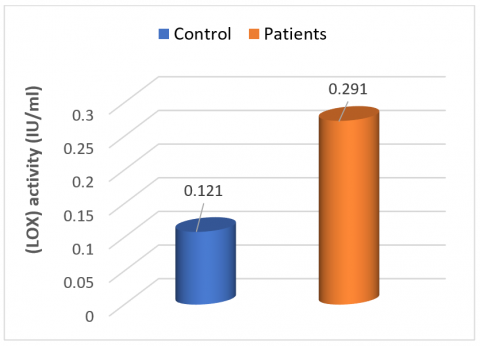
As shown in Table 1, these results reached statistically significant increase in the activity level of the lipoxygenase enzyme found in sera of patients infected with viral Hepatitis B. This elevation is highly statistically significantly elevated at the (P ≤ 0.001). These results suggest that viral Hepatitis B infection is correlated with enhanced lipoxygenase activity. The activity levels of lipoxygenase in the patients' sera are much higher than in comparison with sera of individuals who do not have the infection, the control group. Figure 2 visually demonstrates this finding, by showing how the Hepatitis B-infected group, as a result of this infection, has enzyme activity much higher than the control group. The enzyme is thought to play a role in inflammatory liver damage caused by viral infections, and the increased lipoxygenase activity supports its potential as a biomarker of the severity of liver impairment in Hepatitis B patients.
Table 1. Lipoxygenase (LOX) activity in patients' blood sera, infected with viral hepatitis b and healthy individuals (means ± standard deviations)
|
State |
No. |
LOX Activity Mean ± SD. (UI\ml) |
P Value |
|
Control |
40 |
0.121 ± 0.060 |
0.001 |
|
patients |
40 |
0.291 ± 0.071 |
Figure 2. Level of lipoxygenase (LOX) activity in patients suffering from viral hepatitis B, compared with normal individuals
However, lipoxygenase (LOX) activity in patients with viral Hepatitis B was higher (P ≤ 0.001) compared to the control group. Furthermore, this increased LOX activity is highly associated with Hepatitis B. The large level of statistical significance evokes the notion that the noticed growing in the enzyme activity is not due to chance deviation, and, therefore, evidences that the activity of LOX is very closely associated with the existence of Hepatitis B. This finding leads to the potential that LOX may be a biomarker to assess the sensitivity of the liver to viral Hepatitis B and support its usefulness in elucidating the physiologic basis of the disease.
The results of this study suggest that lipoxygenesses are enhanced to a great degree in sera taken from Viral Hepatitis B patients. These results are in agreement with reports in the literature. Allication of the typical type of lipoxygenase in patients with Hepatitis B was documented in a study of 2017 by Al-Fayyad and Al-lehebe [18] and Natarajan et al. [19] also reported elevated level of lipoxygenase in patients with breast cancer. In support of these observations, Al-Adlee and Al-Guburi [20] reported an elevation in lipoxygenase activity in blood samples taken from colon cancer patients.
In addition, Mahmood and Rashed [21] also stated (in 2013) that a rise in lipoxygenase activity was found in blood sera and tissues of persons with cardiovascular diseases. Further, Jawad et al. [22] found increased enzyme activity in prostate cancer patients, reasoning that the synthesis of eicosanoids and leukotrienes via increased digestion of fatty acids was responsible. This enhanced synthesis has inflammatory effects that promote the generation of cancerous tumors throughout many organs, including the liver. Collectively, these findings highlight the importance of lipoxygenase as a major biomarker for many diseases mediated by inflammation and cancer.
The activity of the lipoxygenase (LOX) was probably elevated in relation to exposure to external factors, the genetic factors, and the occurrence of infections. This creates a boost to the immune system leading the immune system to become triggered and release polyunsaturated fatty acids, including arachidonic acid. As the last group, these acids are fundamental substances for the lipoxygenase [23]. Therefore, metabolism of polyunsaturated fatty acids (such as arachidonic acid, linoleic acid and linolenic acid) is accelerated to yield many compounds, including eicosanoids and leukotrienes. They are closely associated with the pathway of Lipoxygenase (LOX). Moreover, leukotrienes comprise of biological formation lipoxins. Registered as important regulators in resolving and fighting inflammation, Lipoxins are powerful anti-inflammatory lipid mediators [24]. It has also been found to be important for the progression of inflammation and cancer. This could be explained by the fact that the cellular membrane becomes more permeable and because of these higher enzyme actions there is more permeability to chemicals. Membrane permeability increases in instances of inflammation and cancer so that the enzyme is released extracellularly. As a result, the enzyme is elevated in the bloodstream due to this extracellular release [25].
Inhibiting the activity of lipoxygenase has been elucidated to prevent the dissemination of inflammation, secondary growth of tumors and inflammatory response [26, 27].
3.1 Purification of lipoxygenase for individuals with viral hepatitis B
In the initial stages of enzyme purification, proteins are typically concentrated by removing a significant amount of water, which helps to achieve a desired level of purity. Salts, such as ammonium sulfate are used to facilitate this concentration process because they are highly soluble in water. The process is dependent on a method called "salting out" in which salts neutralize the protein's charge. The precipitating effect of this neutralization reduces the solubility of the proteins in the solution. A peptone gel for protein can be prepared by 'external salting out'; the technique, which is often referred to as 'external salting out' works by concentrating protein and separating it from other components in the mixture [28].
Thus, lipoxygenase (LOX) required a multi-step separation and partial purification to obtain. The enzyme was first concentrated by precipitating it 30% ammonium sulfate. In this step, fuel purity of 0.784173 times and enzyme yield equal to 73.68421% were obtained. Membrane dialysis was used to remove excess salt and to further purify enzyme through a buffer solution of 0.2 mol/L and pH of 7. The enzyme was purified to a purity of 0.784173 times after dialysis, with an enzyme yield of 73.68421%. This was achieved by this process to concentrate and partially purify the enzyme to ensure additional purification steps.
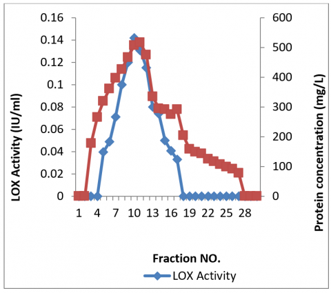
Since then, the LOX enzyme was further purified on by gel filtration chromatography with Sephadex G-100 resin as column-filling material. Application of this technique had one purified enzyme isomer giving a single symmetrical peak (Figure 3). The purification level obtained is 0.57554, yield of the enzyme was 26.10526% as displayed in Table 2. This step was a critical step towards making the enzyme much purer for more accurate analysis and characterization of the properties of the enzyme.
Figure 3. Lipoxygenase (LOX) was purified by gel filtration chromatography from blood sera of patients with viral hepatitis B
The oyster mushroom *Pleurotus ostreatus* was chosen to be extracted and purified its lipoxygenase sucessfully by Mustafa et al. [29]. Likewise, Aanangi et al. [30] got a distinct enzyme purification from mung beans, which ended up a single bundle of lipoxygenase. Lipoxygenase was purified from the human placenta by Mahmoud et al. [31], while Abd Al-Adlee and Al-Gubiri [32], purified a single peak of lipoxygenase from the blood sera of patients with prostate cancer.
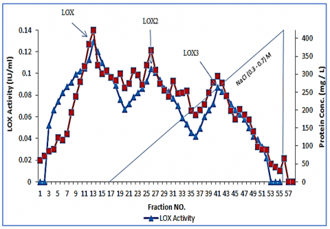
In this study, the isoenzymes of the partially purified lipoxygenase were separated using gel filtration chromatography followed by ion exchange chromatography with DEAE-Cellulose resin as the column-filling material. The process employed gradient solutions of sodium chloride, which are crucial for ion exchange chromatography as it relies on the charge differences among isoenzymes. Three distinct isoenzymes were isolated, as illustrated in Figure 4, each exhibiting varying levels of purity. Specifically, Isoenzyme I achieved a purity level of 0.73133, Isoenzyme II reached 0.67957, and Isoenzyme III attained 0.70723, as detailed in Table 2.
These results are consistent with previous research. Mahmood and Rashed [21] identified three isoenzymes of lipoxygenase during the separation and measurement of enzyme levels in the blood of males with cardiovascular diseases. Additionally, Al-Fayyad and Al-lehebe [18] successfully Kinetic and inhibitory study of partially purified lipoxygenase from epilepsy patients’ serum. These findings collectively underscore the consistency and reproducibility of lipoxygenase purification and characterization across different biological sources and disease contexts.
Figure 4. Separation of lipoxygenase (LOX) isoenzymes from blood sera of patients afflicted with viral hepatitis B using ion exchange chromatography
Table 2. The partial separation and purification of lipoxygenase isomers from the blood sera of patients afflicted with viral hepatitis B
|
Step |
Elute (mI) |
Activity (IU/ml) |
Total Activity (UI) |
Protein Conc. (mg/L) |
Total Protein (mg) |
Specific Activity (IU/mg) |
Purification on (Fold) |
Yield % |
|
Crud serum |
5 |
0.285 |
0.001425 |
684 |
3.42 |
0.000417 |
1 |
100 |
|
Ammonium sulphate |
5 |
0.210 |
0.00105 |
643 |
3.215 |
0.000327 |
0.784173 |
73.68421 |
|
Dialysis |
3.5 |
0.200 |
0.0007 |
595 |
2.0825 |
0.000336 |
0.805755 |
49.12281 |
|
Gel filtation |
3 |
0.124 |
0.000372 |
517 |
1.551 |
0.00024 |
0.57554 |
26.10526 |
|
lon exchange |
|
|
|
|
|
|
|
|
|
LOX1 |
3 |
0.129 |
0.000387 |
423 |
1.269 |
0.000304965 |
0.731330935 |
27.15789474 |
|
LOX2 |
3 |
0.104 |
0.000312 |
367 |
1.101 |
0.000283379 |
0.679565947 |
21.89473684 |
|
LOX3 |
3 |
0.087 |
0.000261 |
295 |
0.885 |
0.000294915 |
0.707230216 |
18.31578947 |
In conclusion, the study highlights a notable increase in the activity of the lipoxygenase enzyme among individuals infected with the B-type hepatitis virus. This heightened enzyme activity is an important marker of the disease's impact on the liver. Furthermore, the purification process of the enzyme proteins revealed distinct patterns during chromatographic analysis: a single peak was observed when using gel chromatography, while three peaks were identified with ion-exchange chromatography utilizing a saline gradient. These findings serve as a diagnostic indicator of liver damage, reflecting the enzyme's altered behavior in response to the viral infection.
[1] Liao, J.L. (2017). Cigarette smoke-induced chronic inflammation leading to COPD and lung cancer: A multiscale modeling study. Journal of Immunobiology, 2(1): 119. https://doi.org/10.4172/2476-1966.1000119
[2] Mohammed, M.J., Awadh, H.A., Gaidan, A.M. (2023). The relationship of serum level of interleukin-4 with the level of anti-hepatitis b antibodies. Journal of Advanced Education and Sciences, 3(3): 01-04.
[3] Hughes, E., Bassi, S., Gilbody, S., Bland, M., Martin, F. (2016). Prevalence of HIV, hepatitis B, and hepatitis C in people with severe mental illness: a systematic review and meta-analysis. The Lancet Psychiatry, 3(1): 40-48.
[4] Upadhyay, R.K., Mattoo, A.K. (2018). Genome-wide identification of tomato (Solanum lycopersicum L.) lipoxygenases coupled with expression profiles during plant development and in response to methyl-jasmonate and wounding. Journal of Plant Physiology, 231: 318-328. https://doi.org/10.1016/j.jplph.2018.10.001
[5] Al-Sarraf, F.S., Hussien, I.A. (2015). Evaluation of the proliferation marker Ki67 as a prognostic factor in patients with breast carcinoma. Journal of the Faculty of Medicine Baghdad, 57(2): 151-155.
[6] Hendrick, R.E., Helvie, M.A. (2011). United States preventive services task force screening mammography recommendations: science ignored. American Journal of Roentgenology, 196(2): W112-W116. https://doi.org/10.2214/AJR.10.5609
[7] Charpin, C., Bonnier, P., Khouzami, A., Andrac, L., Habib, M., Vacheret, H., Lavaut, L.N., Piana, L. (1993). Non palpable breast carcinomas: Histological and immunohistochemical studies of 160 cases. Pathology-Research and Practice, 189(3): 267-274. https://doi.org/10.1016/S0344-0338(11)80509-1
[8] Jawad, Z.N. (2018). The association of urokinase gene 3'-UTR T/C polymorphism with urinary bladder cancer. Biochemical & Cellular Archives, 18(1): 587.
[9] Mastropasqua, M.G. (2013). Breast cancer pathology. In: Oncoplastic and Reconstructive Breast Surgery, pp. 45-53. https://doi.org/10.1007/978-88-470-2652-0_4
[10] Fisher, B., Anderson, S., Bryant, J., Margolese, R.G., Deutsch, M., Fisher, E.R., et al. (2002). Twenty-year follow-up of a randomized trial comparing total mastectomy, lumpectomy, and lumpectomy plus irradiation for the treatment of invasive breast cancer. New England Journal of Medicine, 347(16): 1233-1241. https://doi.org/10.1056/NEJMoa022152
[11] Kim, K.J., Huh, S.J., Yang, J.H., Park, W., et al. (2005). Treatment results and prognostic factors of early breast cancer treated with a breast conserving operation and radiotherapy. Japanese Journal of Clinical Oncology, 35(3): 126-133. https://doi.org/10.1093/jjco/hyi039
[12] Karupusamy, S., Mustafa, M.A., Jos, B.M., Dahiya, P., Bhardwaj, R., Kanani, P., Kumar, A. (2023). Torque control-based induction motor speed control using Anticipating Power Impulse Technique. The International Journal of Advanced Manufacturing Technology, 1-9. https://doi.org/10.1007/s00170-023-10893-5
[13] Wisastra, R., Dekker, F.J. (2014). Inflammation, cancer and oxidative lipoxygenase activity are intimately linked. Cancers, 6(3): 1500-1521. https://doi.org/10.3390/cancers6031500
[14] Li, J., Rao, J., Liu, Y., Cao, Y., Zhang, Y., Zhang, Q., Zhu, D. (2013). 15-Lipoxygenase promotes chronic hypoxia–induced pulmonary artery inflammation via positive interaction with nuclear factor-κB. Arteriosclerosis, Thrombosis, and Vascular Biology, 33(5): 971-979. https://doi.org/10.1161/ATVBAHA.113.301335
[15] Early Breast Cancer Trialists' Collaborative Group. (2000). Favourable and unfavourable effects on long-term survival of radiotherapy for early breast cancer: An overview of the randomised trials. The Lancet, 355(9217): 1757-1770. https://doi.org/10.1016/S0140-6736(00)02263-7
[16] Shastry, B.S., Raghavendra Rao, M.R. (1975). Studies on lipoxygenase from rice bran. Cereal Chemistry, 52(5): 597-603.
[17] Lowry, O.H., Rosebrough, N.J., Farr, A.L., Randall, R.J. (1951). Protein measurement with the Folin phenol reagent. Journal of Biological Chemistry, 193(1): 265-275. https://doi.org/10.1016/S0021-9258(19)52451-6
[18] Al-Fayyad, A., Al-lehebe, N.I. (2021). Kinetic and inhibitory study of partially purified lipoxygenase from epilepsy patients serum. Journal of Education and Science, 30(1): 58-71. https://doi.org/10.33899/edusj.2020.127778.1093
[19] Natarajan, R., Esworthy, R., Bai, W., Gu, J.L., Wilczynski, S., Nadler, J. (1997). Increased 12-Lipoxygenase Expression in Breast Cancer Tissues and Cells. Regulation by Epidermal Growth Factor 1. The Journal of Clinical Endocrinology & Metabolism, 82(6): 1790-1798. https://doi.org/10.1210/jcem.82.6.3990
[20] Al-Adlee, M.A.A., Al-Guburi, N.A.S. (2020). Estimating lipoxygenase and gamma-glutamyl transferase activities in sera of colon cancer patients with partial purification of lipoxygenase. Baghdad Science Journal, 17(2): 481-487. https://doi.org/10.21123/bsj.2020.17.2.0481
[21] Mahmood, A.A., Rashed, R.N. (2013) Separation of lipoxygenase and estimation of its level in blood of males with cardiovascular disease. Rafidain Journal of Science, 24(2): 65-81.
[22] Jawad, Z.N., AR, K., Awad, W. (2020). Association of additive risk of pioglitazone use with the presence of CYP1A1 polymorphisms in the occurrence of bladder cancer. Systematic Reviews in Pharmacy, 11(5): 375. https://doi.org/10.31838/srp.2020.5.53
[23] Siesjö, B.K., Agardh, C.D., Bengtsson, F., Smith, M.L. (1989). Arachidonic acid metabolism in seizures. Annals of the New York Academy of Sciences, 559(1): 323-339. https://doi.org/10.1111/j.1749-6632.1989.tb22619.x
[24] Wächtershäuser, A., Steinhilber, D., Loitsch, S.M., Stein, J. (2000). Expression of 5-lipoxygenase by human colorectal carcinoma Caco-2 cells during butyrate-induced cell differentiation. Biochemical and Biophysical Research Communications, 268(3): 778-783. https://doi.org/10.1006/bbrc.2000.2213
[25] Cole, B.K., Lieb, D.C., Dobrian, A.D., Nadler, J.L. (2013). 12-and 15-lipoxygenases in adipose tissue inflammation. Prostaglandins & Other Lipid Mediators, 104: 84-92. https://doi.org/10.1016/j.prostaglandins.2012.07.004
[26] Samuelsson, B., Dahlen, S.E., Lindgren, J.Å., Rouzer, C.A., Serhan, C.N. (1987). Leukotrienes and lipoxins: Structures, biosynthesis, and biological effects. Science, 237(4819): 1171-1176. https://doi.org/10.1126/science.2820055
[27] Pidgeon, G.P., Lysaght, J., Krishnamoorthy, S., Reynolds, J.V., O’Byrne, K., Nie, D., Honn, K.V. (2007). Lipoxygenase metabolism: Roles in tumor progression and survival. Cancer and Metastasis Reviews, 26(3): 503-524. https://doi.org/10.1007/s10555-007-9098-3
[28] Duong-Ly, K.C., Gabelli, S.B. (2014). Salting out of proteins using ammonium sulfate precipitation. In: Methods in Enzymology, 541: 85-94. https://doi.org/10.1016/B978-0-12-420119-4.00007-0
[29] Mustafa, M.A., Raja, S., Asadi, L.A.A., Jamadon, N.H., Rajeswari, N., Kumar, A.P. (2023). A decision‐making carbon reinforced material selection model for composite polymers in pipeline applications. Advances in Polymer Technology, 2023(1): 6344193. https://doi.org/10.1155/2023/6344193
[30] Aanangi, R., Kotapati, K.V., Palaka, B.K., Kedam, T., Kanika, N.D., Ampasala, D.R. (2016). Purification and characterization of lipoxygenase from mung bean (Vigna radiata L.) germinating seedlings. 3 Biotech, 6(1): 1-8. https://doi.org/10.1007/s13205-016-0427-5
[31] Mahmoud, Z.H., Ajaj, Y., Hussein, A.M., Al-Salman, H.N.K., Mustafa, M.A., et al. (2024). CdIn2Se4@ chitosan heterojunction nanocomposite with ultrahigh photocatalytic activity under sunlight driven photodegradation of organic pollutants. International Journal of Biological Macromolecules, 267: 131465. https://doi.org/10.1016/j.ijbiomac.2024.131465
[32] Abd Al-Adlee, M.A., Al-Guburi, N.A.S. (2020). Estimation, partial purification of lipoxygenase and estimate GGT in the blood patients with prostate cancer. Diyala Journal for Pure Science, 16(1): 84-97. https://doi.org/10.24237/djps.16.01.515B