Saranya Sekar*![]() | Lakshmi Sankaran
| Lakshmi Sankaran![]()
© 2024 The authors. This article is published by IIETA and is licensed under the CC BY 4.0 license (http://creativecommons.org/licenses/by/4.0/).
OPEN ACCESS
The escalating prevalence of genetic disorders in humans underscores the criticality of early detection. Current clinical methodologies, such as cellular activity assessment and chromosomal examination, are widely employed for early-stage prediction of these disorders. However, traditional approaches to chromosomal cell (CC) examination are intricate and labor-intensive. This study proposes a novel deep learning framework (DLF) to overcome these challenges by precisely and efficiently segmenting and classifying CCs. As part of this scheme, images are collected and resized, the DLF is trained using the selected images, segmentation and deep feature extraction of the CC are performed. The proposed methodology involves image collection and resizing, training the DLF with chosen images, CC segmentation and feature extraction and multiclass classification and performance verification. This work implements the VGG-UNet plan to examine the chosen CC images collected from the Biomedical Imaging Laboratory (BioImLab). An experimental inquiry is being carried out on 5474 images and the achieved findings are addressed. The findings of this research suggest that the presented work help to provide a detection accuracy of >98% with the K-Nearest Neighbor supported classifier. This research can be expanded in the future to detect the genetic disorder based on the information obtained from the CC.
chromosome cells, deep-learning, VGG-UNet, segmentation, classification, testimonial.
Recent improvements in science and engineering have led to the automation of numerous examination procedures, particularly in the examination of biological data. The improvements in computing facilities and the accessibility of recent computational tools have empowered us to implement a wide range of diagnostic and detection procedures. This enables accurate evaluation of biological and medical information collected from the human subjects [1, 2].
In the current era, Artificial Intelligence (AI) plans are widely employed to solve a number of complex engineering tasks including medical image processing problems [3]. The advancements in the AI also helps to develop and implement various procedures to examine the complex medical images. As a result of the current research in the biological and medical fields, it has been confirmed that AI frameworks such as heuristic algorithms, Machine-Learning Methods (MLMs) as well as Deep-Learning Mechanisms (DLMs) are being utilized to increase automatic diagnosis accuracy [4, 5].
As part of a genetic disorder examination, chromosome cell (CC) karyotyping is a common biological test. In this task, a person can be examined using the following information; number of available CCs, deletion of a chromosome, duplication of a chromosome, and chromosomes with unusual structures. These analyses play a vital role during the clinical level diagnosis of Edwards syndrome, Turner syndrome, and Down syndrome are examples of genetic illnesses. As an automatic assessment of individual cells, segmentation and classification are involved. As far back as the early cytogeneticists were concerned, the CC was classified as twenty-two pairs of autosomes and one set of sex chromosomes. The automatic classification of the CC is based on its texture and shape features. There are two types of features in a CC: Handcrafted (traditional) Features (HF) as well as Learned Features (LF) derived with the help of a chosen Artificial Intelligence scheme. The past few years, the availability of modern computing facilities has led to the implementation of DLM in a variety of domains, including CC karyotyping, because of the accessibility of the modern computing facility. In order to achieve a higher level of clinical significance, a DLM can be implemented to perform automatic segmentation and classification. As a result, DLM schemes have been widely applied in research works related to CC karyotyping that have been conducted recently [6, 7]. Earlier works on CC karyotyping suggests that the existing procedures provides a satisfactory result on this task and it needs to be improved further with a modified Convolutional Neural Network (CNN) scheme. In this research, we plan to create a framework by integrating the Convolutional Neural Network -supported Segmenting and classification to enhance the results of CC karyotyping by integrating CNN-supported segmentation and classification approaches. A new feature of this work is the implementation of VGG-UNet (VGG19 acts as the backbone) for segmenting the CC region and extracting the HF, including GLCMs and DWTs [8-10]. In this scheme, the pretrained VGG19 functions as the backbone (encoder) while the decoder is constructed based on the encoder structure. In order to form a dominant hybrid feature vector (HF+LF), the HF from the segmented images is combined with the LF from the VGG19 scheme to form a dominant hybrid feature vector (HF+LF). This feature vector will then be used in a cross-validation process for performing the classification task. With the help of different classifiers chosen by the authors of this study, the experimental outcome of this work is verified and compared with the recent work by Al-Kharraz et al. [11]. The proposed scheme has higher levels of classification accuracy than the other approaches in the literature, according to the experimental findings of this research.
The contributions of this research involve;
(i) Application of a CNN segmentation method to extract CCs and compute their shape and texture features based on the extracted CC.
(ii) Improvement of CC karyotyping accuracy through HF and LF classification.
(iii) Evaluation of the scheme's performance and assessment of its clinical relevance through comprehensive performance appraisal and validation.
CC karyotyping is a critical component of clinical-level genetic disorder prediction, prompting various research endeavors as an alternative to the complex and time-consuming conventional methods.
In their work, Pallavoor et al. [12] introduced deep CNN-supported segmentation schemes for CC extraction and evaluation that covered both Q-band and G-band. The presented approach attained a remarkable classification accuracy of 95.75% through the utilization of extracted features, employing a pre-trained SqueezeNet model.
Hernández-Mier et al. [13] presented a machine learning-based automatic karyogram generation method using G-band CC. They employed 119 Q-band images and 24 G-band images, achieving classification accuracy exceeding 90% for two-class classification and over 79% for multiclass classification. The clinical significance of their approach was substantiated with clinically collected images.
Liu et al. [14] advocated a deep learning model (DLM) coupled with the SRAS-net classifier for CC classification in low-resolution images. Their innovative SRAS-net, which integrates InceptionV2 with ResNet, combined with other techniques like the Image Adaptive Module (IAM), SMOTE, and self-adaptive negative feedback network, resulted in an impressive 97.55% accuracy in CC detection.
The study of Al-Kharraz et al. [11] proposed the application of a deep learning algorithm for categorized the CC, and with the VGG19 scheme, they achieved a classification accuracy of >95%. As a result of the results and discussion presented in this paper, we are able to conclude that the proposed approach to CC evaluation is efficient and outperforms more similar existing works in the field.
The earlier works on the CC detection provided an accuracy of 95 to 97.55% and this result need to be improved using an appropriate methodology. To achieve a better detection accuracy of CC, the proposed research work is aimed at developing an integrated deep segmentation and classification technique. As part of this work, VGG-UNet (VGG19 as the backbone) is utilized to extract the chromosomes effectively and shape and texture features are extracted by using handcrafted features (HF) and learned features (LF), and the fusion of these features, denoted as HF+LF, is subsequently deployed in a comprehensive five-fold cross validation framework tailored for the multiclass classification task. Experimental investigations conducted within this study unequivocally establish the superior functionality of the strategy we've suggested when juxtaposed with existing methodologies delineated in the extant literature.
An essential facet of early-stage genetic disease diagnosis is the automatic karyotyping of chromosome cells (CC). This procedure entails the examination of CC using a carefully selected methodology to optimize results. Previous literature has firmly established that deep learning methods (DLM) serve as effective tools for improving the accuracy of CC examinations. Consequently, the proposed scheme incorporates a DLM-supported segmentation and classification system to enhance the evaluation of CC.
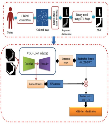
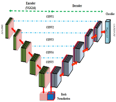
Figure 1 illustrates the procedural framework adopted in this research, encompassing the following pivotal stages: (i) Image acquisition and preprocessing; (ii) Utilization of VGG-UNet for the segmentation of chromosome cells (CCs); (iii) Extraction of handcrafted features (HF) from the segmented images; (iv) Extraction of learned features (LF) from a pre-trained deep learning model (DLM); and (v) Integration and classification of the derived sequential features. Within this study, the assessment of classification accuracy, facilitated by a carefully chosen classifier, serves as the benchmark for evaluating the efficacy of the proposed methodology. A central component of this framework is the VGG-UNet, as depicted in Figure 1(b), which significantly contributes to enhancing the precision of CC mining.
(a) Proposed scheme
(b) VGG-UNet
Figure 1. Structure of the proposed CC detection process
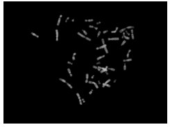
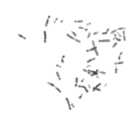
(a) Original image
(b) Complement image
(c) Detected chromosomes
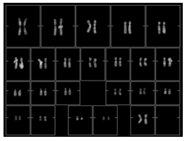
(d) Karyotyping
Figure 2. Sample images available for segmentation task
3.1 Database
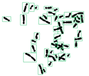
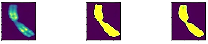
The performance evaluation of the developed chromosome cell (CC) detection tool necessitates rigorous validation using a benchmark database. In this study, we have utilized the test images available from the well-established Biomedical Imaging Laboratory (BioImLab) database, and comprehensive details regarding this dataset can be accessed from Laboratory of Biomedical Imaging [15]. This dataset encompasses two distinct image sets designed to support both segmentation and classification tasks. The images presented in Figure 2 serve a dual purpose, catering to both segmentation and karyotyping. Specifically, Figure 2(a) showcases a Q-band prometaphase image for assessment, while Figure 2(b) represents the complementary image. Figure 2(c) portrays the contrast-adjusted and segmented chromosome images, and Figure 2(d) exhibits the karyotype information included within the dataset.
In the context of this research proposal, we have directed our focus towards the classification database, implementing a systematic approach that includes the following key steps: (i) resizing, (ii) mask generation utilizing ITK-Snap software [16, 17], and (iii) segmentation and classification, guided by the VGG-UNet architecture. For the segmentation task, we have specifically considered 5474 images available in this database for assessment and classification. Within this set, a subset of 500 images, each accompanied by a binary mask, has been selected to perform the segmentation process. These images are then considered for training and validating the proposed VGG-UNet algorithm. By using the trained VGG-UNet to extract the CC section (binary image) from the selected images from all 5474 images, the binary image of the CC is extracted, which is necessary to extract the HF, such as GLCM and DWT. The purpose of this study is to use the pretrained VGG-UNet scheme in order to achieve both
3.2 VGG-UNet
UNet is one of the commonly employed image segmentation practices to extract chosen regions from the digital image. The advantages of the UNet and its variations are supported by earlier research in the literature [18, 19]. The application of VGG-UNet in image extraction can be found in the studies [20, 21]. Khan et al. [9] proposed the VGG-UNet scheme that can be used to extract CC from selected test images. This scheme uses the VGG19 as a backbone (Encoder) and its inverted structure to function as the decoder. There is also the addition of another layer, which is a SoftMax, which extracts the CC section of the test image. The extracted section will be in the form of binary; therefore, it can be used later for the mining of GLCM and DWT features. Information about the VGG-UNet can be found in the studies [22-25]. The initial values for VGG-UNet are assigned as follows; Adam optimizer, MaxPooling layer and ReLu.
The final layer within the decoder is equipped with SoftMax, which plays a pivotal role in extracting the binary representation of the target image section.
A commonly used CNN scheme is image segmentation with UNet (UNet), which can be used to extract parts of a digital image for analysis. Earlier publications in the literature [18, 19] support the UNet and its derivatives. The application of VGG-UNet in image extraction can be found in the studies [20, 21]. Khan et al. [9] proposed the VGG-UNet scheme that can be used to extract CC from selected test images. This scheme uses the VGG19 as a backbone (encoder) and its inverted structure to function as the decoder. There is also the addition of another layer, which is a SoftMax, which extracts the CC section of the test image. The extracted section will be in the form of binary; therefore, it can be used later for the mining of GLCM and DWT features. Information about the VGG-UNet can be found at the studies [22-25]. The following are the initial values for VGG-UNet: Adam optimizer, MaxPoolinglayer, and andReLu. The final section of the decoder is associated with SoftMax to extract the binary image.
3.3 Feature extraction and integration
The proposed scheme emphasizes the importance of feature extraction and the importance of feature supported detection. In this work, both the HF and LF are considered. GLCM [26, 27] and DWT [28, 29] were used so as to obtain the necessary HF. The outcomes of these methods can be obtained by using Eq. (1) and Eq. (2).
$\operatorname{GLCM}_{(1 \times 1 \times 44)}=$ GLCM 1, GLCM $2, \ldots$, GLCM 44 (1)
$D W T_{(1 \times 1 \times 26)}=D W T 1, D W T 2, \ldots, D W T 26$ (2)
This scheme proposes a LF vector of dimension 1×1×1000 that is then reduced by 50% to 1×1×500, resulting in a new hybrid feature vector (HF+LF) of dimension as presented in equation. The classifier is then trained and validated using a five-fold cross-validation procedure based on Eq. (3), which is referred to as the hybrid feature vector (HF+LF).
$\begin{gathered}(H F+L F)_{(1 \times 1 \times 570)}=G L C M_{(1 \times 1 \times 44)}+ D W T_{(1 \times 1 \times 26)}+L F_{(1 \times 1 \times 500)}\end{gathered}$ (3)
3.4 Performance assessment and endorsement
A number of classifiers, such SoftMax, k-NN, Random decision forests (RF), and support-vector networks (SVM), are employed to evaluate the suggested approach, and the results obtained are compared to and justified against those published in the literature. In this study, only classification accuracy is evaluated, and its expression is given in Eq. (4) [30-33].
$Accuracy =A C=\frac{T P+T N}{T P+T N+F P+F N}$ (4)
This portion of the study demonstrates the experimental results obtained using Python software. This experiment is conducted using a work station having Intel i7 processor, 16GB of RAM, and 4GB of video memory.
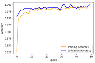
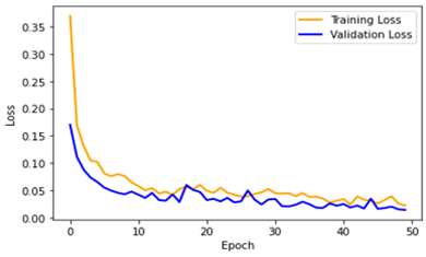
Training and testing the VGG-UNet algorithm with properly picked images accompanied by binary masks is the first step in verifying the reliability of the segmentation process. We continuously monitor the system's efficiency during the training phase by analyzing accuracy and loss statistics. It's worth noting that this research uses an CNN architecture for both separation and classification.
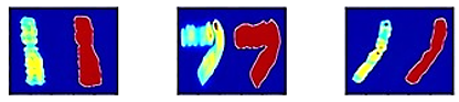
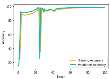
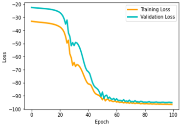
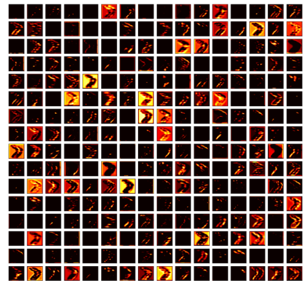
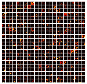
Our research investigates accuracy and loss values as critical verification measures in beneficial to thoroughly appraise the efficiency of the chosen method. Figure 3 depicts these metrics graphically, with Figure 3(a) displaying the training images, which include both the original picture and its associated mask, and Figures 3(b-c) displaying the accuracy and loss values, respectively. Figure 3(d) showcases the resulting segmented binary image.
(a) Training Images
(b) Accuracy
(c) Loss
(d) Validation
Figure 3. The outcome of VGG-UNet on the given database
From the observations made in Figure 3(d), it becomes evident that the predicted chromosome closely aligns with the provided mask, serving as compelling evidence for the efficacy of the segmentation achieved through the VGG-UNet model. Subsequently, this segmented image is employed for the extraction of essential shape features, further enhancing the depth of our analysis.
As a result, the proposed segmentation model achieves an accuracy of 94.27% and accurately extracts the binary image of CC from the selected test image. As discussed in Eq. (3), handcrafted features (HFs) such as GLCM and DWT are extracted from these images and combined with learned features (LFs). The proposed scheme is subsequently tested using a multiclass classifier to validate the accuracy of this one-dimensional feature vector.
(a) Conv1
(b) Conv2
(c) Conv3
(d) Conv4
Figure 4. Intermediate layer result obtained from VGG19
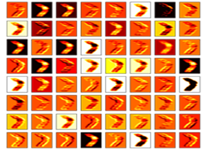
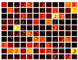
The VGG19-supported image classification task is initially executed using the SoftMax classifier. The necessary outcomes from the CNN scheme are obtained during this operation, as presented in Figures 4 and 5. Figure 4 shows the images collected from various convolutional layers of VGG19, and Figures 4(a-d) present the outcomes of convolutional layer 1 (Conv1) to convolutional layer 4 (Conv4), confirming that the chosen image is transformed into features during this process. Figure 5 depicts the accuracy and loss values computed during the training and validation processes for 50 epochs.
The proposed scheme achieves a classification accuracy of 96.93% with SoftMax, 98.04% with KNN, 96.36% with RF, and 97.28% with the SVM classifier. The achieved accuracy is then verified against other results found in the literature, confirming the merit of VGG-UNet.
(a) Accuracy
(b) Loss
Figure 5. Training and validation performance of VGG19 for the chosen database
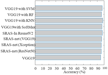
The data presented in Table 1 unequivocally demonstrate that the outcomes achieved in this study surpass those of alternative methods documented in the literature [11, 14]. This substantiates the excellence of our proposed approach relative to existing methodologies. Figure 6 provides a visual comparison of accuracy between our proposed method and the methodologies under consideration, with the combined approach of our proposed scheme and K-Nearest Neighbor (KNN) yielding the most favorable results.
Through the integration of VGG-UNet with the VGG19 backbone in this research, we have successfully implemented a unified segmentation and classification method, resulting in enhanced chromosome cell (CC) segmentation and detection accuracy. Looking forward, there is potential for further improvements by exploring the fusion of learned features (LF) with an ensemble of LF-based methods, as this scheme capitalizes on the synergy between both handcrafted features (HF) and LF to achieve heightened classification accuracy.
The findings derived from this research unequivocally validate the effectiveness of the proposed technique when applied to the selected database. In the future, the performance of this scheme can be extended to include the evaluation of clinically collected CC images, offering prospects for even more robust validation and application.
Table 1. Multiclass classification accuracy during chromosome detection
|
Reference |
Scheme |
Accuracy (%) |
|
Al-Kharraz et al. [11] |
VGG19 |
95.04 |
|
Liu et al. [14] |
SRAS-net (ResNet50) |
95.92 |
|
SRAS-net (Xception) |
96.01 |
|
|
SRAS-net (VGG19) |
96.56 |
|
|
SRAS-net (inception_resnet_V2) |
97.55 |
|
|
Proposed |
VGG19 with SoftMax |
96.88 |
|
VGG19 with KNN |
98.11 |
|
|
VGG19 with RF |
96.28 |
|
|
VGG19 with SVM |
97.19 |
Figure 6. Comparison of classification accuracy of the proposed work with existing methods
This research introduces a novel approach for the automated recognition of chromosome cells (CC) through the integration of Convolutional Neural Networks (CNNs) for segmentation and classification tasks. Specifically, the VGG19 architecture is harnessed to execute these essential functions. The initial phase of the proposed tool employs the VGG-UNet model to effectively segment the requisite CC section, while the subsequent phase leverages the VGG19 model for classification. The CNN-based segmentation process results in a binary representation of the CC, which is subsequently utilized for the extraction of Handcrafted Features (HF) such as Gray-Level Co-occurrence Matrix (GLCM) and Discrete Wavelet Transform (DWT).
To enhance the discriminative power of our approach, this research generates a hybrid feature vector by sequentially combining Deep Features (DF), GLCM, and DWT-derived features. This feature vector is then employed to provide a binary classification outcome with the aid of a chosen classifier. The empirical findings of this study unequivocally establish the superiority of the K-Nearest Neighbor (KNN) classifier, achieving classification accuracy surpassing 98%, which outperforms existing methods documented in the literature.
However, it's important to acknowledge a limitation inherent in this scheme, which primarily relies on CNN-based segmentation and classification. In future investigations, alternative approaches for CC classification, distinct from segmentation, could be explored to further enhance the versatility and robustness of the proposed methodology.
The images utilized in this study are accessible at the following link: https://bioimlab.dei.unipd.it/Chromosome%20Data%20SSe%204Class.htm.
The authors affirm that there are no conflicts of interest to disclose.
[1] Collins, R.L., Brand, H., Karczewski, K.J., Zhao, X.F., Alföldi, J., Francioli, L.C., Khera, A.V., Lowther, C., Gauthier, L.D., Wang, H., Watts, N.A., Solomonson, M., O’Donnell-Luria, A., Baumann, A., Munshi, R., Walker, M., Whelan, C.W., Huang, Y.Q., Brookings, T., Sharpe, T., Stone, M.R., Valkanas, E., Fu, J., Tiao, G., Laricchia, K.M., Ruano-Rubio, V., Stevens, C., Gupta, N., Cusick, C., Margolin, L., Team, G.A.D.P., Consortium, G.A.D., Taylor, K.D., Lin, H.J., Rich, S.S., Post, W.S., Chen, Y.D.I., Rotter, J.I., Nusbaum, C., Philippakis, A., Lander, E., Gabriel, S., Neale, B.M., Kathiresan, S., Daly, M.J., Banks, E., MacArthur, D.G., Talkowski, M.E. (2020). A structural variation reference for medical and population genetics. Nature, 581(7809): 444-451. https://doi.org/10.1038/s41586-020-2287-8
[2] Luo, H.M., Li, M., Yang, M.Y., Wu, F.X., Li, Y.H., Wang, J.X. (2021). Biomedical data and computational models for drug repositioning: a comprehensive review. Briefings in Bioinformatics, 22(2): 1604-1619. https://doi.org/10.1093/bib/bbz176
[3] Mahmud, M., Kaiser, M.S., McGinnity, T.M., Hussain, A. (2021). Deep learning in mining biological data. Cognitive Computation, 13: 1-33. https://doi.org/10.1007/s12559-020-09773-x
[4] Ganeshkumar, M., Ravi, V., Sowmya, V., Gopalakrishnan, E.A., Soman, K.P. (2021). Explainable deep learning-based approach for multilabel classification of electrocardiogram. IEEE Transactions on Engineering Management, 70(8): 2787-2799. https://doi.org/10.1109/TEM.2021.3104751
[5] Jawahar, M., Ravi, V., Prassanna, J., Jasmine, S.G., Manikandan, R., Sekaran, R., Kannan, S. (2022). CovMnet-deep learning model for classifying coronavirus (COVID-19). Health and Technology, 12(5): 1009-1024. https://doi.org/10.1007/s12553-022-00688-1
[6] Menaka, D., Vaidyanathan, S.G. (2022). Chromenet: A CNN architecture with comparison of optimizers for classification of human chromosome images. Multidimensional Systems and Signal Processing, 33(3): 747-768. https://doi.org/10.1007/s11045-022-00819-x
[7] Remani Sathyan, R., Chandrasekhara Menon, G., Thampi, R., Duraisamy, J.H. (2022). Traditional and deep‐based techniques for end‐to‐end automated karyotyping: a review. Expert Systems, 39(3): e12799. https://doi.org/10.1111/exsy.12799
[8] Daniel, J., Rose, J.T.A., Vinnarasi, F.S.F., Rajinikanth, V. (2022). VGG-UNet/VGG-SegNet supported automatic segmentation of endoplasmic reticulum network in fluorescence microscopy images. Scanning, 2022. https://doi.org/10.1155/2022/7733860
[9] Khan, M.A., Rajinikanth, V., Satapathy, S.C., Taniar, D., Mohanty, J.R., Tariq, U., Damaševičius, R. (2021). VGG19 network assisted joint segmentation and classification of lung nodules in CT images. Diagnostics, 11(12): 2208. https://doi.org/10.3390/diagnostics11122208
[10] Abunadi, I., Senan, E.M. (2021). Deep learning and machine learning techniques of diagnosis dermoscopy images for early detection of skin diseases. Electronics, 10(24): 3158. https://doi.org/10.3390/electronics10243158
[11] Al-Kharraz, M.S., Elrefaei, L.A., Fadel, M.A. (2020). Automated system for chromosome karyotyping to recognize the most common numerical abnormalities using deep learning. IEEE Access, 8: 157727-157747. https://doi.org/10.1109/ACCESS.2020.3019937
[12] Pallavoor, A.S., TS, S., Pallavoor, S.K. (2022). Chromosome segmentation analysis using image processing techniques and autoencoders. arXiv Preprint arXiv: 2209.05414. https://doi.org/10.48550/arXiv.2209.05414
[13] Hernández-Mier, Y., Nuño-Maganda, M.A., Polanco-Martagón, S., García-Chávez, M.D.R. (2020). Machine learning classifiers evaluation for automatic karyogram generation from G-banded metaphase images. Applied Sciences, 10(8): 2758. https://doi.org/10.3390/app10082758
[14] Liu, X.B., Fu, L.J., Lin, J.C.W., Liu, S. (2022). SRAS‐net: Low‐resolution chromosome image classification based on deep learning. IET Systems Biology, 16(3-4): 85-97. https://doi.org/10.1049/syb2.12042
[15] BioImLab - Laboratory of Biomedical Imaging. https://bioimlab.dei.unipd.it/Data%20Sets.htm, accessed on May. 30, 2020.
[16] Yushkevich, P.A., Piven, J., Hazlett, H.C., Smith, R.G., Ho, S., Gee, J.C., Gerig, G. (2006). User-guided 3D active contour segmentation of anatomical structures: significantly improved efficiency and reliability. Neuroimage, 31(3): 1116-1128. https://doi.org/10.1016/j.neuroimage.2006.01.015
[17] itk-SNAP. http://www.itksnap.org/pmwiki/pmwiki.php, accessed on Apr. 26, 2022.
[18] Ronneberger, O., Fischer, P., Brox, T. (2015). U-net: convolutional networks for biomedical image segmentation. In Medical Image Computing and Computer-Assisted Intervention-MICCAI: 18th International Conference, Springer International Publishing, 234-241. https://doi.org/10.1007/978-3-319-24574-4_28
[19] Jain, P.K., Dubey, A., Saba, L., Khanna, N.N., Laird, J.R., Nicolaides, A., Fouda, M.M., Suri, J.S., Sharma, N. (2022). Attention-based UNet deep learning model for plaque segmentation in carotid ultrasound for stroke risk stratification: an artificial intelligence paradigm. Journal of Cardiovascular Development and Disease, 9(10): 326. https://doi.org/10.3390/jcdd9100326
[20] Krishnamoorthy, S., Zhang, Y.X., Kadry, S., Yu, W.F. (2022). Framework to segment and evaluate multiple sclerosis lesion in MRI slices using VGG-UNet. Computational Intelligence and Neuroscience. https://doi.org/10.1155/2022/4928096
[21] Nawaz, A., Akram, U., Salam, A.A., Ali, A.R., Rehman, A.U., Zeb, J. (2021). VGG-UNET for brain tumor segmentation and ensemble model for survival prediction. In 2021 International Conference on Robotics and Automation in Industry (ICRAI), Rawalpindi, Pakistan, pp. 1-6. https://doi.org/10.1109/ICRAI54018.2021.9651367
[22] Fradi, M., Zahzah, E., Machhout, M. (2022). Real-time application based CNN architecture for automatic USCT bone image segmentation. Biomedical Signal Processing and Control, 71: 103123. https://doi.org/10.1016/j.bspc.2021.103123
[23] Le, N.A., Moon, J., Lowe, C.G., Kim, H., Choi, S. (2022). An automated framework based on deep learning for shark recognition. Journal of Marine Science and Engineering, 10(7): 942. https://doi.org/10.3390/jmse10070942
[24] Kadry, S., Srivastava, G., Rajinikanth, V., Rho, S., Kim, Y.S. (2022). Tuberculosis detection in chest radiographs using spotted hyena algorithm optimized deep and handcrafted features. Computational Intelligence and Neuroscience, 2022. https://doi.org/10.1155/2022/9263379
[25] Shi, J.Y., Dang, J., Cui, M., Zuo, R.Z., Shimizu, K., Tsunoda, A., Suzuki, Y. (2021). Improvement of damage segmentation based on pixel-level data balance using vgg-unet. Applied Sciences, 11(2): 518. https://doi.org/10.3390/app11020518
[26] Tavus, B., Kocaman, S., Gokceoglu, C. (2022). Flood damage assessment with sentinel-1 and sentinel-2 data after Sardoba dam break with GLCM features and random forest method. Science of the Total Environment, 816: 151585. https://doi.org/10.1016/j.scitotenv.2021.151585
[27] Hussain, L., Alsolai, H., Hassine, S.B.H., Nour, M.K., Duhayyim, M.A., Hilal, A.M., Salama, A.S., Motwakel, A., Yaseen, I., Rizwanullah, M. (2022). Lung cancer prediction using robust machine learning and image enhancement methods on extracted gray-level co-occurrence matrix features. Applied Sciences, 12(13): 6517. https://doi.org/10.3390/app12136517
[28] Varuna Shree, N., Kumar, T.N.R. (2018). Identification and classification of brain tumor MRI images with feature extraction using DWT and probabilistic neural network. Brain Informatics, 5(1): 23-30. https://doi.org/10.1007/s40708-017-0075-5
[29] Vidya, K.S., Ng, E.Y.K., Acharya, U.R., Chou, S.M., San Tan, R., Ghista, D.N. (2015). Computer-aided diagnosis of myocardial infarction using ultrasound images with DWT, GLCM and HOS methods: A comparative study. Computers in Biology and Medicine, 62: 86-93. https://doi.org/10.1016/j.compbiomed.2015.03.033
[30] Khan, M.A., Arshad, H., Damaševičius, R., Alqahtani, A., Alsubai, S., Binbusayyis, A., Nam, Y.Y., Kang, B.G. (2022). Human gait analysis: A sequential framework of lightweight deep learning and improved moth-flame optimization algorithm. Computational Intelligence and Neuroscience, 2022. https://doi.org/10.1155/2022/8238375
[31] Arshad, H., Khan, M.A., Sharif, M.I., Yasmin, M., Tavares, J.M.R.S., Zhang, Y.D., Satapathy, S.C. (2022). A multilevel paradigm for deep convolutional neural network features selection with an application to human gait recognition. Expert Systems, 39(7): e12541. https://doi.org/10.1111/exsy.12541
[32] Ali, M., Shah, J.H., Khan, M.A., Alhaisoni, M., Tariq, U., Akram, T., Kim, Y.J., Chang, B. (2022). Brain tumor detection and classification using PSO and convolutional neural network. Computers, Materials & Continua, 73(3): 4501-4518. https://doi.org/10.32604/cmc.2022.030392
[33] Priya, S.S., Premkumar, M., Arun, M., Sachan, V. (2022). Artificial neural networks oriented testbed for multiantenna wireless application. Instrumentation, Mesures, Métrologies, 21(1): 7-12. https://doi.org/10.18280/i2m.210102