Tamal Kumar Kundu![]() | Dinesh Kumar Anguraj*
| Dinesh Kumar Anguraj*![]() | Debnath Bhattacharyya
| Debnath Bhattacharyya![]()
© 2024 The authors. This article is published by IIETA and is licensed under the CC BY 4.0 license (http://creativecommons.org/licenses/by/4.0/).
OPEN ACCESS
Malaria, a mosquito-borne blood infection, is caused by the Plasmodium genus. The traditional diagnostic approach relies on the manual examination of stained blood cells under a microscope. However, this labour-intensive and time-consuming process can be significantly improved with machine learning techniques to analyze microscopic images of blood smears for parasite detection. This paper reviews the various methodologies previously employed, focusing on the different strategies used for imaging. A comprehensive summary is provided, detailing the work conducted on both thin and thick blood smear images. Emerging developments in deep learning, coupled with contemporary mobile technologies, are also highlighted as potential future tools for malaria diagnosis. The paper further explores recent advancements in machine learning techniques for malaria detection and identification in images, emphasizing challenges associated with image processing. In addition, a detailed comparison is made between various machine learning approaches to provide a comprehensive overview. The application of these advanced machine learning and deep learning techniques holds the potential to revolutionize the process of malaria detection and control.
malaria parasitic blood smear, microscopy for object detection, feature extraction and parasite identification, machine learning & hybrid machine learning classification, deep learning technique, malaria diagnosis using mobile microscopy
Malaria is a global health concern with far-reaching implications. It is a parasitic infection transmitted by the Anopheles mosquito species, resulting in a life-threatening disease that poses a significant public health risk worldwide. The Plasmodium parasite carries out a multistage lifecycle, inducing periodic fevers characteristic of the disease. Four Plasmodium species can infect and be transmitted by humans, causing a range of symptoms that, with prompt treatment, can be rapidly managed. However, in the absence of timely intervention, individuals might experience severe symptoms such as cerebral malaria, severe malarial Aneamia, and coma, or it may even be fatal. Severe malaria Aneamia (SMA) is a leading cause of infant mortality in malaria-endemic regions. Infection is a primary driver of morbidity and mortality in these areas, imposing considerable social and economic burdens [1].
Despite being declared a fully preventable and treatable disease by the World Health Organization (WHO) in 2016 [2], malaria remains a critical global health challenge. Swift and effective treatment is crucial to significantly reduce malaria-related mortality. According to the WHO (2020), between 2000 and 2019, malaria caused over 7.6 million deaths from approximately 1.5 billion infections annually [3]. The extent of malaria mortality worldwide is depicted in Figures 1 and 2.
The examination of Giemsa-stained blood films via microscopy remains the gold standard for Plasmodium detection, as reported by studies like Tangpukdee et al. [4]; Yang et al. [5]; Mavandadi et al. [6]; and Holmström et al. [7]. The microscope's cost-effectiveness, simplicity, and adaptability make it particularly suitable for low-resource, high-disease-burden regions. Tangpukdee et al. [4] demonstrated the enhanced malaria diagnosis via traditional microscopy compared to various other methods. However, there is a growing demand for alternative diagnostic methods that are rapid, highly sensitive, require minimal or no blood samples, and allow for self-testing.
Under a microscope, the four stages of malaria infection in the blood—ring, trophozoite, schizont, and gametocyte—can be clearly distinguished. Early detection is crucial for reducing the mortality rate associated with malaria. Therefore, prompt diagnosis and effective treatment are key to mitigating the high mortality rates related to this disease.
This study aims to categorize the diverse imaging methodologies used in previous research. A summary table is presented detailing the work conducted on both thin and thick blood smear images. Recent advancements in deep learning and mobile technologies, which hold promise for future malaria diagnosis, are also highlighted. The paper further explores the challenges associated with image processing, focusing on recent developments in machine-learning algorithms for malaria detection and identification in images. The strategies discussed range from basic image processing and feature extraction to the application of machine learning for malarial parasite detection. The implementation of these strategies plays a critical role in optimizing cost, time, and accuracy in malaria diagnosis.
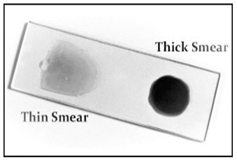
Bloodstains are usually taken from a finger prick and serve as the most solid precedent for conducting tests for malaria. To determine malaria, two distinct viscosities of blood smears are used. Thick blood spreads are valuable for distinguishing the presence of parasites whereas thin blood smears help examiners find what types of malaria are responsible for the contamination. Both smear imagery is displayed in Figure 1 on a glass slide.
Figure 1. Thin and thick blood smear
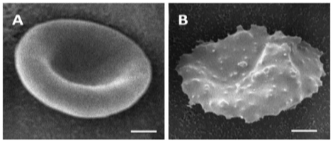
Because they contain a significant amount of hemoglobin, which has an 8-micron diameter, erythrocytes have a distinctive pink colour. If anomalous parasites are present in red blood cells, if shape deformation occurs, the presence of plasmodium is identified. Likewise, the proportion of erythrocytes infected by the plasmodium parasite (density) is determined in Figure 2. In addition, Ahirwar et al. [8] portrayed the morphological elements between ordinary RBC and malaria parasite-contaminated RBC.
Figure 2. Human erythrocytes infected with P. falciparum and not parasitized. In comparison to a human erythrocyte membrane infected with P. falciparum, smooth surface topography was seen with non-parasitized erythrocytes in (a) and (b) [9]
The standard microscope is the central apparatus for malaria diagnosis from blood smear images for normal and malarial cases in the interior area. Figure 3 depicts the flow diagram of the malaria parasite detection process.
Figure 3. Process flow diagram for finding malaria parasites
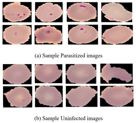
For the different stages of the intelligent malaria parasite detection and classification system, we present an overview of several dataset types here. Even though there are still certain restrictions in some phases of standard or benchmark public datasets, such as gesture detection and pothole identification, they are necessary for validating the approach in these phases. Using artificial intelligence with digital image processing methods on patient blood smear samples is necessary for the identification of malaria. A few sample images are shown in Figure 4(a) indicates the sample Parasitized images and 4(b) indicates the sample uninfected images which are also shown below. Each patient's blood smear sample is used to produce images of thick and thin blood smears. Blood smear images are commonly employed in machine detection studies and have been used by researchers [10].
Figure 4. Visualization collection of the dataset
5.1 Availability of standard public dataset
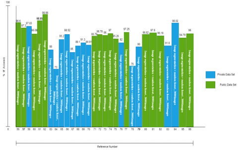
Table 1 further furnishes an enumeration of myriad datasets generated through disparate light microscopy methodologies employed by diverse researchers. The tally of images or sequences, along with the diverse types of discernible objects, is delineated alongside the dimensions of the datasets. In the subsequent stages outlined in Table 2, an analysis is presented of the deployment of private datasets within the framework of an intelligent system for malaria parasite detection and classification, as undertaken by various researchers.
Steps used during pre-processing, such as image background correction, noise reduction, and contrast augmentation, might occasionally assist in refreshing the concept. Besides, misusing the slides and image-capturing device settings brings commotions into the infinitesimal image. The captured images are inherently containing noises. Staining blood is significant for other developed perceptions of hematological problems. Pre-processing is a quality improvement technique and noise deportation technique from the image to process the image into useful sections, without retaining the parasite's specific details. This article focused on pre-processing images applied to overhaul malarial bloodstain images. With this method, problems like uneven lighting, blurring, and the appearance of undesirable features may be resolved.
Table 1. Standard public dataset table
|
Dataset Type |
Used Dataset |
Number of Images |
Dataset Link |
Classification Type |
|
Public |
Malaria Cell images |
27,558 |
https://ceb.nlm.nih.gov/proj/malaria/cell images.zip |
Binary Classification |
|
Public |
Malaria Cell images |
27,558 |
https://www.kaggle.com/datasets/iarunava/cell-images-for detecting-malaria |
Binary Classification |
|
Public |
Blood cells images |
12444 |
https://www.kaggle.com/paultimothymooney/blood |
Multi Classification |
Table 2. Standard private dataset table
|
Dataset Type |
Used Dataset |
Number of Images |
Dataset |
Phase |
Classification Type |
|
Private |
Malaria Cell images |
200 |
Chittagong Medical College 150P. falciparum & 50 uninfected |
Malaria Detection |
- |
|
Private |
Malaria Cell images |
2703 |
Employing a 1000x magnification, |
Detection and classification of Malaria |
Binary Classification |
|
Private |
Blood cells images |
13000 |
National Institute of Health |
|
Multi Classification |
6.1 Thick smear
In 2009 Frean [11] proposed reducing noise from an image using the median filter for solid specification to detect malaria in thick Giemsa stain blood. Two crucial steps were carried out by Elter et al. [12] to identify the parasites. The initial step of pre-processing was underway. As indicated by this exploration, the extent of red and green parts for identifying things that contain chromatin, a thick blood smear is the most useful component. May and Aziz [13] proposed a way to consequently measure and order erythrocytes contaminated by plasmodium and utilized a wiener filter to eliminate the undesirable noise. Arco et al. [14] performed on thick blood smear and in case of pre-processing, which comprises two tasks; image filtering to decrease the noise with the help of a Gaussian low-pass filter and further developing image quality utilizing histogram equalization.
Presented as an approach to analyzing and processing images by Memon et al. [15] utilizing the median filter to reduce unwanted noise from images and the algorithm under consideration exhibited a heightened degree of efficiency, coupled with a markedly superior level of precision, in contrast to the NCC and Fuzzy classifier methodologies recently employed by contemporary researchers. Dave and Upla [16] proposed a technique that worked on thick stains to identify malaria with the help of converted image RGB to HSV. In 2018, Poostchi et al. [17] depicted various image analyses with the assistance of artificial intelligence to distinguish malaria parasites from microscopic blood smear images. A reliable segmented approach was put out by Salamah et al. [18] whereas Fatima and Farid [19] utilized a bilateral filter to eliminate undesirable commotion from minuscule images.
6.2 Thin smear
In 2007, Diaz et al. [20] published an automatic detection of microscopic imaging acquired from thinner blood smears from malaria. Ghosh et al. [21] proposed Leishman-stained images captured through the light microscope of peripheral blood smears characterized by malarial parasites and their type using a machine learning approach. They used erythrocyte segmentation utilizing marker-controlled watershed transformation and a few created a completely computerized technique in 2012 for identifying blood stains contaminated with malarial parasites and determining the parasite's life stage and used Laplacian filter for unwanted noise removal from gathered pictures and proposed a way to distinguish the types of malaria. Erythrocyte segmenting was suggested by Sharif et al. [22] with the help of red platelets with the assistance of a watershed algorithm.
Sheikhhosseini et al. [23] utilized a nonlinear diffusion filter to remove undesirable noise and proposed an automatic malaria diagnosis-based algorithm. Rakshit and Bhowmik [24] used a wiener filter to eliminate the blurring portion in images. Maiseli et al. [25] proposed a programmed and low-cost parasitemia distinguishing for low-end microscopy imaging gadgets. Razzak [26] proposed a strategy for the programmed identification of falciparum and vivax plasmodium with the help of RBC segmentation with RNN. Makkapati and Rao [27] proposed a segmentation technique that segments the RBC over HSV colour space recognizes malaria parasitic infection in peripheral blood smear images and calculates the optimal saturation threshold value.
From the study, we found distinctive detection and segmentation methods that are used for thin smears and thick smears described below.
7.1 Thin smear
From previous papers, we found distinctive segmentation methods which are used for thin spots. By far, most of the procedures are thresholding methods. Cell segmentation should be precise to figure out the right parasitemia. Be that as it may, contacting cells specifically convolute the distinguishing proof and division of individual cells. To solve these kinds of problems, we use active contour watershed methods. In the case of Hough transformation, we assume the shape of the blood cells.
Zou et al. [28] worked on a diagnosis technique within a large Field of View. In the year 2011, An approach to determine the malarial parasite was proposed by Damahe et al. [29] where parasites in bloodstain images are segmented by the Zack thresholding technique. Nasir et al. [30] showed a contemporary segmentation method in their paper to detect malaria in the thin stain. Panchbhai et al. [31] proposed a segmentation technique of RBC in thin stains. Suryawanshi and Dixit [32] used Poisson distribution for image segmentation utilizing minimum error thresholding in giemsa stained blood images Gatc et al. [33] proposed a double thresholding technique to detect malaria parasite in erythrocytes for exactness of recognition. Das et al. [34] worked on microscopic images utilizing a machine-learning algorithm on peripheral bloodstains for erythrocytic cell segmentation using the marker-controlled watershed method. Tulsani et al. [35] proposed segmentation using morphological watershed transformation.
Kusworo [36] proposed a colour segmentation to detect malarial contamination in infected thin blood smears. Using morphological characteristics, recommended utilizing digital image analysis to automatically itemize malaria parasites. Gopakumar et al. [37] proposed identifying malaria with the help of bloodstain sample images using a focus stack with the help of a slide scanner and Convolution Neural Network (CNN). Maitra et al. [38] suggested a counting technique for the erythrocytes using the Hough transform with the help of an automatic segmentation technique. Halim et al. [39] segregate the healthy and parasite-infected blood images utilizing the help of matching patterns with parameter optimization and cross-validation methods.
7.2 Thick smear
The segmentation strategies for thick stains are unmistakable in that white platelets and parasites should be portioned. We know that white platelets are always greater than red platelets and have more surface, making their identification easier. The division strategies for thick spreads are unmistakable in that white platelets and parasites should be portioned. Moreover, white platelets should be recognized and won't be handled or arranged further. When in doubt, parasites are pretty much nothing, and their trustworthy conspicuous evidence is, for the most part, critical. In this way, affirming these things is more essential than their division in each pragmatic sense, which may clarify the strength of thresholding strategies and morphological exercises again. Thick bloodstain is assessed for malaria parasites in 1 microliter of blood.
That means malarial parasites are enumerated in more than 100 high-force fields Moody and Chiodini [40]. To count the number of malarial parasites, present in each digital image, Toha and Ngah [41] suggested examining blood-thickening smear images. Mandal et al. [42] proposed a normalized cut strategy for dividing RBCs tainted with malarial parasites utilizing blood films which are thicker. Park et al. [43] used AGNES+and morphological gradient techniques to detect parasites in thick blood film segmentation, whereas, in the same year, Park proposed an electronic technique for identifying and arranging red platelets tainted by malarial parasite falciparum at trophozoite or schizont stage for unstained cells. A machine-learning approach that uses smartphones to identify malaria parasites in thick blood smears was proposed by Yang et al. [44] and Kassim et al. [45] also suggested the same approach for thick smears where patients infected with malaria in instances of P. Falciparum and p. Vivax.
When extracting features, tissues on pathology slides are examined for structure and other properties as well as for the targeted discriminant artefacts or regions of interest. Appropriate subgroup selection is important for enhancing type accuracy, reducing complexities, and ensuring that the right capabilities are optimized for study. Different features from different arrays are used to split normal and abnormal erythrocytes, and new variables are computed. Some features like Morphology, texture, strength, etc., are the simplest examples utilized for distinguishing erythrocytes. The histogram-based options for varied colour channels like the hue, intensity and saturation are used to distinguish the conventional and infected erythrocytes. In the case of colour and the statistical features, options comprise space, perimeter, grayscale histogram, and saturation histogram, the metric is especially utilized for protozoal (malaria) infection detection.
Parasites were stained, colouration capabilities are most common, and plenty of papers use them. Di Ruberto et al. [46] proposed another morphological way to distinguish the malarial contamination from Giemsa-stained blood image and in the year 2012 he proposed a diverse order method. One is an automatic thresholding-based morphological methodology, and one is more dependent on colour histogram likenesses. Nguyen et al. presented a new technique for adequately dividing amassed cells utilizing esteem in distance image change Nguyen et al. [47]. Ghosh et al. [48] proposed another procedure that further develops the image quality and threshold-based segmentation of microscopic images to recognize the presence of Plasmodium. Lee et al. [49] worked on an optofluidic microscope to diagnose malaria in a high-resolution blood image. von Mühlen [50] demonstrated the recognition of malarial infection in smears with the help of morphological features. Mehrjou et al. [51] proposed a programmed system for recognizing parasites using an automated complex microscope. Khatri et al. [52] implemented a quick and precise method to distinguish malaria parasites with the assistance of morphological features. Chavan and Sutkar [53] also proposed disease recognition and utilizing image analysis with the help of feature extraction. Inside the erythrocyte has been described the usage of quite a few textures and morphological capabilities. The thought is that those capacities will mimic the presence of ring structure with recognizable cytoplasm and distinctive special parasite characteristics in infected cells. The trademark decision methodologies used to decrease trademark dimensionality comprise PCA, F-statistic, etc.
Morphological features signify the dimension and shape of an erythrocyte while not considering the density, and profoundly in the instance of different parasites, the erythrocytes are broadened, but they stay the same as before. Various parameters like the area, perimeter, eccentricity, compactness ratio, roundness ratio, and bending energy are important features that are important with morphological features. Kusworo [54] proposed malarial parasite recognizable proof in tainted red platelets at the advancement stage utilizing colour segmentation and examined the roundness ratio. Other features like the Hu moment, colour histogram, colour auto-correlogram, and relative shape measurement that depend on portable help for diagnosing transferable diseases in remote locations were organized. Devi et al. [55] proposed work, k-Nearest Neighbor including various elements. The ratio isolates and compounds erythrocytes present in thin blood smear for a diagnostic blood disorder. Bibin et al. [56] proposed a technique utilizing a deep belief network to recognize the existence of malaria parasites in human bloodstain images. Eshel et al. [57] proposed two studies: Diagnostics Parasite and species identification and parasite quantification. Kareem et al. [58] proposed a computerized determination strategy for malaria parasite recognition dependent on morphology, relative size, and intensity variation.
Most of the papers use the conventional RGB colour space, whereas a few papers use the HSV colour space and a couple utilize the green channel of RGB to remove staining-related shading data in images. We consider there may be a compelling case for the usage of a colouration area. This is a better ideal for extracting the same old staining colours. Kumar et al. [59] proposed an image grouping framework to emphatically distinguish malaria parasites present in thin blood spreads. For segmenting contaminated red platelets in peripheral blood stain images, Mandal et al. [60] used the normalized cut approach. and checked the efficiency of RGB, YCbCr, and HSV utilizing the Rand’s Index. Abbas and Mohamad [61] worked on RGB colour image improvement for red platelets segmentation in the YCbCr colour space. SURF, a local feature extraction technique, was presented by Thung et al. as an approach to locate malarial parasites in blood. Vink et al. [62] worked on a quantitative cartridge scanner for low-density malarial parasite detection.
With the use of a computer vision algorithm, Linder et al. [63] developed a decision support system for identifying malaria parasites that considered test areas with the highest potential of contamination with the disease. As of 2014, Annaldas et al. [64] constructed a processing method for images with morphological and thresholding strategies that can be utilized to recognize erythrocytes to mechanize the analysis of malaria on thin blood spreads and discover the potential parasite count. Chayadevi and Raju [65] worked on effective fuzzy base colour segmentation with the assistance of fractal feature extraction. In 2013, by using an image analysis technique to distinguish between live and dead parasites in samples that had previously received pharmacological treatment, Moon et al. [66] proposed the quantitative detection and categorization of the Parasite lifecycle. Muralidharan proposed automatic malarial cell recognition based on feature selection. In 2016 Jan et al. [67] worked on a method to identify malarial parasites in blood tests with giemsa stained.
Not many papers have made order advancements unequivocally for cell separation or parasite identification. Ross et al. [68] worked on peripheral blood stains with the help of a computerized image-processing technique to diagnose malaria and its type. To identify the infecting species and life-cycle stages of these parasites in images of fragile blood and to discriminate between them, Tek et al. [69] recommended using computer technology. Ghosh et al. proposed Leishman-stained images captured through the light microscope of peripheral blood smears characterized malarial parasites and their type utilizing a machine learning approach and they used erythrocyte segmentation operating marker-controlled watershed transformation. A fully programmed framework was developed by Savkare and Narote [70] for classifying and counting erythrocytes that have been infected with malaria parasites and identifying the parasites' life phase.
To discriminate between the stages of parasites in humans, Nugroho et al. [71] developed an image-processing approach. Parasite cells are recovered as features using histogram-based texture analysis, which also includes extracted features. To differentiate between falciparum and vivax, Preißinger et al. [72] an innovative neural network-driven framework has been devised, boasting the capability to swiftly classify red blood cells into four distinct categories. These encompass healthy cells and three distinct groups of infected cells, stratified based on the age of the parasitic infestation. Erythrocytes and the malaria parasite were categorized using geometric characteristics by Kumarasamy et al. [73]. Charpe and Bairagi [74] proposed computerized malarial parasite and their stage discovery, which continues in advances like picture procurement, division, highlight extraction, and afterwards arrangement. The highlights: shading, shape, size, in-strained quality, the surface will be extricated, and arrangement is finished utilizing SVM. The parasite contaminated RBC, and their count is also found in this work. Vijayalakshmi et al. [75] postulated an experimental design aimed at elucidating the aberrations in texture feature metrics through the manipulation of image luminance via the modulation of intensity control settings. Computerized identification of malaria parasites in images of thin blood films stained with Giemsa was investigated by Preedanan et al. [76]. Methods for automated segmentation, feature extraction, and classification are essential for understanding parasitemia. Segmentation depends on versatile thresholding and watershed strategies. Measurable highlights are then figured out for every cell and grouped utilizing an SVM binary classifier. Peñas et al. [77] proposed a distinguishing malaria parasite in peripheral blood spreads utilizing AI. With an accuracy of 92.4%, an affectability of 95.2%, and a precision of 87.9%, the article used a convolutional neural network to identify malaria parasites and characterize the two species, Plasmodium falciparum and Plasmodium vivax. Using images of stained thin blood smears, Bashir et al. [78] suggested an accurate, speedy, and acceptable model of malaria analysis. Using images of stained thin blood smears, Bashir et al. suggested an accurate, speedy, and acceptable model of malaria analysis. The tactic made use of the erythrocyte and Plasmodium parasite intensity characteristics. They proposed many features based on intensity. The presentation of these features on the red platelet tests from the made data set has been assessed utilizing an Artificial Neural Network (ANN) classifier.
To differentiate malaria parasites in thin blood spreads and classify them into one of the four unique kinds of malaria, Ndour et al. [79] suggested an image-processing approach. The preprocessing stage involved several techniques to improve the photos. In the system's first segment, red blood cells (RBC) are taken out of blood pictures using morphological handling. They suggested a brand-new algorithm that detected questionable areas and located the parasites in the photographs. The distribution of RBCs in each image is calculated after they are divided into tainted and untainted cells.
In the year 2012, Kaewkamnerd et al. [80] proposed a system for automatically identifying and categorizing different types of the malaria parasite in thick bloodstains whereas Zhang et al. [81] analyzed red blood cells for unlabeled malaria parasites in the year 2016. Khan et al. [82] suggested a vision-based way to distinguish the malarial parasite from thick smear with the help of light microscopy images and Leishman stain by utilizing Kapur segmentation to categorize contaminated blood smears [83].
An approach for locating malaria parasites MLP network was used by Seman et al. [84] to classify the detection of malaria parasites on thin blood smears. Shekar et al. [85] suggested using CNN-based deep learning to automatically categorize and forecast the spread of tainted blood. The performance in recognizing Plasmodium falciparum-tainted platelets was estimated by normal accuracy, and execution on distinguishing parasitic disease at the image level by Kuo et al. [86] in the year 2020.
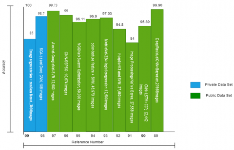
The difficulties (comprehension as a result) and the growing problems with robustness force us to reevaluate how artificial intelligence can be used to do computations that are easy to grasp, analyze, or explain. Also necessary are secure, dazzling structures. New algorithms that give precision, ability, etc. are necessary to be developed in response to the rising demand for ability. With the hybrid model, easily we get more optimal results. Research on combining machine learning methods into various hybrid systems has recently come to light. Because of the preliminary nature of this study, additional research is required. Diker [87] developed a machine-learning model based on Deep Residual CNN with a Bayesian optimization approach for the visual classification of malaria cells. A public database of 27558 images of malaria cells is available from the NIH. Canonical Correlation Analysis is a deep learning technique proposed by the authors Patil et al. [88] The CCA approach concurrently extracts, learns, and trains numerous nuclei patches to observe how overlapping nuclei impact the model. Blood cell pictures overlap, reducing the size of the input image, accelerating classification, and resulting in more precise weight values as the network converges. 12,442 blood cell pictures make up the dataset, of which 2487 are test photographs and 9957 are training images. An algorithm for detecting malarial illness was reported by Soomro et al. [89]. To detect malaria, they used image processing methods and filters with the Naive Bayes Classifier. The dataset utilized in their investigation was made up of one of the 27,558 pictures from the Kaggle public datasets pool. Because the photographs in the collection arrive in different sizes, it is essential to scale each image to the same standard measurement of 300x300. To prevent misleading findings brought on by human error, Somasekar and Reddy [90] developed a transfer learning model and applied it to 27,560 cell photos. Mohammed and Abdelrahman [91] developed a model that combines the benefit of automatically extracting features offered by deep models with the higher classification accuracy offered by traditional machine learning classifiers. Bashar [92] first described a color-texture feature (YCbCr HOG). Sahlol et al. [93] extracted attributes from WBC images using VGGNet. Cat Swarm Optimization (SSPSO) algorithm-based optimized Convolutional neural networks (SSPSO-CNN) technique and the Salp Swarm Algorithm were included, according to Kumar et al. [94]. In the year 2021, Arowolo et al. [95] suggested using a fresh, optimized hybrid investigation strategy. Convolutional neural networks were also suggested by Mitrović and Milošević [96] for the detection of malaria based on the categorization of thin blood smear images of the possibly infected cells.
The trained Alex Net and Google Net designs served as the foundation for an algorithm described by Thompson et al. [97] The feature vector for the last pooling layer of both CNN architectures has been combined, and the SVM classifies the resulting feature vector (Google Net-SVM). Using deep learning and images of blood smears, Jha et al. [98] developed a leukaemia diagnosis module. The detection phase also involves pre-processing, segmentation, feature extraction, and classification. Based on mutual information, the proposed hybrid model combines the segmentation outcomes of the active contour model and the fuzzy C means technique (MI). In Figure 5, we elucidate individuals involved in the development of a hybrid machine-learning model designed for malaria parasite identification.
Figure 5. Identification of malaria using hybrid machine learning from 2018 to 2022
After that, statistical and Local Directional Pattern (LDP) data are taken from the segmented pictures and put into the suggested Deep CNN classifier that is based on the Chronological Sine Cosine Algorithm (SCA). Abbas et al. [99] employed a two-step method that involved picture segmentation before moving on to life stage categorization.
Digital Pathology has been coordinated to finish off with deep learning strategies. The deep learning strategy is broadly utilized. AI strategy with patterns has effectively improved proficiency in various non-clinical fields. Deep learning is supervised learning that necessitates a large amount of training data. Deep learning techniques can be considered as a further developed form of the notable multi-facet neural network classifiers prepared with back-spread, with a few additional layers. This improvement in learning strategy has radically decreased the heap of microscopic analysts and let down the downsides of microscopic examination. According to LeCun et al. [100], deep learning made significant advances in taking care of issues. It has ended up being genuinely adept at finding complex designs in high-dimensional information and is accordingly material to numerous areas of science like beating records in image recognition. Convolution neural networks and autoencoders, which can aid in accurately identifying different phases of malaria infection, have been improved by Mallat [101] and Krizhevsky and Hinton [102].
Recent analyses of deep learning have shown that pattern classification models based on the deep learning viewpoint may fundamentally defeat models learnt based on traditional classifiers [103].
This is the explanation clinical execution has been among the last to execute deep learning as clarified getting ready images is harder to snag considering master experience measures and various factors. A convolutional neural organization, a deep neural network, is essentially utilized. CNN's biggest strength is its deep architecture. The CNN's extracting essential and secret features. Features that are removed are passed into a wholly connected neural network that uses them to classify images.
In the wake of completing a customary level-set cell division strategy, Liang et al. [104] used a convolutional neural network to isolate tainted and uninfected cells in thin blood spreads which is the principal paper to apply deep figuring out how to malarial sickness findings. Dong et al. [105] applied deep learning, figuring out how to perform cell segmentation. Prakash et al. [106] suggested a new Convolution Neural Network based system which detects the malaria parasite in thin blood smear microscopic images. Hung et al. introduced an end-to-end framework utilizing a quicker region-based convolutional neural network.
To identify the malaria parasite in red blood cells, Sayyed et al. [107] examined the efficacy of convolutional and capsule networks together. Shah et al. [108] made a customized three-connected layers convolution model, which gives us a faster and cheaper malaria parasite detection technique. Even though a few more studies by Pagès-Zamora et al. [109] used a similar classification-based methodology to distinguish the parasites from other stained parts, the accuracy of parasite identification has not been high enough to support an automated framework for determining malarial infection. Hung et al. [110] depicted a Faster R-CNN strategy for object identification of individual contaminated platelets. Recent research in deep learning indicates a significant increment in the utilization of DBNs attributed to the progressions in the effective layer-by-layer learning method demonstrated by Hinton [111]. Rajaraman et al. [112] proposed preprepared CNN-based DL models as component extractors toward characterizing parasitized and uninfected cells to support further developed infection screening Computerized malaria screening utilizing DL procedures made as a powerful symptomatic guide. Vijayalakshmi [113] introduced a deep neural network model for identifying malaria parasites in blood smear images utilizing a transfer learning approach with the assistance of the Visual Geometry Group (VGG) network and support vector machine. In Figure 6, we clarify the people engaged in creating a deep learning model specifically intended for the identification of malaria parasites.
Figure 6. Identification of malaria using deep learning from 2018 to 2022
Masud et al. [114] worked on a productive deep-learning technique that identifies the infected and uninfected cells from thin blood smears and assesses the viability of different existing DL models for malaria recognition. Masud et al. [14] proposed a deep learning architecture, for example, CNN, which can be helpful progressively malaria recognition viably and precisely from input images and to reduce physical work with a portable mobile application and the end, they assessed the performance of a custom CNN model utilizing a repeating Stochastic Gradient Descent (SGD) analyzer with a programmed learning rate locater and acquired an exactness of 97.30% in characterizing sound and tainted cell images with a serious level of accuracy and affectability.
For the multiplexed DNA detection of malaria, Guo et al. [115] described a mobile phone-based start-to-end stage. The system makes use of a low-cost paper-based microfluidic symptom test, which is combined with a deep learning algorithm to assist with neighborhood selection and blockchain technology to store and network-protected data.
Chakradeo et al. [116] worked on a VGG-based model for recognizing malarial parasites and comparing it with a recent model. The model outperforms most already created models in the scope of precision measurements. The model enjoys the benefit of being built from a moderately modest number of layers. Our model outperforms most already created models in the scope of precision measurements. This decreases the PC assets and computational time. Davidson et al. [117] described an ML technique that accomplishes the identification of red platelets, separating tainted or uninfected cells, and life stages of parasite categorization from natural, heterogeneous smear pictures. In the case of the pre-trained Faster Region-based Convolutional Neural Networks (R-CNN) model for RBC identification, their model performs precisely, with a normal accuracy of 0.99. Irmak [118] presented a new deep learning-based malaria detection strategy. A convolutional neural network with 20 weighted layers is planned and proposed to recognize parasitized microscopic images from uninfected images. An aggregate of 27,558 peripheral blood smear images was utilized to prepare and test the CNN model, and 95.28% exactness was acquired. In addition to a Deep Neural Network (DNN) based three-dimensional modelling algorithm that renders 3D models of parasitic cells in Augmented Reality (AR), Ramesh [119] worked on a convolutional neural network that considers the fast computerized analysis of malaria and achieved high classification accuracy 98%.
Poostchi et al. [120] proposed a computerized system to recognize and segment red platelets and distinguish contaminated cells in Wright-Giemsa-stained thin blood smears. Utilizing image analysis and ML procedures, they processed computerized images of peripheral blood smears to decide the parasitemia in each smear. They also utilized a cell extraction strategy to portion RBCs, specifically covering cells, and showed that a common Bination of RGB tone and surface highlights beats different elements.
Molina et al. [121] proposed a Deep Convolution Neural Network with the assistance of VGG 16 architecture by utilizing transfer learning to recognize malarial parasites in peripheral blood smears and Loh et al. proposed an automated deep mask R-CNN to identify malarial parasites from blood smear images and characterize those Loh et al. [122]. Hossain et al. [123] proposed a framework for malaria recognizable proof utilizing Variational Quantum Circuit (VQC), which is a half-and-half quantum-traditional (QC) AI strategy around the same time. Jena et al. [124] additionally proposed a discovery procedure for malaria parasites utilizing a deep neural network. Dey et al. [125] proposed another way to distinguish malaria parasites by utilizing the Deep Greedy Network with transfer learning.
Cell phones have carried colossal accommodation and sizable effect on present-day culture, with efficient processing abilities and easy-to-understand provisions, for example, individual data the executive’s application, minimal computerized cameras, and worldwide situating framework [GPS] route. Because of these incredible underlying sensors, the cell phone is setting its foundations in the clinical field as an option in contrast to diagnostics purposes. Eze et al. [126] proposed another plan compromise for an AI model to help malaria case viewing in remote and asset-guided regions.
Nowadays, the increased quality of cellular networks with mobile phones inbuilt superior digital cameras has turned into an optimal stage for some undeniable level imaging and recognizing mobile Health (mHealth) applications coming about in a few convenient fields prepared Point of Care (POC) devices. Because they are so adaptable, POC stages give medical services the freedom to be enhanced all around the world by offering excellent alternatives to current imaging modalities that are low-cost, small, and energy-efficient. Using the current portable foundation results in a huge decrease in the value and size of cell primarily based plans when dissimilar from a normal magnifying lens. A realistic optical cellphone-based transmission polarized light microscopic system was put out by Zhu et al. [127] for imaging the malaria pigment known as hemozoin.
Hur et al. [128] examined parallel flow cytometry whereas Seo et al. [129] proposed a holographic image for cytometry. Zhu et al. [130] worked on fluorescent imaging with the assistance of a mobile device. Breslauer et al. [131] constructed a mobile phone with a light magnifying device and showed that it is practical for medical applications. True optical magnification, as opposed to merely digital zooming, is possible with the microscope that can be attached to a contemporary mobile phone's camera. Still, the main drawback is that those gadgets produce inferior image quality. So, a more functional methodology is to just join the cell phone with a connector to a regular microscope eyepiece enabling blood slide images to be captured with the cell phone's camera. Rosado et al. [132] presented an assorted framework approach for picture handling of malaria-contaminated thick blood spread acquired with negligible expense and accessible gadgets, such as cellular phones.
By visualizing sickle-red and P. falciparum-tainted platelets in bright fields, Agbana et al. [133] demonstrated the light microscope's potential for analytical usage. The necessity to detect platelets consistently underperformed the target. Dallet et al. [134] provide a flexible application platform for Android smartphones that can analyze malaria from a Giemsa-stained thin blood film image. The malaria pigment known as hemozoin was imaged by Pirnstill and Coté [135] using a practical optical transmission polarized light microscope system based on a mobile phone.
A bespoke mobile phone magnifying lens was developed by Skandarajah et al. [136]. It can be used with phones from many manufacturers. A mobile phone-based RDT assessment stage that can be used with different parallel stream immuno-chromatographic measurements and comparison tests was demonstrated by Mudanyali et al. [137]. To check for the presence of white blood cells and falciparum trophozoites in thick smears stained with Giemsa, an image handling and evaluation approach utilizing a controlled arrangement. In contrast, Sio et al. [138] created an automated image analysis-based method for the rapid and accurate determination of parasitemia.
Using smartphone technology and image-based analysis software, Herrera et al. [139] experimented with the analytical execution of a device for the computerized interpretation of RDTs. To link a cell phone to a magnifying glass, Quinn et al. [140] presented their 3D-printable connection design in 2007. All images for their analysis, however, were captured using a specific magnifying microscope camera, which had a better pixel objective than their mobile phone camera.
In their 2017 paper, Oliveira et al. [141] developed a computerized, mobile phone-based symptomatic paradigm for malaria. An easy-to-use UI was proposed by Moallem et al. [142] for a mobile phone-based system that examines blood smear pictures for malaria screening. And that cell phone is used in conjunction with a connection for a magnifying glass. With the use of mobile technology and deep learning. To analyze malaria in 100 high-resolution TBF FoVs, Manescu et al. [143] presented the pitifully guided Multiple Objects Features Fusion (MOFF) technique. This method relies just on the clinical-microscopy conclusion of the example (powerless management with names provided by routine clinical-microscopy) and doesn't call for any difficult item-level explanations for preparation (complete supervision). The method is also capable of identifying specific parasites in photos, which helps determine the severity of the infection and how well it is responding to therapy.
In 2020, Pattanaik et al. [144] proposed an effective multi-magnification deep Residual Neural Network (MM-ResNet), completed naturally ordering the minute blood smear images as infected/nontainted at various amplifications. They had tentatively assessed their methodology by utilizing it to prepare more proficient variations of various reduced deep CNNs, assessed on smartphone datasets. The MM-ResNet start-to-finish system shows comparative or predominant precision to the pattern designs, as estimated by GPU timings on the freely accessible microscopic blood smear cellphone images. This methodology mainly utilizes an MMResNet for malaria-contaminated erythrocyte ID in infinitesimal blood smear images. They proposed an original broadened MM-ResNet structure characterizing the blood smear pictures into contaminated and noninfected erythrocyte images with a normal precision of 98.08%.
To identify malaria parasites in TBF, several methods have been used, but none have yielded accurate negative and positive prescient execution suitable for clinical application and revealed a novel way to overcome these challenges and provide a therapeutically useful framework. It creates a Deep Malaria Convolutional Neural Network classifier (DeepMCNN) for computerized malaria conclusion using common clinical microscopy names from our quality-controlled malaria centres.
Manescu et al. [145] tell us while malaria parasite object location is a mediator step in accomplishing patient analysis, preparing deep learning object finders requires a huge number of expert manpower for training deep learning object detectors of marking a huge dataset of digitized TBF.
Hung and Carpenter [146] suggested a smartphone-based, semi-automated system that examines blood smear pictures for malaria screening and has a straightforward user interface. An Android mobile app integrates several features, including picture collecting, image filtering, and the executives of the information collected. The cell phone is mixed with a magnifying instrument connector as displayed, which is a reasonable arrangement by the plan. Android cell phones and magnifying instruments are generally accessible in intestinal sickness centres. Furthermore, a connector is normally cheap.
For reliable parasitemia assurance, Nakasi et al. [147] developed a start-to-finish deep learning method to computerize the containment and count of P. falciparum parasites and White Blood Cells (WBCs). On a collection of images with thick blood smears that had been remarked on, the approach involved building PC vision models. These deep learning models, such as Single Shot MultiBox Detector (SSD) models and Faster Regional Convolutional Neural Network (Faster R-CNN), which help manage the acquired computerized pictures, were used in their construction. Additionally, an adaptable mobile phone-based derivation application was used to quantify and transmit the suggested SSD model to identify in situ parasites that cause intestinal illness from WBCs. Yunda et al. [148] proposed a method for detecting Malaria parasites in dense blood film images is elucidated, integrating AGNES and Morphological Gradient techniques during image segmentation. Following wavelet-based feature extraction, a neural network classification phase is applied, with Principal Component Analysis (PCA) utilized to reduce the feature set and optimize neural network efficiency.
Tables 3 and 4 show mobile devices used to detect malaria in thick and thin smears. The results show that the devices to use for image acquisition with the help of mobile devices, the objectives, depend on those objectives, their observations, different parameters, and the corresponding analysis. In 2019, Aris et al. [149] scrutinized various chromatic constituents with the intent of ameliorating the efficacy of parasite enumeration predicated upon images of densely layered blood smears. Within the confines of this research inquiry, we have undertaken an exhaustive exploration of five discrete colour spaces, namely YCbCr, RGB, CMY, HSV, and HSL. Furthermore, we have diligently dissected and extracted eight distinct chromatic constituents, denoted as Y, Cb, R, G, C, M, S, and L, to ascertain the optimal chromatic attribute for the quantification of malaria parasites. It still needs work to create a deep learning model that can distinguish between actual parasites, pollutants, and artefacts in a way that is comparable to a human expert. In most cases, the technique does, nevertheless, allow for reliable case identification. If a blood cell is very likely to have a parasite, it may be said to be in the first stage of AI-based analysis, which is quicker and more precise than manual testing. The ability of a deep learning algorithm to distinguish between genuine parasites, pollutants, and artefacts in the same way as a human expert would require additional development. In most cases, the technique does, nevertheless, allow for reliable case identification. It may be described as the first artificial intelligence-based procedure that, faster and more accurately than manual testing, can indicate whether a blood cell is highly likely to contain a parasite. Kundu et al. [150] inaugurated the OML-AMPDC methodology, integrating a bifold pre-processing strategy utilizing adaptive filtering and CLAHE for the dual purposes of noise eradication and contrast augmentation. The feature extraction entails the utilization of Local Derivative Radial Patterns (LDRP), while a random forest classifier is enlisted for the categorization of blood smear images. The fine-tuning of pivotal parameters through particle swarm optimization serves to refine and amplify the comprehensive classification efficacy of the random forest modality.
Razin et al. [151] executed a Convolutional Neural Network (CNN) alongside the YOLOv5 algorithm to discern and categorize instances of malaria. Fuhad et al. [152] present a fully automated model grounded in Convolutional Neural Networks (CNN) for the diagnostic assessment of malaria using microscopic blood smear images. Employing a diverse array of methodologies, encompassing knowledge distillation, data augmentation, Autoencoder, CNN-based feature extraction, and subsequent classification using a Support Vector Machine (SVM) or K-Nearest Neighbors (KNN), the model undergoes meticulous training through three distinct procedures-termed general training, distillation training, and autoencoder training—to refine and elevate both accuracy and inference performance.
Table 3. Identification of malaria utilizing mobile devices with microscopes in a thin blood smear
|
Image Acquisition Device |
Author |
Sample Size |
AI Method |
Thin Strain |
Objective |
Sensitivity |
Specificity |
Accuracy |
F Score |
Observation |
|
Tensor Flow light model-based mobile phone |
Fuhad et al. [152] |
26161 |
Automated CNN |
Gaimsa |
Leveraging a blood smear to automatically recognize the malaria parasite |
- |
- |
99.23% |
- |
Making a quick and viable mobile app for the automated detection of malaria parasite |
|
Light microscope with a smartphone with adapter (USPA 2) |
Moallem et al. [142] |
2967 |
Customized CNN |
Gaimsa |
Cellphone to screen both thick and thin smear |
|
|
|
|
The screening process is quicker, more predictable, and less reliant upon human aptitude. |
|
94.70% |
99.20% |
95.90% |
- |
|||||||
|
Microscope with webcam Logitech C 270, Samsung tab 2 &Sony DSC H1 3D printers |
Oliveira et al. [141] |
1332 |
- |
Gaimsa |
Distinguish the Plasmodium falciparum species in the ring transformative phase |
- |
- |
91% |
- |
Minimal expense demonstrative apparatuses specially created for cell phones. |
|
CUDA enables a single 16 GB NVIDAI-----P100 GPU with an Android malaria detection app |
Masud et al. [114] |
27558 |
Deep CNN with SGD |
Gaimsa |
Malaria identification using deep learning strategies in mobile healthcare |
- |
- |
97.30% |
90.70% |
Effectively & and accurately malaria parasite detection utilizing Convolution Neural Network |
|
Olympus CX 31 with Canon EOS 500 D |
Kaewkamnerd et al. [80] |
20 |
- |
Giemsa |
creating a module for image capture and analysis |
- |
- |
82.20% |
- |
model is equipped with automated units to precisely regulate the stage and objective focus point placements. |
|
Nokia N73 mobile,3.2-megapixel CMOS camera, an emission interference filter (Chroma D550/50m) |
Breslauer et al. [131] |
- |
- |
Gaimsa |
Construct a microscope operated by cell phone to image P. falciparum-contaminated red blood cells in brightfield |
- |
- |
- |
- |
concentrate on the design and improvement of cell phones for capturing images |
|
|
|
|
|
|
|
|
|
|
|
Table 4. Identification of Malaria utilizing mobile devices with microscopes in a thin blood smear
|
Image Acquisition Device |
Author |
Sample Size |
AI Method |
Thick Strain |
Objective |
Sensitivity |
Specificity |
Accuracy |
F Score |
Observation |
|
Light microscope, Smartphone, adapter (USPA2) |
Moallem et al. [142] |
2967 |
Customized CNN |
Giemsa |
Cellphone to screen both thick and thin smear |
|
|
|
Focus on developing and designing cell phones that can take images. |
|
|
74.00% |
79.33% |
78.00% |
||||||||
|
Microscope with Android phone version 7 |
Yang et al. [44] |
1819 |
Customized CNN |
Giemsa |
Malaria parasite recognition in thick blood spread with the help of cell phone |
92.59% |
94.33% |
93.46% |
93.40% |
Screening with intensity-based ICMS and classifying each with the help of CNN |
|
Microscope with a 3D adapter with mobile phone |
Quinin et al. [140] |
2903 |
Deep CNN |
Field |
Images of interactions using deep learning techniques on a server for remote discovery |
- |
- |
- |
- |
Pay close attention to how cell phone cameras are designed and improved |
|
Optical magnification printer type with smartphone |
Yunda et al. [148] |
WBC-1935, Tropozyte-352 |
- |
Giemsa |
Create a mobile microscope that can capture brightfield images of P. falciparum-contaminated red blood cells. |
93.8% (WBC) |
80.5% (WBC) |
- |
- |
Utilizing mobile devices to take images and analyze them |
|
98.2% (Tropo) |
72% (Tropo) |
|||||||||
|
Nokia N73 mobile,3.2 megapixel CMOS camera |
Breslauer et al. [131] |
- |
- |
Giemsa |
Build a smartphone microscope to take brightfield images of P. falciparum-contaminated red blood cells. |
- |
- |
- |
- |
Images of interactions using deep learning techniques on a server for remote discovery |
From previous research work, we find some performance analyses. On the premise of those performances, we make a table named Table 5 which is displayed underneath. In the context of parameterization, we have constructed a comparative analysis tableau as delineated in Table 6 herein, thereby facilitating a discernment of superior attributes with relative ease.
Table 5. Result and performance analysis
|
Blood Smear and Staining Type |
Methodology |
Classification Method |
Performance Statistics |
|
Thin Giemsa [Light microscope] |
Preprocessing applied filtering [64, 116], Median filtering [9, 63, 92, 93, 111, 127], SUSAN [33, 78], Gaussian low pass filter [138], Morphological operation [96, 100, 147], Laplacian [93, 96, 145], Local histogram equalization [95, 96, 98, 127], Forward discrete curve [98], Low pass filter [99], linear model [109], Gray world colour normalization [62] |
K-means [67], Quaternion Fourier Transform (QFT) [44] Thresholding [84, 92, 109, 129] Bayesian classifier [40, 100], Euclidean distance classifier [114], K-nearest Neighbors [99, 102, 147], K Mean [61], SVM [153], Decision tree [150], Template matching [130], Genetic algorithm [130], Support vector machine [9, 96, 97, 102], Normalized cross-correlation [91], Deep learning [105], Nural network [131] |
Min sensitivity-72.37, Max sensitivity-100 [139], Min specificity-50 [129], Max specificity-99.74 [152] |
|
Segmentation technique-Otsu thresholding [96, 111, 127], Histogram [92, 95, 126, 131], Morphological [63, 100, 141, 147], Lookup table [99], Hough transform [110], K-means [34], Watershade [6], template matching [109], Active contour model [67, 143], Nural network [75], Fuzzy rule-based segmentation & Fuzzy divergence [96] |
|||
|
Feature Type- Color, Morphological, Texture RGB [7, 8, 96, 110, 147,], HSV[126, 127, 138, 141], LAB [102, 138], Intensity[45, 67, 111, 128], Color co-occurrence matrix [40, 109], Shape [32, 40, 92, 94, 96, 98, 103, 129, 133, 134, 139, 147] Moments [40, 46, 64], Roundness ratio[131], Area granulometry[97, 147], Haralick [142], GLCM [103], LBP [142], Fractal [138], Gradient Texture [8, 69, 145], Gray level co-occurrence matrix [142, 103, 139], Entropy [64, 129], HoG [9] |
|||
|
Thin Leishman [Light microscope] |
Pre-processing applied-grey world color normalization [12, 48, 71], Geometric mean [12, 61] |
Thresholding [144], Bayesian classifier [12, 61], Naive Bayes Tree [72, 103], Logistic regression [36] SVM [61], Nural network [71, 103], Fuzzy Interface System [101] |
Min sensitivity-96.62, Max sensitivity-100, Min specificity-84.39 [71], Max specificity-98.64 [103] |
|
Feature Type-Color, Morphological, Texture-RGB [48], Intensity [103], Color co-occurrence matrix [72], Moments [12, 71], Haralick [36], GLRLM (Gray level run length matrices) [71], GLCM [12], LBP [12, 71, 138], Fractal [138], Gray level co-occurrence matrix [61], Entropy [12, 71] |
|||
|
Thick Giemsa [Light microscope] |
Preprocessing-Median- [66, 86], Laplacian spatial filter [41] |
K-Mean clustering [120], owed source games [8], SVM [130], Neural Networks [149] |
Minsensitivity-80.5 [86], Max sensitivity-97.60 [120], Min specificity-93.8 [86], Max specificity-99.4 [8] |
|
Segmentation technique thresholding [86], Histogram thresholding [41, 65, 115], Morphological operator [76] |
|||
|
Feature Type- Color, Morphological, Texture- RGB [86], HSV [41], Intensity [115, 121], Haralick [130], Shape [65, 86, 130], moment [65, 130] |
|||
|
Thick Leishman [Light microscope] |
Feature Type- Color, Morphological, Texture-LAB [82] |
K-Mean clustering [82] Naiive Bayes tree [31], Nearest mean classifier [82] |
Min sensitivity-69% [152] Max sensitivity-100 [31] Min Specificity-Max specificity-98.64 [31] |
|
Year |
Author |
Method Applied |
Accuracy |
Specificity |
Sensitivity |
F1 Score |
Precision |
|
2013 |
Das et al. [34] |
SVM with Beslan Classifier |
.840 |
.981 |
.689 |
- |
- |
|
2016 |
Devi et al. [55] |
SVM, Naive Bayes, k-NN, ANN |
.9632 |
.9287 |
.9679 |
.8531 |
- |
|
2016 |
Liang et al. [104] |
Machine learning based CNN |
.9737 |
.9699 |
.9775 |
.9736 |
.9736 |
|
2017 |
Bibin et al. [56] |
Deep Belief Network |
.963 |
.976 |
.959 |
- |
- |
|
2018 |
Gopakumar et al. [37] |
CNN |
.977 |
.971 |
.985 |
- |
- |
|
2019 |
Rajaraman et al. [112] |
VGG-19 and Squeeze Net |
.995 |
.971 |
.985 |
- |
- |
|
2020 |
Fuhad et al. [152] |
Automated CNN |
.9923 |
.9952 |
.9917 |
.9922 |
.9892 |
|
2021 |
Irmak et al. [119] |
CNN |
.9528 |
.9550 |
.9500 |
- |
.9550 |
Many individuals have died from the terrible disease malaria, and it poses a serious threat to many more. Beyond simply humans, it affects a variety of other animals. This particular illness even has the World Health Organization worried. To preserve a person's life, malaria must be discovered early. It may be possible to use this idea to help in malaria prevention. A big advancement in the current industrial revolution and digitalization might be made by this method of illness diagnosis and healthcare applications. AI approaches may be able to assist in some of the most challenging scenarios when used in conjunction with guidelines for society's advancement. Here we have discussed in this review article the most recent development in automated malaria identification with the help of machine learning techniques. This article benefits all by giving different procedures for malaria parasite detection to achieve a better and faster identification and detection mechanism. This is a unique area of examination that has seen a broad number of distributions somewhat recently. Nowadays, exact malarial parasite analysis has a high worth. Precise and quick conclusions are essential in combating malarial identification. Precise and quick conclusions is essential in combating malarial identification. The strategies mentioned here range from a fundamental level of image processing, feature extraction, and machine learning and smartphones with machine learning techniques involved in malarial parasite detection. Given the comprehensive acknowledgement of deep learning, the significance of a gigantic, explained data, image repository for training is widely comprehended, bringing about a decent help of data-obtaining endeavours.
We also focus on current deep-learning techniques that give us a clearer view of the identification and detection of malaria parasites in blood smears. Very few articles have been published till now. Most of the paper explored thin blood smears utilizing deep learning techniques, but it is without any doubt that we’ll see articles for a thick blood smear. Automatic recognition of malaria infection has turned into an honest answer for the arrangement to beat different difficulties undifferentiated from emotional expectation and analysis.
The innovation of the cell phone is held promise for medical diagnosis. An accurate and fast-paced examination of parasitic infections in a simplistic design is slated to receive widespread acceptance soon. Most of the papers explored thin blood smears utilizing deep learning techniques with mobile phones, but it is awful without any doubt that we’ll see articles for thick blood smears also very soon. Till now, laboratory detection of malaria parasites has been dependent on the standard microscope. In most cases in rural areas, modern microscopes are not available there. That’s why they rely upon conventional microscopes. In a conventional microscope, the lance quality is not up to mark. In that situation, if we attach a powerful mobile camera to the eyepiece of the microscope, which is far better than the standard microscope camera, we can get a better-quality image. From that, we easily identified parasite detection. Further investigations are needed to investigate the helpfulness of such devices for finding malaria in field conditions and clinical examples. Given these events, robotized microscopy is especially important in the race toward a modest, basic, and dependable technique for diagnosing malaria parasites. The difficulty of malaria detection in blood cells may be addressed using one of the most current AI-based approaches called deep learning. Deep learning helps to build reliable, usable, and effective solutions. An overview of the numerous methods and tactics that may be used to detect malaria accurately and effectively is provided by this study. A variety of different approaches and models have been used in multiple expert systems. This article also discusses the effectiveness of models put out by other researchers and offers a quicker, less costly, and more accurate method of detecting malaria than microscopic analysis. As part of the survey, many approaches are examined and understood. Examples of these techniques include machine learning, image processing, and deep learning. One of the best methods for obtaining data accuracy and cost-effectiveness is deep learning. Deep neural network technologies help to bring the demonstrated capabilities of learning computations closer to the level of human touch, transforming the way that people think about CAD from a tool for "moment conclusion" to one that is more collaborative. In this study, to identify and diagnose malaria, we carefully investigated and listed the unique deep-learning methodologies. Deep learning has achieved enormous success in many other fields, but its true promise in medical imaging has not yet been released. Ahead of clinical use, it is necessary to address fascinating issues associated with deep learning-based techniques for malaria detection and diagnosis.
The lack of communication between the clinical and research professionals and data processing is a significant problem. Throughout the research phase, maintaining a solid working relationship between clinical and research engineers can boost the likelihood of success. Clinical experts may offer advice on how to use current technology in clinical settings, and a team of computer engineers can creatively analyze research results using their specific knowledge. To create functional pictures of specific patients, researchers can connect the anatomical data acquired using state-of-the-art imaging technologies to computer models. The cross-disciplinary cooperation with clinical management or marketing experts that might arise from malaria engineering and diagnostics could be advantageous.
When using methods, we must consider factors like the data provided to us for the application since they will help us decide which strategies to utilize. The ability to apply deep learning models for their provided performance has considerably grown with the acquisition of labelled data. To prevent mistakes, noise, and missing numbers, labelling is carried out by a team of professionals. Therefore, human (experts) annotated data is necessary for a successful supervised learning strategy.
Data Dependent, a key element is that deep learning models work well with large data sets. These models cannot learn from tiny datasets since it is impossible to train and evaluate them on such a small quantity of data. Therefore, with additional training data, these techniques may become successful. According to much research, data enrichment will be a solution to having a lot of data. However, it can need sampling overlapping picture patches or introduce noise to the images. Overfitting can occur if the enhanced samples are strongly correlated with one another. A common issue in statistical modelling and machine learning is overfitting. Additionally, errors come from local patches' inability to consider the image's overall spatial context.
It is advised to compare these procedures using public datasets which is far better than ones gathered from clinics and hospitals if this study decides to compare methods to determine which is the most successful. A problem with the hospital-generated datasets that can't be utilized as input for deep learning methods. It might be difficult to use data from different institutions because of institutional variations in how patients are treated and how data is collected and coded. These models are hence rigid. This topic of normalization is what we're talking about. The main outcome of this investigation was the absence of general deep-learning models.
The complexity and time trade-offs that various treatments considerably entail in the two primary treatment stages, the learning phase and the testing phase, are crucial to take into account. The application of deep learning models in medicine faces several difficulties due to their extensive parameterization, which makes it challenging to identify the precise factors that contribute to predictions and exacerbates the notion of model overfitting. It is practically hard to comprehend and describe more complex models. Machine learning algorithms are frequently employed to make judgements about a problem, but they are frequently unable to articulate the elements that impact their choice. Most deep learning and machine learning approaches are not particularly effective at explaining.
Powerful and productive malaria parasite identification and Machine learning procedures in minute blood smear images are, by and large, still needing impromptu creation and necessitate avoidance as well. In this situation, exact malarial parasitemia analysis and evaluating research has high worth. Precise and quick conclusions are essential in combating malarial sicknesses that cause a large number of passing around the world. Computerized recognition of malaria infection has turned into an open answer for an arrangement to beat different difficulties undifferentiated from emotional expectation and analysis. Most cell phone-based demonstrative gadgets have been tried in all-around controlled lab conditions. Further investigations are needed to investigate the helpfulness of such devices for the finding of malaria in field conditions and clinical examples. Imprecise findings and reviewing have caused enormous passing rates especially in kids worldwide and in tropical nations that prompt millions and more than 1,000,000 passing every year. The eventual fate of cell phones helped indicative innovations are promising and the far and wide reception of such advances is expected sooner rather than later for the precise and fast analysis of parasitic illnesses in a simple to-utilize design.
The history of medicine has been significantly altered by machine learning and deep learning. The way that individuals save, handle, and analyze data to make better decisions has substantially altered as a result of these paradigms. These have also sparked studies towards the creation of technology that transforms quickly throughout time. Complex real-world issues can be solved using machine learning techniques. The significance of machine learning, in particular deep learning, is emphasized in this essay while discussing how to solve the difficult difficulties of malaria detection and categorization. This essay has provided a thorough analysis of each of these new paradigms by highlighting how they have improved the healthcare system. We provide some information about the various deep learning architectures, particularly convolutional neural networks. Additionally, we foresaw difficulties that hinder the effectiveness of deep learning models. In our proposed article we will aggressively advocate for the use of artificial intelligence-based programs, particularly deep learning, to effectively combat malaria by preventing human deaths while using fewer resources. Future research may find it necessary to construct a pre-trained algorithm that has been modified and tailored on a benchmark dataset to simplify the default models and get the best results. If necessary, a clinical evaluation should compare how well clinicians perform using the newly developed system (CAD) to how well they use an existing instrument. As deep learning algorithms become more widely used, hardware support increases, and appealing deep learning method characteristics like robustness, generalizability, and expert-level accuracies become more apparent, we anticipate seeing an increase in the number of CAD systems for malaria detection and diagnosis.
[1] Zekar, L., Sharman, T. (2020). Plasmodium falciparum malaria. https://europepmc.org/article/NBK/nbk555962.
[2] WHO global malaria programme: World malaria report. (2016). https://www.mmv.org/newsroom/news-resources-search/world-malaria-report-2016?gclid=CjwKCAiA5L2tBhBTEiwAdSxJX3mI4SHN2BngVBFCpTkxNQX1TnCHEXM6jwO29OtjXu2BH2OwVZZCvRoCNKYQAvD_BwE, accessed on 23 October 2023.
[3] World Health Organization. (2020). World malaria report. https://www.mmv.org/newsroom/news-resources-search/world-malaria-report-2020?gclid=CjwKCAiA5L2tBhBTEiwAdSxJX-8aqrwGOg7hcYgwtx3dWDBpmGCq_cMkFXPwjEsbzrIMTCWbctYh1BoCAvAQAvD_BwE, accessed on 23 October 2023.
[4] Tangpukdee, N., Duangdee, C., Wilairatana, P., Krudsood, S. (2009). Malaria diagnosis: A brief review. The Korean Journal of Parasitology, 47(2): 93-102. https://doi.org/10.3347/kjp.2009.47.2.93
[5] Yang, D., Subramanian, G., Duan, J., Gao, S., Bai, L., Chandramohana das, R., Ai, Y. (2017). A portable image-based cytometer for rapid malaria detection and quantification. PloS One, 12(6): e0179161. https://doi.org/10.1371/journal.pone.0179161
[6] Mavandadi, S., Dimitrov, S., Feng, S., Yu, F., Sikora, U., Yaglidere, O., Padmanabhan, S., Nielsen, K., Ozcan, A. (2012). Distributed medical image analysis and diagnosis through crowd-sourced games: A malaria case study. PloS One, 7(5): e37245. https://doi.org/10.1371/journal.pone.0037245
[7] Holmström, O., Stenman, S., Suutala, A., Moilanen, H., Kücükel, H., Ngasala, B., Mårtensson, A., Mhamilawa, L., Aydin-Schmidt, B., Lundin, M., Diwan, V., Linder, N., Lundin, J. (2020). A novel deep learning-based point-of-care diagnostic method for detecting Plasmodium falciparum with fluorescence digital microscopy. PloS One, 15(11): e0242355. https://doi.org/10.1371/journal.pone.0242355
[8] Ahirwar, N., Pattnaik, S., Acharya, B. (2012). Advanced image analysis-based system for automatic detection and classification of malarial parasites in blood images. International Journal of Information Technology and Knowledge Management, 5(1): 59-64.
[9] Hayakawa, E.H., Matsuoka, H. (2016). Detailed methodology for high-resolution scanning electron microscopy (SEM) of murine malaria parasitized erythrocytes. Parasitology International, 65(5): 539-544. https://doi.org/10.1016/j.parint.2016.03.006
[10] https://lhncbc.nlm.nih.gov/LHCBpublications/pubs/MalariaDatasets.html, accessed on 1 July 2021.
[11] Frean, J.A. (2009). Reliable enumeration of malaria parasites in thick blood films using digital image analysis. Malaria Journal, 8(1): 1-8. https://doi.org/10.1186/1475-2875-8-218
[12] Elter, M., Haßlmeyer, E., Zerfaß, T. (2011). Detection of malaria parasites in thick blood films. In 2011 Annual International Conference of the IEEE Engineering in Medicine and Biology Society, pp. 5140-5144. https://doi.org/10.1109/IEMBS.2011.6091273
[13] May, Z., Aziz, S.S.A.M. (2013). Automated quantification and classification of malaria parasites in thin blood smears. In 2013 IEEE International Conference on Signal and Image Processing Applications, pp. 369-373. https://doi.org/10.1109/ICSIPA.2013.6708035
[14] Arco, J.E., Górriz, J.M., Ramírez, J., Álvarez, I., Puntonet, C.G. (2015). Digital image analysis for automatic enumeration of malaria parasites using morphological operations. Expert Systems with Applications, 42(6): 3041-3047. https://doi.org/10.1016/j.eswa.2014.11.037
[15] Memon, M.H., Khanzada, T.J.S., Memon, S., Hassan, S.R. (2019). Blood image analysis to detect malaria using filtering image edges and classification. TELKOMNIKA (Telecommunication Computing Electronics and Control), 17(1): 194-201. https://doi.org/10.12928/TELKOMNIKA.v17i1.11586
[16] Dave, I.R., Upla, K.P. (2017). Computer-aided diagnosis of malaria disease for thin and thick blood smear microscopic images. In 2017 4th International Conference on Signal Processing and Integrated Networks (SPIN), Noida, India, pp. 561-565. https://doi.org/10.1109/SPIN.2017.8050013
[17] Poostchi, M., Silamut, K., Maude, R.J., Jaeger, S., Thoma, G. (2018). Image analysis and machine learning for detecting malaria. Translational Research, 194: 36-55. https://doi.org/10.1016/j.trsl.2017.12.004
[18] Salamah, U., Sarno, R., Arifin, A.Z., Nugroho, A.S., Rozi, I.E., Asih, P.B.S. (2019). A robust segmentation for malaria parasite detection of thick blood smear microscopic images. International Journal of Advanced Science, Engineering and Information Technology, 9(4): 1450-1459.
[19] Fatima, T., Farid, M.S. (2020). Automatic detection of Plasmodium parasites from microscopic blood images. Journal of Parasitic Diseases, 44(1): 69-78. https://doi.org/10.1007/s12639-019-01163-x
[20] Díaz, G., Gonzalez, F., Romero, E. (2007). Infected cell identification in thin blood images based on colour pixel classification: Comparison and analysis. In Progress in Pattern Recognition, Image Analysis and Applications: 12th Euromerican Congress on Pattern Recognition, CIARP, Valparaiso, Chile, November 13-16. Springer Berlin Heidelberg, 12: 812-821. https://doi.org/10.1007/978-3-540-76725-1_84
[21] Ghosh, M., Das, D., Chakraborty, C., Ray, A.K. (2011). Plasmodium vivax segmentation using modified fuzzy divergence. In 2011 International Conference on Image Information Processing, Shimla, India, pp. 1-5. https://doi.org/10.1109/ICIIP.2011.6108873
[22] Sharif, J.M., Miswan, M.F., Ngadi, M.A., Salam, M.S.H., bin Abdul Jamil, M.M. (2012). Red blood cell segmentation using masking and watershed algorithm: A preliminary study. In 2012 International Conference on Biomedical Engineering (ICoBE), Penang, Malaysia, pp. 258-262. https://doi.org/10.1109/ICoBE.2012.6179016
[23] Sheikhhosseini, M., Rabbani, H., Zekri, M., Talebi, A. (2013). Automatic diagnosis of malaria based on a complete circle-ellipse fitting search algorithm. Journal of Microscopy, 252(3): 189-203. https://doi.org/10.1111/jmi.12081
[24] Rakshit, P., Bhowmik, K. (2013). Detection of the presence of parasites in human RBC in case of diagnosing malaria using image processing. In 2013 IEEE Second International Conference on Image Information Processing (ICIIP-2013), Shimla, India, pp. 329-334. https://doi.org/10.1109/ICIIP.2013.6707610
[25] Maiseli, B., Mei, J., Gao, H., Yin, S. (2014). An automatic and cost-effective parasitemia identification framework for low-end microscopy imaging devices. In 2014 International Conference on Mechatronics and Control (ICMC), Jinzhou, China, pp. 2048-2053. https://doi.org/10.1109/ICMC.2014.7231926
[26] Razzak, M.I. (2015). Malarial parasite classification using recurrent neural network. International Journal of Image Processing, 9: 69-79.
[27] Makkapati, V.V., Rao, R.M. (2009). Segmentation of malaria parasites in peripheral blood smear images. In 2009 IEEE International Conference on Acoustics, Speech and Signal Processing, pp. 1361-1364. https://doi.org/10.1109/ICASSP.2009.4959845
[28] Zou, L.H., Chen, J., Zhang, J., Garcia, N. (2010). Malaria cell counting diagnosis within a large field of view. In 2010 International Conference on Digital Image Computing: Techniques and Applications, Sydney, NSW, Australia, pp. 172-177. https://doi.org/10.1109/DICTA.2010.40
[29] Damahe, L.B., Krishna, R., Janwe, N., Thakur, N.V. (2011). Segmentation-based approach to detect parasites and RBCs in blood cell images. International Journal Computing Science and Applications, 4(2): 71-81.
[30] Nasir, A.A., Mashor, M.Y., Mohamed, Z. (2012). Segmentation-based approach for detection of malaria parasites using moving k-means clustering. In 2012 IEEE-EMBS Conference on Biomedical Engineering and Sciences, Langkawi, Malaysia, pp. 653-658. https://doi.org/10.1109/IECBES.2012.6498073
[31] Panchbhai, V.V., Damahe, L.B., Nagpure, A.V., Chopkar, P.N. (2012). RBCs and parasites segmentation from thin smear blood cell images. International Journal of Image, Graphics and Signal Processing, 4(10): 54-60. https://doi.org/10.5815/ijigsp.2012.10.08
[32] Suryawanshi, M.S., Dixit, V. (2013). Improved technique for detection of malaria parasites within the blood cell images. International Journal of Scientific & Engineering Research, 4: 373-376.
[33] Gatc, J., Maspiyanti, F., Sarwinda, D., Arymurthy, A.M. (2013). Plasmodium parasite detection on red blood cell image for the diagnosis of malaria using double thresholding. In 2013 International Conference on Advanced Computer Science and Information Systems (ICACSIS), Sanur Bali, Indonesia, pp. 381-385. https://doi.org/10.1109/ICACSIS.2013.6761605
[34] Das, D.K., Chakraborty, C., Mitra, B., Maiti, A.K., Ray, A.K. (2013). Quantitative microscopy approach for shape-based erythrocytes characterization in Aneamia. Journal of Microscopy, 249(2): 136-149. https://doi.org/10.1111/jmi.12002
[35] Tulsani, H., Saxena, S., Yadav, N. (2013). Segmentation using morphological watershed transformation for counting blood cells. International Journal of Computer Applications & Information Technology, 2(3): 28-36.
[36] Kusworo, A. (2015). Identification of plasmodium falciparum development phase in malaria-infected red blood cells using adaptive colour segmentation and decision tree-based classification. International Journal of Applied Engineering Research, 10(2): 4043-4055.
[37] Gopakumar, G.P., Swetha, M., Sai Siva, G., Sai Subrahmanyam, G.R.K. (2018). Convolutional neural network‐based malaria diagnosis from a focus stack of blood smear images acquired using a custom‐built slide scanner. Journal of Biophotonics, 11(3): e201700003. https://doi.org/10.1002/jbio.201700003
[38] Maitra, M., Gupta, R.K., Mukherjee, M. (2012). Detection and counting of red blood cells in blood cell images using the Hough transform. International Journal of Computer Applications, 53(16): 18-22.
[39] Halim, S., Bretschneider, T.R., Li, Y., Preiser, P.R., Kuss, C. (2006). Estimating malaria parasitemia from blood smear images. In 2006 9th International Conference on Control, Automation, Robotics and Vision, Singapore, pp. 1-6. https://doi.org/10.1109/ICARCV.2006.345381
[40] Moody, A.H. Chiodini, P.L. (2000). Methods for the detection of blood parasites. Clinical & Laboratory Hematology, 22(4): 189-201.
[41] Toha, S.F., Ngah, U.K. (2007). Computer-aided medical diagnosis for the identification of malaria parasites. In 2007 International Conference on Signal Processing, Communications and Networking, Chennai, India, pp. 521-522. https://doi.org/10.1109/ICSCN.2007.350655
[42] Mandal, S., Kumar, A., Chatterjee, J., Manjunatha, M., Ray, A.K. (2010). Segmentation of blood smear images using normalized cuts for detection of malarial parasites. In 2010 Annual IEEE India Conference (INDICON), Kolkata, India, pp. 1-4. https://doi.org/10.1109/INDCON.2010.5712739
[43] Park, H.S., Rinehart, M.T., Walzer, K.A., Chi, J.T.A., Wax, A. (2016). Automated detection of P. falciparum using machine learning algorithms with quantitative phase images of unstained cells. PloS One, 11(9): e0163045. https://doi.org/10.1371/journal.pone.0163045
[44] Yang, F., Poostchi, M., Yu, H., Zhou, Z., Silamut, K., Yu, J., Maude R.J., Jaeger S., Antani, S. (2019). Deep learning for smartphone-based malaria parasite detection in thick blood smears. IEEE Journal of Biomedical and Health Informatics, 24(5): 1427-1438. https://doi.org/10.1109/JBHI.2019.2939121
[45] Kassim, Y.M., Yang, F., Yu, H., Maude, R.J., Jaeger, S. (2021). Diagnosing malaria patients with plasmodium falciparum and vivax using deep learning for thick smear images. Diagnostics, 11(11): 1994. https://doi.org/10.3390/diagnostics11111994
[46] Di Ruberto, C., Dempster, A., Khan, S., Jarra, B. (2000). Automatic thresholding of infected blood images using granulometry and regional extrema. In Proceedings 15th International Conference on Pattern Recognition. ICPR-2000. IEEE, 3: 441-444. https://doi.org/10.1109/ICPR.2000.903579
[47] Nguyen, N.T., Duong, A.D., Vu, H.Q. (2010). A new method for splitting clumped cells in red blood images. In 2010 Second International Conference on Knowledge and Systems Engineering, Hanoi, Vietnam, pp. 3-8. https://doi.org/10.1109/KSE.2010.27
[48] Ghosh, P., Bhattacharjee, D., Nasipuri, M., Basu, D.K. (2011). Medical aid for automatic detection of malaria. In Computer Information Systems-Analysis and Technologies: 10th International Conference, CISIM 2011, Kolkata, India, December 14-16. Springer Berlin Heidelberg, pp. 170-178. https://doi.org/10.1007/978-3-642-27245-5_22
[49] Lee, K.S., Cox-Singh, J., Singh, B. (2009). Morphological features and differential counts of Plasmodium knowlesi parasites in naturally acquired human infections. Malaria Journal, 8(1): 1-10. https://doi.org/10.1186/1475-2875-8-73
[50] von Muḧlen, A. (2004). Computer image analysis of malarial Plasmodium vivax in human red blood cells. California Polytechnic State University.
[51] Mehrjou, A., Abbasian, T., Izadi, M. (2013). Automatic malaria diagnosis system. In 2013 First RSI/ISM International Conference on Robotics and Mechatronics (ICRoM), Tehran, Iran, pp. 205-211. https://doi.org/10.1109/ICRoM.2013.6510106
[52] Khatri, K.M., Ratnaprabha, V.R., Agrawal, S.S., Bhalchandra, A.S. (2013). Image processing approach for malaria parasite identification. International Journal of Computer Applications®(IJCA).
[53] Chavan, S.N., Sutkar, A.M. (2014). Malaria disease identification and analysis using image processing. International Journal Latest Trends Engineering Technology, 3(3): 218-223.
[54] Kusworo, A. (2016). Identifying the developmental phase of plasmodium falciparum in malaria-infected red blood cells using adaptive colour segmentation and back propagation neural network. International Journal of Applied Engineering Research, 11(15): 8754-8759.
[55] Devi, S.S., Roy, A., Sharma, M., Laskar, R.H. (2016). kNN classification-based erythrocyte separation in microscopic images of thin blood smear. In 2016 2nd International Conference on Computational Intelligence and Networks (CINE), Bhubaneswar, India, pp. 69-72. https://doi.org/10.1109/CINE.2016.19
[56] Bibin, D., Nair, M.S., Punitha, P. (2017). Malaria parasite detection from peripheral blood smear images using deep belief networks. IEEE Access, 5: 9099-9108. https://doi.org/10.1109/ACCESS.2017.2705642
[57] Eshel, Y., Houri-Yafin, A., Benkuzari, H., Lezmy, N., Soni, M., Charles, M., Swaminathan, J., Solomon, H., Sampathkumar, P., Premji, Z., Mbithi, C., Nneka, Z., Onsongo, S., Maina, D., Levy-Schreier, S., Cohen, C.L., Gluck, D., Pollak, J.J., Salpeter, S.J. (2017). Evaluation of the Parasite platform for malaria diagnosis. Journal of Clinical Microbiology, 55(3): 768-775. https://doi.org/10.1128/jcm.02155-16
[58] Kareem, S., Kale, I., Morling, R.C. (2012). Automated malaria parasite detection in thin blood films: -A hybrid illumination and colour constancy insensitive, morphological approach. In 2012 IEEE Asia Pacific Conference on Circuits and Systems, Kaohsiung, Taiwan, pp. 240-243. https://doi.org/10.1109/APCCAS.2012.6419016
[59] Kumar, A., Choudhary, A., Tembhare, P.U., Pote, C.R. (2012). Enhanced identification of malarial-infected objects using the Otsu algorithm from thin smear digital images. International Journal of Latest Research in Science and Technology, 1(2): 159-163.
[60] Mandal, S., Sarkar, R.R., Sinha, S. (2011). Mathematical models of malaria-a review. Malaria Journal, 10(1): 1-19. https://doi.org/10.1186/1475-2875-10-202
[61] Abbas, N., Mohamad, D. (2013). Microscopic RGB colour image enhancement for blood cell segmentation in YCBCR colour space for k-means clustering. Journal of Theoretical and Applied Information Technology, 55(1): 117-125.
[62] Vink, J.P., Laubscher, M., Vlutters, R., Silamut, K., Maude, R.J., Hasan, M.U., De Haan, G. (2013). An automatic vision‐based malaria diagnosis system. Journal of Microscopy, 250(3): 166-178. https://doi.org/10.1111/jmi.12032
[63] Linder, N., Turkki, R., Walliander, M., Mårtensson, A., Diwan, V., Rahtu, E., Pietikäinen, M., Lundin, M., Lundin, J. (2014). A malaria diagnostic tool based on computer vision screening and visualization of Plasmodium falciparum candidate areas in digitized blood smears. PLoS One, 9(8): e104855. https://doi.org/10.1371/journal.pone.0104855
[64] Annaldas, S., Shirgan, S.S., Marathe, V.R. (2014). Automatic identification of malaria parasites using image processing. International Journal of Emerging Engineering Research and Technology, 2(4): 107-112.
[65] Chayadevi, M., Raju, G. (2014). Usage of art for automatic malaria parasite identification based on fractal features. International Journal of Video & Image Processing and Network Security IJVIPNS-IJENS, 14: 7-15.
[66] Moon, S., Lee, S., Kim, H., Freitas-Junior, L.H., Kang, M., Ayong, L., Hansen, M.A. (2013). An image analysis algorithm for malaria parasite stage classification and viability quantification. PloS One, 8(4): e61812. https://doi.org/10.1371/journal.pone.0061812.
[67] Jan, Z., Khan, A., Sajjad, M., Muhammad, K., Rho, S., Mehmood, I. (2018). A review on automated diagnosis of malaria parasite in microscopic blood smears images. Multimedia Tools and Applications, 77: 9801-9826.
[68] Ross, N.E., Pritchard, C.J., Rubin, D.M., Duse, A.G. (2006). Automated image processing method for the diagnosis and classification of malaria on thin blood smears. Medical and Biological Engineering and Computing, 44: 427-436. https://doi.org/10.1007/s11517-006-0044-2
[69] Tek, F.B., Dempster, A.G., Kale, I. (2010). Parasite detection and identification for automated thin blood film malaria diagnosis. Computer Vision and Image Understanding, 114(1): 21-32. https://doi.org/10.1016/j.cviu.2009.08.003
[70] Savkare, S.S., Narote, S.P. (2011). Automatic detection of malaria parasites for estimating parasitemia. International Journal of Computer Science and Security (IJCSS), 5(3): 310-315.
[71] Nugroho, H.A., Akbar, S.A., Murhandarwati, E.E.H. (2015). Feature extraction and classification for detection of malaria parasites in thin blood smear. In 2015 2nd International Conference on Information Technology, Computer, and Electrical Engineering (ICITACEE). Semarang, Indonesia, pp. 197-201. https://doi.org/10.1109/ICITACEE.2015.7437798
[72] Preißinger, K., Kellermayer, M., Vértessy, B.G., Kézsmárki, I., Török, J. (2022). Reducing data dimension boosts neural network-based stage-specific malaria detection. Scientific Reports, 12(1): 16389.
[73] Kumarasamy, S.K., Ong, S.H., Tan, K.S. (2011). Robust contour reconstruction of red blood cells and parasites in the automated identification of the stages of malarial infection. Machine Vision and Applications, 22: 461-469. https://doi.org/10.1007/s00138-010-0284-x
[74] Charpe, K.C., Bairagi, V.K. (2015). Automated malaria parasites and their stage detection in microscopic blood images. In 2015 IEEE 9th International Conference on Intelligent Systems and Control (ISCO), Coimbatore, India pp. 1-4. https://doi.org/10.1109/ISCO.2015.7282336
[75] Vijayalakshmi, A., Rajesh Kanna, B., Banukumar, S. (2018). Estimation of texture variation in malaria diagnosis. In Computational Signal Processing and Analysis: Select Proceedings of ICNETS2, Singapore, pp. 325-334. https://dx.doi.org/10.1007/978-981-10-8354-9_30
[76] Preedanan, W., Phothisonothai, M., Senavongse, W., Tantisatirapong, S. (2016). Automated detection of plasmodium falciparum from Giemsa-stained thin blood films. In 2016 8th International Conference on Knowledge and Smart Technology (KST), Chiang Mai, Thailand, pp. 215-218. https://doi.org/10.1109/KST.2016.7440501
[77] Peñas, K.E.D., Rivera, P.T., Naval, P.C. (2017). Malaria parasite detection and species identification on thin blood smears using a convolutional neural network. In 2017 IEEE/ACM International Conference on Connected Health: Applications, Systems and Engineering Technologies (CHASE), Philadelphia, PA, USA, pp. 1-6. https://doi.org/10.1109/CHASE.2017.51
[78] Bashir, A., Mustafa, Z.A., Abdelhameid, I., Ibrahim, R. (2017). Detection of malaria parasites using digital image processing. In 2017 International Conference on Communication, Control, Computing and Electronics Engineering (ICCCCEE), Khartoum, Sudan, pp. 1-5. https://doi.org/10.1109/ICCCCEE.2017.7867644
[79] Ndour, P.A., Larréché, S., Mouri, O., Argy, N., Gay, F., Roussel, C., Jauréguiberry, S., Perillaud, C., Langui, D., Biligui, S., Chartrel, N., Mérens, A., Kendjo, E., Ghose, A., Hassan, M.U., Hossain, A., Kingston, H.W.F., Plewes, K., Dondorp, A.M., Danis, M., Houzé, S., Bonnefoy, S., Thellier, M., Woodrow, C.J., Buffet, P.A., French Artesunate Working Group. (2017). Measuring the Plasmodium falciparum HRP2 protein in blood from artesunate-treated malaria patients predicts post-artesunate delayed hemolysis. Science Translational Medicine, 9(397): eaaf9377. https://doi.org/10.1126/scitranslmed.aaf9377
[80] Kaewkamnerd, S., Uthaipibull, C., Intarapanich, A., Pannarut, M., Chaotheing, S., Tongsima, S. (2012). An automatic device for detection and classification of malaria parasite species in thick blood film. In Bmc Bioinformatics. BioMed Central, 13(17): 1-10. https://doi.org/10.1186/1471-2105-13-S17-S18
[81] Zhang, Z., Ong, L.S., Fang, K., Matthew, A., Dauwels, J., Dao, M., Asada, H. (2016). Image classification of unlabeled malaria parasites in red blood cells. In 2016 38th Annual International Conference of the IEEE Engineering in Medicine and Biology Society (EMBC), Orlando, FL, USA, pp. 3981-3984. https://doi.org/10.1109/EMBC.2016.7591599
[82] Khan, N.A., Pervaz, H., Latif, A.K., Musharraf, A. (2014). Unsupervised identification of malaria parasites using computer vision. In 2014 11th International Joint Conference on Computer Science and Software Engineering (JCSSE), Chon Buri, Thailand, pp. 263-267. https://doi.org/10.1109/JCSSE.2014.6841878
[83] Abimala, T., Rini, R.J. (2019). Classification of distinct plasmodium species in thin blood smear images using Kapur segmentation strategy. Research Journal of Pharmacy and Technology, 12(5): 2309-2316. http://dx.doi.org/10.5958/0974-360X.2019.00385.8
[84] Seman, N.A., Isa, N.A.M., Li, L.C., Mohamed, Z., Ngah, U.K., Zamli, K.Z. (2008). Classification of malaria parasite species based on thin blood smears using multilayer perceptron network. International Journal of the Computer, the Internet and Management, 16(1): 46-52.
[85] Shekar, G., Revathy, S., Goud, E.K. (2020). Malaria detection using deep learning. In 2020 4th International Conference on Trends in Electronics and Informatics (ICOEI), Tirunelveli, India, pp. 746-750. https://doi.org/10.1109/ICOEI48184.2020.9143023
[86] Kuo, P.C., Cheng, H.Y., Chen, P.F., Liu, Y.L., Kang, M., Kuo, M.C., Hsu S.F, Lu H.J, Hong S., Su C.H, Liu D.P., MSc, Y.C.T., Chuang, J.H. (2020). Assessment of expert-level automated detection of Plasmodium falciparum in digitized thin blood smear images. JAMA Network Open, 3(2): e200206. https://doi.org/10.1001/jamanetworkopen.2020.0206
[87] Diker, A. (2022). An efficient model of residual-based convolutional neural network with Bayesian optimization for the classification of malarial cell images. Computers in Biology and Medicine, 148: 105635. https://doi.org/10.1016/j.compbiomed.2022.105635
[88] Patil, A.M., Patil, M.D., Birajdar, G.K. (2021). White blood cells image classification using deep learning with canonical correlation analysis. Irbm, 42(5): 378-389. https://doi.org/10.1016/j.irbm.2020.08.005
[89] Soomro, A., Baloch, E.R.B., Jawaid, M.M., Zehra, Q., Babar, N. (2021). Malaria cells image analysis using image processing filters and naïve Bayes classifier. Engineering Science and Technology International Research Journal, 5(1): 18-23.
[90] Somasekar, J., Reddy, B.E. (2015). Segmentation of erythrocytes infected with malaria parasites for the diagnosis using microscopy imaging. Computers & Electrical Engineering, 45: 336-351. https://doi.org/10.1016/j.compeleceng.2015.04.009
[91] Mohammed, H.A., Abdelrahman, I.A.M. (2017). Detection and classification of malaria in thin blood slide images. In 2017 International Conference on Communication, Control, Computing and Electronics Engineering (ICCCCEE), Khartoum, Sudan, pp. 1-5. https://doi.org/10.1109/ICCCCEE.2017.7866700
[92] Bashar, M.K. (2019). Improved classification of malaria parasite stages with support vector machine using combined colour and texture features. In 2019 IEEE Healthcare Innovations and Point of Care Technologies, (HI-POCT), Bethesda, MD, USA, pp. 135-138. https://doi.org/10.1109/HI-POCT45284.2019.8962686
[93] Sahlol, A.T., Kollmannsberger, P., Ewees, A.A. (2020). Efficient classification of white blood cell leukaemia with improved swarm optimization of deep features. Scientific Reports, 10(1): 2536. https://doi.org/10.1038/s41598-020-59215-9
[94] Kumar, A., Choudhary, A., Tembhare, P., Pote, C. (2012). Enhanced identification of malarial infected objects using an otsu algorithm from thin smear digital images. International Journal of Latest Research in Science and Technology ISSN, 1(2): 2278-5299.
[95] Arowolo, M.O., Adebiyi, M.O., Adebiyi, A.A., Olugbara, O. (2021). Optimized hybrid investigative-based dimensionality reduction methods for malaria vector using KNN classifier. Journal of Big Data, 8(1): 1-14. https://doi.org/10.1186/s40537-021-00415-z
[96] Mitrović, K., Milošević, D. (2021). Classification of malaria-infected cells using convolutional neural networks. In 2021 IEEE 15th International Symposium on Applied Computational Intelligence and Informatics (SACI), pp. 000323-000328. https://doi.org/10.1109/SACI51354.2021.9465636
[97] Thompson, J.M., Eick, S.M., Dailey, C., Dale, A.P., Mehta, M., Nair, A., Cordero, J.F., Welton, M. (2020). Relationship between pregnancy-associated malaria and adverse pregnancy outcomes: A systematic review and meta-analysis. Journal of Tropical Pediatrics, 66(3): 327-338. https://doi.org/10.1093/tropej/fmz068
[98] Jha, K.K., Das, P., Dutta, H.S. (2020). FAB classification-based leukaemia identification and prediction using machine learning. In 2020 International Conference on System, Computation, Automation and Networking (ICSCAN), pp. 1-6. https://doi.org/10.1109/ICSCAN49426.2020.9262388
[99] Abbas, N., Saba, T., Mohamad, D., Rehman, A., Almazyad, A.S., Al-Ghamdi, J.S. (2018). Machine-aided malaria parasitemia detection in Giemsa-stained thin blood smears. Neural Computing and Applications, 29: 803-818.
[100] LeCun, Y., Bengio, Y., Hinton, G. (2015). Deep learning. Nature, 521(7553): 436-444. https://doi.org/10.1038/nature14539
[101] Mallat, S. (2016). Understanding deep convolutional
networks. Philosophical Transactions of the Royal Society A: Mathematical, Physical and Engineering Sciences, 374(2065): 20150203. https://doi.org/10.1098/rsta.2015.0203
[102] Krizhevsky, A., Hinton, G.E. (2011). Using very deep autoencoders for content-based image retrieval. In ESANN, 1: 2.
[103] Simard, P.Y., Steinkraus, D., Platt, J.C. (2003). Best practices for convolutional neural networks applied to visual document analysis. In Icdar, 3(2003).
[104] Liang, Z., Powell, A., Ersoy, I., Poostchi, M., Silamut, K., Palaniappan, K., Guo, P., Hossain, A., Sameer, A., Maude, R.J., Huang, J.X., Jaeger, S., Thoma, G. (2016). CNN-based image analysis for malaria diagnosis. In 2016 IEEE International Conference on Bioinformatics and Biomedicine (BIBM), Shenzhen, China, pp. 493-496. https://doi.org/10.1109/BIBM.2016.7822567
[105] Dong, Y., Jiang, Z., Shen, H., Pan, W.D. (2017). Classification accuracies of malaria-infected cells using deep convolutional neural networks based on decompressed images. In SoutheastCon 2017, Concord, NC, USA, pp. 1-6. https://doi.org/10.1109/SECON.2017.7925268
[106] Prakash, S.S., Kovoor, B.C., Visakha, K. (2020). Convolutional neural network-based malaria parasite infection detection using thin microscopic blood smear samples. In 2020 Second International Conference on Inventive Research in Computing Applications (ICIRCA), Coimbatore, India, pp. 308-313. https://doi.org/10.1109/ICIRCA48905.2020.9182944
[107] Sayyed, A.S., Saha, D., Hossain, A.R., Shahnaz, C. (2019). Effectiveness of convolutional and capsule network in malaria parasite detection. In 2019 IEEE International Conference on Signal Processing, Information, Communication & Systems (SPICSCON), Dhaka, Bangladesh, pp. 68-73. https://doi.org/10.1109/SPICSCON48833.2019.9065074
[108] Shah, D., Kawale, K., Shah, M., Randive, S., Mapari, R. (2020). Malaria parasite detection using deep learning: Beneficial to humankind. In 2020 4th International Conference on Intelligent Computing and Control Systems (ICICCS), Madurai, India, pp. 984-988. https://doi.org/10.1109/ICICCS48265.2020.9121073
[109] Pagès-Zamora, A., Cabrera-Bean, M., Diaz-Vilor, C. (2019). Unsupervised online clustering and detection algorithms using crowdsourced data for malaria diagnosis. Pattern Recognition, 86: 209-223.
[110] Hung, J., Goodman, A., Ravel, D., Lopes, S.C., Rangel, G.W., Nery, O.A., Malleret, B., Nosten, F., Lacerda, M.V.G., Ferreira, M.U., Rénia, L., Duraisingh, M.T., Costa, F.T.M., Marti, M., Carpenter, A.E. (2020). Keras R-CNN: Library for cell detection in biological images using deep neural networks. BMC Bioinformatics, 21(1): 1-7. https://doi.org/10.1186/s12859-020-03635-x
[111] Hinton, G.E. (2002). Training products of experts by minimizing contrastive divergence. Neural Computation, 14(8): 1771-1800. https://doi.org/10.1162/089976602760128018
[112] Rajaraman, S., Antani, S.K., Poostchi, M., Silamut, K., Hossain, M.A., Maude, R.J., Jaeger S, Thoma, G.R. (2018). Pre-trained convolutional neural networks as feature extractors toward improved malaria parasite detection in thin blood smear images. PeerJ, 6: e4568. https://doi.org/10.7717/peerj.4568
[113] Vijayalakshmi, A. (2020). Deep learning approach to detect malaria from microscopic images. Multimedia Tools and Applications, 79: 15297-15317. https://doi.org/10.1007/s11042-019-7162-y
[114] Masud, M., Alhumyani, H., Alshamrani, S.S., Cheikhrouhou, O., Ibrahim, S., Muhammad, G., Hossain, M.S., Shorfuzzaman, M. (2020). Leveraging deep learning techniques for malaria parasite detection using mobile application. Wireless Communications and Mobile Computing, 2020: 1-15. https://doi.org/10.1155/2020/8895429
[115] Guo, X., Khalid, M.A., Domingos, I., Michala, A.L., Adriko, M., Rowel, C., Ajambo D, Garrett A, Kar S, Yan X, Reboud, J., Tukahebwa, E.M., Cooper, J.M. (2021). Smartphone-based DNA diagnostics for malaria detection using deep learning for local decision support and blockchain technology for security. Nature Electronics, 4(8): 615-624. https://doi.org/10.1038/s41928-021-00612-x
[116] Chakradeo, K., Delves, M., Titarenko, S. (2021). Malaria parasite detection using deep learning methods. International Journal of Computer and Information Engineering, 15(2): 175-182.
[117] Davidson, M.S., Andradi-Brown, C., Yahiya, S., Chmielewski, J., O’Donnell, A.J., Gurung, P., Jeninga, M.D., Prommana, P., Andrew, D.W., Petter, M., Uthaipibull, C., Boyle, M.J., Ashdown, G.W., Dvorin, J.D., Reece, S.E., Wilson, D.W., Cunningham, K.A., Ando, D.M., Dimon, M., Baum, J. (2021). Automated detection and staging of malaria parasites from cytological smears using convolutional neural networks. Biological Imaging, 1: e2. https://doi.org/10.1017/S2633903X21000015
[118] Irmak, E. (2021). A novel implementation of deep-learning approach on malaria parasite detection from thin blood cell images. Electrica, 21(2): 216-224.
[119] Ramesh, V. (2021). End-to-end malaria diagnosis and 3D cell rendering with deep learning. arXiv Preprint arXiv: 2108.04220. https://doi.org/10.48550/arXiv.2108.04220
[120] Poostchi, M., Ersoy, I., McMenamin, K., Gordon, E., Palaniappan, N., Pierce, S., Maude R.J., Bansal A., Srinivasan P., Miller L., Palaniappan K., Thoma, G., Jaeger, S. (2018). Malaria parasite detection and cell counting for human and mouse using thin blood smear microscopy. Journal of Medical Imaging, 5(4): 044506. https://doi.org/10.1117/1.JMI.5.4.044506
[121] Molina, A., Rodellar, J., Boldú, L., Acevedo, A., Alférez, S., Merino, A. (2021). Automatic identification of malaria and other red blood cell inclusions using convolutional neural networks. Computers in Biology and Medicine, 136: 104680. https://doi.org/10.1016/j.compbiomed.2021.104680
[122] Loh, D.R., Yong, W.X., Yapeter, J., Subburaj, K., Chandramohan Das, R. (2021). A deep learning approach to the screening of malaria infection: Automated and rapid cell counting, object detection and instance segmentation using Mask R-CNN. Computerized Medical Imaging and Graphics, 88: 101845. https://doi.org/10.1016/j.compmedimag.2020.101845
[123] Hossain, M.M., Rahim, M.A., Bahar, A.N., Rahman, M.M. (2021). Automatic malaria disease detection from blood cell images using the variational quantum circuit. Informatics in Medicine Unlocked, 26: 100743. https://doi.org/10.1016/j.imu.2021.100743
[124] Jena, B., Thakar, P., Nayak, V., Nayak, G.K., Saxena, S. (2021). Malaria parasites detection using deep neural network. In Deep Learning Applications in Medical Imaging. IGI Global, pp. 209-222. https://doi.org/10.4018/978-1-7998-5071-7.ch009
[125] Dey, S., Nath, P., Biswas, S., Nath, S., Ganguly, A. (2021). Malaria detection through digital microscopic imaging using a deep greedy network with transfer learning. Journal of Medical Imaging, 8(5): 054502. https://doi.org/10.1117/1.JMI.8.5.054502
[126] Eze, I.C., Bassa, F.K., Essé, C., Koné, S., Acka, F., Laubhouet-Koffi, V., Kouassi, D., Utzinger, J., Bonfoh, B., Goran, E.K.N., Probst-Hensch, N. (2019). Epidemiological links between malaria parasitemia and hypertension: Findings from a population-based survey in rural Côte d’Ivoire. Journal of Hypertension, 37(7): 1384-1392. https://doi.org/10.1097/HJH.0000000000002071
[127] Zhu, H., Yaglidere, O., Su, T.W., Tseng, D., Ozcan, A. (2011). Cost-effective and compact wide-field fluorescent imaging on a cell phone. Lab on a Chip, 11(2): 315-322. https://doi.org/10.1039/C0LC00358A
[128] Hur, S.C., Tse, H.T.K., Di Carlo, D. (2010). Sheathless inertial cell ordering for extreme throughput flow cytometry. Lab on a Chip, 10(3): 274-280. https://doi.org/10.1039/B919495A
[129] Seo, S., Su, T.W., Tseng, D.K., Erlinger, A., Ozcan, A. (2009). Lens-free holographic imaging for on-chip cytometry and diagnostics. Lab on a Chip, 9(6): 777-787. https://doi.org/10.1039/B813943A
[130] Zhu, H., Yaglidere, O., Su, T.W., Tseng, D., Ozcan, A. (2011). Cost-effective and compact wide-field fluorescent imaging on a cell phone. Lab on a Chip, 11(2): 315-322. https://doi.org/10.1039/C0LC00358A
[131] Breslauer, D.N., Maamari, R.N., Switz, N.A., Lam, W.A., Fletcher, D.A. (2009). Mobile phone-based clinical microscopy for global health applications. PloS One, 4(7): e6320. https://doi.org/10.1371/journal.pone.0006320
[132] Rosado, L., Da Costa, J.M.C., Elias, D., Cardoso, J.S. (2016). Automated detection of malaria parasites on thick blood smears via mobile devices. Procedia Computer Science, 90: 138-144. https://doi.org/10.1016/j.procs.2016.07.024
[133] Agbana, T.E., Diehl, J.C., van Pul, F., Khan, S.M., Patlan, V., Verhaegen, M., Vdovin, G. (2018). Imaging and identification of malaria parasites using a cellphone microscope with a ball lens. PloS One, 13(10): e0205020. https://doi.org/10.1371/journal.pone.0205020
[134] Dallet, C., Kareem, S., Kale, I. (2014). Real-time blood image processing application for malaria diagnosis using mobile phones. In 2014 IEEE International Symposium on Circuits and Systems (ISCAS), Melbourne, VIC, Australia, pp. 2405-2408. https://doi.org/10.1109/ISCAS.2014.6865657
[135] Pirnstill, C.W., Coté, G.L. (2015). Malaria diagnosis using a mobile phone polarized microscope. Scientific Reports, 5(1): 13368. https://doi.org/10.1038/srep13368
[136] Skandarajah, A., Reber, C.D., Switz, N.A., Fletcher, D.A. (2014). Quantitative imaging with a mobile phone microscope. PloS One, 9(5): e96906. https://doi.org/10.1371/journal.pone.0096906
[137] Mudanyali, O., Dimitrov, S., Sikora, U., Padmanabhan, S., Navruz, I., Ozcan, A. (2012). Integrated rapid-diagnostic-test reader platform on a cellphone. Lab on a Chip, 12(15): 2678-2686. https://doi.org/10.1039/C2LC40235A
[138] Sio, S.W., Sun, W., Kumar, S., Bin, W.Z., Tan, S.S., Ong, S.H., Kikuchi, H., Oshima, Y., Tan, K.S. (2007). Malaria Count: An image analysis-based program for the accurate determination of parasitemia. Journal of Microbiological Methods, 68(1): 11-18. https://doi.org/10.1016/j.mimet.2006.05.017
[139] Herrera, S., Vallejo, A.F., Quintero, J.P., Arévalo-Herrera, M., Cancino, M., Ferro, S. (2014). Field evaluation of an automated RDT reader and data management device for Plasmodium falciparum/Plasmodium vivax malaria in endemic areas of Colombia. Malaria Journal, 13(1): 1-10. https://doi.org/10.1186/1475-2875-13-87
[140] Quinin, J.A., Andama, A., Munabi, I., Kiwanuka, F.N. (2014). Automated blood smear analysis for mobile malaria diagnosis. Mobile Point-of-Care Monitors and Diagnostic Device Design, 31: 115.
[141] Oliveira, A.D., Prats, C., Espasa, M., Serrat, F.Z., Sales, C.M., Silgado, A., Codina D.L., Arruda M.E., i Prat, J.G., Albuquerque, J. (2017). The malaria system microapp: A new, mobile device-based tool for malaria diagnosis. JMIR Research Protocols, 6(4): e6758. https://doi.org/10.2196/resprot.6758
[142] Moallem, G., Poostchi, M., Yu, H., Silamut, K., Palaniappan, N., Antani, S., Hossain, M.A., Maude, R.J., Jaeger, S., Thoma, G. (2017). Detecting and segmenting white blood cells in microscopy images of thin blood smears. In 2017 IEEE Applied Imagery Pattern Recognition Workshop (AIPR), Washington, DC, USA, pp. 1-8. https://doi.org/10.1109/AIPR.2017.8457970
[143] Manescu, P., Bendkowski, C., Claveau, R., Elmi, M., Brown, B.J., Pawar, V., Shaw M.J., Fernandez-Reyes, D. (2020). A weakly supervised deep learning approach for detecting malaria and sickle cells in blood films. In Medical Image Computing and Computer Assisted Intervention-MICCAI 2020: 23rd International Conference, Lima, Peru, pp. 226-235. https://doi.org/10.1007/978-3-030-59722-1_22
[144] Pattanaik, P.A., Mittal, M., Khan, M.Z., Panda, S.N. (2022). Malaria detection using deep residual networks with mobile microscopy. Journal of King Saud University-Computer and Information Sciences, 34(5): 1700-1705. https://doi.org/10.1016/j.jksuci.2020.07.003
[145] Manescu, P., Shaw, M.J., Elmi, M., Neary‐Zajiczek, L., Claveau, R., Pawar, V., Kokkinos, I., Oyinloye, G., Bendkowski, C., Oladejo, O.A., Oladejo, B.F., Clark, T., Timm, D., Shawe-Taylor, J., Srinivasan, M.A., Lagunju, I., Sodeinde, O., Brown, B.J., Fernandez‐Reyes, D. (2020). Expert‐level automated malaria diagnosis on routine blood films with deep neural networks. American Journal of Hematology, 95(8): 883-891. https://doi.org/10.1002/ajh.25827
[146] Hung, J., Carpenter, A. (2017). Applying faster R-CNN for object detection on malaria images. In Proceedings of the IEEE Conference on Computer Vision and Pattern Recognition Workshops, pp. 56-61.
[147] Nakasi, R., Mwebaze, E., Zawedde, A. (2021). Mobile-aware deep learning algorithms for malaria parasites and white blood cell localization in thick blood smears. Algorithms, 14(1): 17. https://doi.org/10.3390/a14010017
[148] Yunda, L., Ramirez, A.A., Millán, J. (2012). Automated image analysis method for p-vivax malaria parasite detection in thick film blood images. Sistemas & Telemática, 10(20): 9-25.
[149] Aris, T.A., Nasir, A.S.A., Chin, L.C., Jaafar, H., Mohamed, Z. (2020). Fast k-Means clustering algorithm for malaria detection in thick blood smear. In 2020 IEEE 10th International Conference on System Engineering and Technology (ICSET), Shah Alam, Malaysia, pp. 267-272. https://doi.org/10.1109/ICSET51301.2020.9265380
[150] Kundu, T.K., Anguraj, D.K. (2023). Optimal machine learning based automated malaria parasite detection and classification model using blood smear images. Traitement du Signal, 40(1): 91-99. https://doi.org/10.18280/ts.400108
[151] Razin, W.R.W.M., Gunawan, T.S., Kartiwi, M., Yusoff, N.M. (2022). Malaria parasite detection and classification using CNN and YOLOv5 architectures. In 2022 IEEE 8th International Conference on Smart Instrumentation, Measurement and Applications (ICSIMA), Melaka, Malaysia, pp. 277-281. http://doi.org/10.1109/ICSIMA55652.2022.9928992
[152] Fuhad, K.M.F., Tuba, J.F., Sarker, M.R.A., Momen, S., Mohammed, N., Rahman, T. (2020). Deep learning based automatic malaria parasite detection from blood smear and its smartphone based application. Diagnostics, 10(5):329. https://doi.org/10.3390/diagnostics10050329