Prathipati Silpa Chaitanya*![]() | Susanta Kumar Satpathy
| Susanta Kumar Satpathy![]()
© 2023 IIETA. This article is published by IIETA and is licensed under the CC BY 4.0 license (http://creativecommons.org/licenses/by/4.0/).
OPEN ACCESS
Brain tumors, the second leading cause of mortality as identified by numerous health agencies, constitute a significant health challenge. Given the integral role of the brain in governing essential functionalities, such as memory, vision, learning, and problem-solving, early detection of malignant brain tumors is critical for effective medical intervention. Magnetic Resonance Imaging (MRI), demonstrating superior precision and reliability over Computed Tomography (CT), is a preferred modality for brain cancer identification. This study introduces an innovative approach to brain tumor detection and segmentation, utilizing fragmentation. Fragmentation, a promising method for brain cancer analysis, involves the differentiation of cancerous tissue from other brain components, such as fatty tissue, edema, normal brain matter, and cerebrospinal fluid. The critical role of denoising in this process, which entails iterative thresholding until the tumor is appropriately segregated, is examined. Further, the use of image classification for distinguishing abnormal regions indicative of a brain tumor in MRI scans is outlined. The proposed Multilevel De-noising model with Precision Edge-based Fragmentation for Tumor Size Diagnosis (MD-PES-TSD) is presented as a comprehensive framework for the detection and segmentation of MRI images. The MD-PES-TSD model is designed to effectively reduce image noise, identify structural brain edges, differentiate between abnormal and normal brain regions, and ultimately determine the size of the tumor. An evaluation of MRI data scans segregated into gray matter (GM), white matter (WM), and cerebrospinal fluid (CSF) regions, following the implementation of early-stage denoising in the pre-processing phase and feature extraction through segmentation, is conducted. The MD-PES-TSD model is shown to outperform existing models in comparative analysis, signifying its potential as an effective solution for brain tumor detection and segmentation.
multilevel denoising, segmentation, denoising, precision edge detection, pre-processing, tumor size detection, feature extraction
Brain tumors are among the most perilous forms of cancer, affecting individuals of all age groups, including children and adults. Gliomas, originating from glial cells in the brain and spinal cord, are the most common primary brain tumors [1]. Among them, glioblastoma exhibits a wide range of histologic and aggressive grades, with an average survival span of just over 14 months for affected patients [2]. To identify brain malignancies, non-invasive Magnetic Resonance Imaging (MRI) has gained popularity due to its ability to provide diverse tissue contrasts in various imaging modalities [3].
Currently, the thorough separation and evaluation of brain cancer structures in MRI images heavily rely on skilled neuroradiologists, making the process time-consuming [4]. Therefore, there is a pressing need for robust and automated brain tumor segmentation methods that can significantly impact the diagnosis and treatment of brain tumors [5]. Such advancements could also lead to early detection and treatment of conditions like Alzheimer's disease (AD), schizophrenia, and dementia [6]. Automating brain tumor segmentation can aid radiologists in conveying critical information about tumor volume, location, and shape, thereby facilitating more effective and meaningful treatment decisions [7].
However, medical imaging analysis faces challenges due to disparities between tumor and normal adjacent tissue (NAT) concerning size, bias field, location, and shape [8]. The process of locating and segmenting brain tumors on MRI scans is vital but complex, necessitating advanced medical applications [8]. Brain imaging modalities like T1c, T2c, T2, and FLAIR have been commonly used for brain tumor analysis, providing critical information about different tumor sections [9]. While encouraging segmentation results have been achieved with the BRATS 2018 dataset, the complexity of brain tumor structures requires extensive training and testing [10].
Image segmentation, the process of dividing a digital image [11-14] into distinct sections or components, plays a crucial role in medical imaging analysis [15]. It aids in detecting anomalies and boundaries present in digital images, facilitating analysis by removing unnecessary obstacles [16]. The thresholding approach is commonly used for image segmentation due to its simplicity. However, this approach can only generate two classes and is not suitable for multi-channel images and may introduce noise during the segmentation process [17, 18].
To address these challenges, it is imperative to develop an efficient image segmentation method that can handle multi-channel images and effectively remove noise [19-22]. In this context, the denoising of MRI images is of paramount importance, as it enhances image clarity and aids in accurate disease detection [23]. Manual segmentation, while an alternative option, is time-consuming, tiresome, and prone to errors [24]. Therefore, the development of computer-based segmentation methods that accurately define the boundaries of brain tissue is crucial [25]. Figure 1 shows an example of image denoising.
Figure 1. Normal and denoised image
Image processing methods are employed to determine the contrast between two pixels in digital images. Edges in an image refer to abrupt transitions, which can be categorized into three types: horizontal, vertical, and diagonal edges [26]. Often, these edges can obscure critical image features, but filters incorporating edge detection technologies enhance image sharpness and facilitate edge detection [27]. However, Edge-based segmentation models may encounter challenges when dealing with images containing numerous edges, affecting segmentation accuracy [28]. Nonetheless, edges play a crucial role in defining the perimeters of objects, and their identification relies on differences in intensity levels.
Early and accurate diagnosis of brain tumors is of utmost importance in planning an effective treatment strategy. Denoising is a vital technique employed to remove artifacts from digital photographs, and it proves especially valuable in medical imaging, enhancing image clarity and aiding in disease detection [29]. In medical image analysis, image segmentation holds significant importance, particularly in brain tumor segmentation, a complex and time-consuming process with results varying among experts [30]. Image segmentation involves partitioning an image into non-overlapping regions based on factors like grey level and color. This process helps distinguish different structures, and in the context of brain tumor imaging, it utilizes the grey-level value of pixels [30]. Figure 2 illustrates the segmentation of brain MRI images and its various types.
MRI image using patients, on the other hand, confront a number of obstacles due to the type and content of these pictures [31]. It's difficult for doctors and computer-aided tools to interpret photos if there is a lot of noise, which decreases the image clarity. MRI image segmentation is a critical issue in medical image analysis and visualization due to noise [31]. A noisy environment makes it difficult to separate the region of concern. Furthermore, the boundary borders can be complicated, absent, or weak in some circumstances. Because of these difficulties, it is challenging to produce reliable MRI segmentation in practice. It is in this context that methods for image processing and analysis are being developed in order to provide computer tools, specifically software, that can assist medical experts and researchers in their utilization of MRI pictures. To further process an image, numerous algorithms [32, 33] are required for image processing. By safeguarding the boundaries and other features of the image, image De-noising can be accomplished. To extract useful information from an image, several algorithms are required. Remove noise from an image by safeguarding its edges and other components, which is what is meant by image De-noising.
Figure 2. Segmentation process
In this comprehensive review, we explore cutting-edge denoising and preprocessing techniques designed for MRI brain tumor images. These techniques represent the forefront of image processing research, aiming to enhance the accuracy and efficiency of brain tumor analysis and diagnosis.
Chen et al. [1] have pioneered the development of feed-forward denoising convolutional neural networks (DnCNNs), leveraging the power of deep architectures and learning algorithms. Unlike traditional discriminative denoising models, DnCNNs can handle Gaussian denoising with uncertain noise levels, a critical advantage for real-world MRI images. Khosravanian et al. [2] introduced the Empirical Wavelet Technique (EWT) with fuzzy means computing and SVM classifiers, making it highly adaptable for evolving image processing research. Tang et al. [3], on the other hand, innovatively explored the Modified Winnow Algorithm (MWA), a data-driven approach that excels in precise brain tumor segmentation without any prior assumptions about classes.
Ehrhardt et al. [5] proposed a sophisticated approach that combines intensity, texture, and Gradient Vector Flow, providing distinct tumor boundaries while preserving essential details. Meanwhile, Kollem et al. [6] achieved significant noise reduction using Haar & Daubechies Transforms, elevating the quality of medical images, particularly for speckle noise reduction. Further advancements emerged with Orea-Flores et al. [7], who masterfully integrated denoising and resolution development techniques, presenting a formidable strategy for improving overall image quality. Chauhan and Choi [9] impressively adopted a wavelet-based denoising approach with low-complexity local thresholding, demonstrating exceptional noise-to-detail ratio results.
Kala and Deepa [11] cleverly combined image denoising and contrast stretching, amplifying the contrast in images for enhanced diagnostic accuracy. Additionally, Xu and Noo [14] explored a diverse range of filtering methods for noise removal, including median, mean, Wiener, anisotropic diffusion, and non-local means, augmenting preprocessing effectiveness. To address the intricacies of noise reduction, Song et al. [16] ingeniously struck a balance between bias and variance, paving the way for noise reduction without compromising structural features. Moreover, Hashimoto et al. [17] employed a thresholding approach with wavelet coefficients, delivering impressive results in function recovery from noisy data.
Pushing the boundaries of edge-aware spatial denoising, Mathew et al. [31] introduced a signal-dependent window selection technique to transform traditional filters into nonlocal filters, drastically improving mean square error, peak signal-to-noise ratio, and structural similarity index. In the realm of glioblastoma tumor preprocessing, Mzoughi et al. [32] orchestrated a novel combination of denoising and contrast-enhancement techniques, showcasing competitive performance within the Multimodal Brain Tumor Segmentation dataset (BraTS 2015). However, the generalization of their approach to other datasets and computational complexity requires further investigation.
Collectively, these state-of-the-art denoising and preprocessing techniques represent a giant leap forward in the field of medical image processing, demonstrating their potential to revolutionize brain tumor analysis, diagnosis, and treatment planning.
Glioblastoma tumors in the brain can benefit from the proposed methods noise reduction and contrast enhancement when used in conjunction with magnetic resonance imaging (MRI). Such a procedure could be viewed as a difficult one. Denoising can sometimes result in the loss of picture features and edges that are critical to the success of other MR data post processing approaches, such as classification and registration. To avoid over-enhancement while lowering noise in uniform areas and keeping the actual image's details is likewise a difficult problem. By selecting and optimising the best parameters, the proposed method addresses these issues by combining multiple well-established techniques for denoising and segmentation.
An effective diagnostic technique for the brain is MRI. It's difficult to make an accurate diagnosis because of the noise created during the capture of these MR images. Pre-processing medical photographs is a vital part of the process, and there are a variety of ways to remove noise. MRI is one of the most effective methods for detecting brain tumors. Radiologists utilize MRI scans to help diagnose disease and treat patients who are having problems because of it. MRI images, on the other hand, are never free of noise. The problem of removing noise from photographs is one of fundamental importance. It is necessary to perform preprocessing before moving on to the next task if the images have been collected with noise. Filtering methods are employed to remove unwanted background noise. Existing denoising methods have the issue of causing blurring in the final image because of the filtering effect on the image's edges. The proposed model framework is depicted in Figure 3.
Figure 3. Proposed model framework
The essential information, such as edges, is maintained in the proposed model while the extraneous information is omitted. This principle states that a reduction in overall signal variation towards the original signal is achieved as a result. The advantage of the provided noise removal strategy over other methods is that linear smoothness or median filtering reduces noise and smoothed edges more effectively or limitedly. On the other hand, variational denoising is extremely effective in preserving the edges while smoothing out the presence of noise in the flat sections. This research work proposes a Multilevel Denoising model with Precise Edge based Segmentation for tumor Size Detection (MD-PES-TSD) model for accurate segmentation and denoising of images. The process of segmentation and denoising is discussed clearly in the algorithm.
Algorithm MD-PES-TSD
{
Input: Brain MRI Image Dataset {BMRIDS}
Output: Denoised Image
Step-1: Initially the MRI image dataset is considered to train the model. The MRI image will be considered for segmentation that splits the image into multiple partitions and the segmentation process is performed as
$\operatorname{Im} g(i)=\sqrt{|\delta(x, y)|^2+R(x)}$
$\operatorname{Iseg}(\operatorname{Im} g(i))=\frac{\delta}{2 * \lambda} \sum_{i=1}^N\|\mu(x)+\mu(y)\|+L(i)$
Here δ represents the process of extraction of pixels that are considered as x and y, λ is the number of segments to be divided in the image considered and µ is the poor-quality pixels extracted. R is the extracted pixel range extracted from a range of 0 to 255. L is the maximum intensity level of the pixel group considered.
Step-2: The segmentation divides the image into multiple portions and each portion is used for detecting the precise edge of the brain structure. The edges will be identified so that pixel extraction can be performed inside the precise edges of the image segments. The precise edge detection is performed as
$\begin{gathered}\operatorname{Edge}(\operatorname{Iseg}(i))=\sum_{i=1}|G(\operatorname{Iseg}(x, y))-G(x+1, y+1)| \\ \text { PedgeSet }[M]=\lambda * \delta(x) / \sqrt{2 \pi}+\operatorname{sim}(\operatorname{Iseg}(\delta(x)), \operatorname{Iseg}(\delta(x+1)))\end{gathered}$
Here G is the pixel intensity values of a segment from an image. x, x+1, y and y+1 are the pixels and neighbour pixels.
Step-3: The initial denoising model is performed the image segments after edge detection. The denoising process is used to eliminate the noise content from the images for accurate processing. The initial level image denoising is performed as
$\operatorname{IID}(\operatorname{ISeg}(i))=\sum_{i=1}^M\left[\left\|x^{\mu+1}+y^{\mu+1}\right\|^2-\lambda \tau+(\delta-\tau)+L^{\prime}(x, y)+T h\right]$
Here τ is the noise pixel attributes that will be removed from the image and the poor intensity pixels are added with the threshold intensity Th. L' is the maximum intensity level of the pixel group considered.
Step-4: The pixel extraction is performed on the images and the pixel vector set is generated that holds only relevant information of the image. The pixel extraction is performed as
$\begin{gathered}\operatorname{PixSet}(I I D(i))=\frac{1}{\lambda}+\sum_{i=1}^\lambda \delta(i, i+1) \times \mu^* R\left(x_i, y_i\right) \in I \text { seg } \\ P V[M]=\sum_{i=0}^\lambda V\left(\delta(x+i, x+i+1)+\frac{\operatorname{PedgeSet}(x, y)}{\lambda}\right)\end{gathered}$
V is the value that is assigned based on the extracted intensity range.
Step-5: The weight allocation to the features extracted from the pixel vector is performed. The weights are allocated to the features that are highly correlated. The weight allocation is performed as
$\begin{aligned} \text { WallocSet }[\delta]= & \max _{1 \leq \lambda \leq M} P V(\mu-i)+\min (P V(M, M-i)) +\sum_{i=1}^\lambda \max (\text { PedgeSet }(i, \lambda-i))\end{aligned}$
Step-6: The multi-level denoising is applied on the finalized vector set for improving the image quality so that detection of tumor will be accurate. The multi level denoising is applied as
$\operatorname{MLD}($ WallocSet $(i))=\sum_{i=1}^2\left[\left\|x^{\mu+1}+y^{\mu+1}\right\|^2-\lambda \tau+(\delta-\tau)-\min (\operatorname{PedgeSet}(x, y))]\right.$
$M L D S e t[M]=\sum_{i=1}^\lambda \min (\delta(x, y), \delta(x+1, y+1))+\frac{\tau^2}{\mu}$
}
Noise in MRI is a big concern since it can lead to incorrect diagnosis of patients and mislead clinicians. An additional problem with MRI quantitative imaging is that pictures can be visually corrupted by noise. It is less useful if a specific area or tissue has a low signal to noise ratio. Denoising methods for noisy pictures are needed in order to improve MRI's qualitative and quantitative measurements; hence an efficient MRI reconstructing process is essential. The proposed model concentrates on image denoising and segmentation for accurate detection of brain tumor. The suggested model is developed in py and performed in Google Colab. The dataset is considered from the URL https://www.kaggle.com/datasets/sartajbhuvaji/brain-tumor-classification-mri. The suggested Multi-tiered Denoising models using Exact Augment Fragmentation for Tumor Size Identification (MD-PES-TSD) models is compared with the existing models.
Figure 4. Image segmentation accuracy
Figure 4 showcases the image segmentation accuracy of existing Mathew et al. [31], Mzoughi et al. [32], and the proposed model. Each model's accuracy is evaluated against varying numbers of images. The figure is evident that the proposed model consistently outperforms the existing models, achieving higher segmentation accuracy across all image quantities. Furthermore, the accuracy of the proposed model continues to improve as the number of images increases, demonstrating its robustness and scalability with large datasets. In contrast, the existing models show limited improvements in segmentation accuracy as the number of images grows, indicating their inferior performance compared to the proposed model.
Figure 5 illustrates the image pixel quality levels of the existing Mathew et al. [31], Mzoughi et al. [32], and the proposed model, concerning various numbers of images. The results indicate that the proposed model consistently achieves higher image pixel quality levels compared to both Mathew et al. [31] and Mzoughi et al. [32] models. As the number of images increases, the proposed model's image quality levels continue to improve, showcasing its superior denoising capabilities for larger datasets. On the other hand, the image quality levels of existing models tend to plateau, limiting their ability to effectively enhance image pixel quality in scenarios with a substantial number of images.
Figure 5. Image pixel quality enhancement levels
Figure 6. Edge detection time levels
Figure 7. Edge detection accuracy levels
Figure 6 presents the edge detection time of the existing Mathew et al. [31], Mzoughi et al. [32], and the proposed model, concerning varying numbers of images. Importantly, the proposed model consistently achieves lower edge detection times compared to both Mathew et al. [31] and Mzoughi et al. [32] models, indicating faster processing speed. As the number of images increases, the proposed model's edge detection time remains relatively low, ensuring efficient real-time edge detection capabilities. In contrast, both Mathew et al. [31] and Mzoughi et al. [32] models require longer edge detection times, limiting their practicality for real-time edge detection applications.
Figure 7 presents the edge detection accuracy of the existing models Mathew et al. [31], Mzoughi et al. [32], and the proposed model, with varying numbers of images. Notably, the proposed model consistently achieves higher edge detection accuracy compared to both Mathew et al. [31] and Mzoughi et al. [32] models, especially in challenging scenarios with complex edges. As the number of images increases, the proposed model's edge detection accuracy continues to improve, demonstrating its capability to handle diverse edge structures effectively. In contrast, the edge detection accuracy of existing models remains relatively constant, indicating limitations in their ability to cope with complex edge patterns.
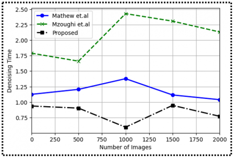
Figure 8. Denoising time levels
Figure 8 illustrates the denoising time of the state-of-art models Mathew et al. [31], Mzoughi et al. [32], and the proposed model, concerning varying numbers of images. Remarkably, the proposed model consistently achieves lower denoising times compared to both Mathew et.al and Mzoughi et al. [32] models, indicating faster and more efficient denoising performance. As the number of images increases, the proposed model's denoising time remains low, making it suitable for real-time denoising applications. In contrast, both Mathew et al. [31] and Mzoughi et al. [32] models require longer denoising times, impacting their usability for real-time denoising tasks.
Figure 9. Denoising accuracy levels
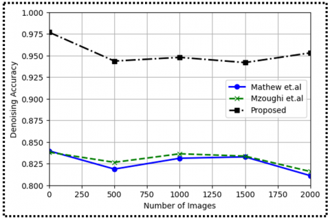
Figure 9 highlights the denoising accuracy of the existing models Mathew et al. [31], Mzoughi et al. [32], and the proposed model, concerning varying numbers of images. The results indicate that the proposed model consistently achieves higher denoising accuracy compared to both Mathew et al. [31] and Mzoughi et al. [32] models, effectively reducing noise in denoised images. Additionally, as the number of images increases, the proposed model's denoising accuracy improves, showcasing its adaptability to different noise patterns and its ability to provide better denoising results than the existing models.
Figure 10. MSE
Figure 11. PSNR
Figure 10 demonstrates the Mean Squared Error (MSE) of the existing models Mathew et al. [31], Mzoughi et al. [32], and the proposed model, with varying numbers of images. Notably, the proposed model consistently achieves lower MSE values compared to both Mathew et al. [31] and Mzoughi et al. [32] models, indicating more accurate reconstruction of denoised images. As the number of images increases, the proposed model's MSE values decrease, showcasing its capability to reconstruct denoised images with higher fidelity. In contrast, the MSE values of the existing models remain relatively high and do not show significant improvement with an increase in the number of images, highlighting their inferior denoising performance compared to the proposed model.
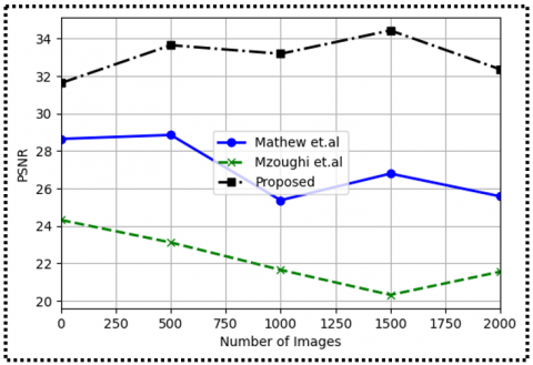
Figure 11 displays the Peak Signal-to-Noise Ratio (PSNR) of the existing models Mathew et al. [31], Mzoughi et al. [32], and the proposed model, concerning varying numbers of images. The results indicate that the proposed model consistently achieves higher PSNR values compared to both Mathew et al. [31] and Mzoughi et al. [32] models, indicating superior image quality in denoised images. As the number of images increases, the proposed model's PSNR values improve, showcasing its ability to enhance image quality with larger datasets. In contrast, the PSNR values of existing models do not show significant improvement with an increase in the number of images, revealing limitations in their ability to provide high-quality denoised images.
In this research addresses the critical challenge of Gaussian noise in MR images, which can significantly impact image quality and reliability in clinical diagnosis and analysis tasks. MRI is a vital medical imaging technique, offering high-contrast, multidimensional images without radiation exposure, making it indispensable for visualizing organs and tissues within the body. However, the presence of random noise poses a hurdle for accurate image segmentation and visualization, necessitating effective MR image denoising solutions. The primary goal of image denoising is to remove distracting background noise while preserving essential details for accurate diagnosis. In this study, various denoising techniques, including Wiener filtering, bilateral filtering, and total variation filtering, were explored and evaluated. The results showed that the proposed Multilevel Denoising model with Precise Edge-based Segmentation for Tumor Size Detection achieved outstanding denoising accuracy of up to 97%, effectively reducing noise without compromising image sharpness.
Moreover, the novel Edge Direction algorithm was introduced, which not only denoises the image but also preserves its edges, addressing the issue of blurring that is often encountered with traditional denoising methods. By incorporating edge component extraction during processing, the proposed algorithm demonstrated superior denoising results, producing images of enhanced quality and accuracy. The new brain tumor segmentation architecture showcased in this research takes full advantage of the detailed information provided by MRI images. By accurately segmenting brain images and performing denoising, the proposed model contributes to precise tumor detection. This achievement holds great promise in revolutionizing the field of medical image processing, empowering healthcare professionals with reliable and accurate tools for improved brain tumor diagnosis and treatment planning.
The obtained results demonstrate the effectiveness and potential of the proposed model in achieving remarkable denoising accuracy. As future work, exploring pixel-to-pixel analysis could further enhance image quality and advance accuracy levels. Continued research and development in the field of MR image denoising hold significant potential for enhancing patient care and improving outcomes in brain tumor diagnosis and treatment. With further refinements, the proposed model has the potential to make a significant impact in the medical field, ultimately benefiting patients worldwide by enabling early and precise detection of brain tumors.
[1] Chen, G., Li, Q., Shi, F., Rekik, I., Pan, Z. (2020). RFDCR: Automated brain lesion segmentation using cascaded random forests with dense conditional random fields. Neuroimage, 211: 116620. https://doi.org/10.1016/j.neuroimage.2020.116620
[2] Khosravanian, A., Rahmanimanesh, M., Keshavarzi, P., Mozaffari, S. (2020). Fast level set method for glioma brain tumor segmentation based on superpixel fuzzy clustering and lattice boltzmann method. Computer Methods and Programs in Biomedicine, 198: 105809. https://doi.org/10.1016/j.cmpb.2020.105809
[3] Tang, Z., Ahmad, S., Yap, P.T., Shen, D. (2018). Multi-atlas segmentation of MR tumor brain images using low-rank based image recovery. IEEE Transactions on Medical Imaging, 37(10): 2224-2235. https://doi.org/10.1109/TMI.2018.2824243
[4] Bakas, S., Akbari, H., Sotiras, A., et al. (2017). Segmentation labels and radiomic features for the pre-operative scans of the TCGA-LGG collection. The Cancer Imaging Archive, 286. https://doi.org/10.7937/K9/TCIA.2017.GJQ7R0EF
[5] Ehrhardt, M.J., Markiewicz, P., Schönlieb, C.B. (2019). Faster PET reconstruction with non-smooth priors by randomization and preconditioning. Physics in Medicine & Biology, 64(22): 225019. https://doi.org/10.1088/1361-6560/ab3d07
[6] Kollem, S., Reddy, K.R.L., Rao, D.S. (2020). Modified transform‐based gamma correction for MRI tumor image denoising and segmentation by optimized histon‐based elephant herding algorithm. International Journal of Imaging Systems and Technology, 30(4): 1271-1293. https://doi.org/10.1002/ima.22429
[7] Orea-Flores, I.Y., Gallegos-Funes, F.J., Arellano-Reynoso, A. (2019). Local complexity estimation based filtering method in wavelet domain for magnetic resonance imaging denoising. Entropy, 21(4): 401. https://doi.org/10.3390/e21040401
[8] Mzoughi, H., Njeh, I., Slima, M.B., Hamida, A.B., Mhiri, C., Mahfoudh, K.B. (2019). Denoising and contrast-enhancement approach of magnetic resonance imaging glioblastoma brain tumors. Journal of Medical Imaging, 6(4): 044002. https://doi.org/10.1117/1.JMI.6.4.044002
[9] Chauhan, N., Choi, B.J. (2019). Denoising approaches using fuzzy logic and convolutional autoencoders for human brain MRI image. International Journal of Fuzzy Logic and Intelligent Systems, 19(3): 135-139. https://doi.org/10.5391/IJFIS.2019.19.3.135
[10] Gupta, D., Ahmad, M. (2018). Brain MR image denoising based on wavelet transform. International Journal of Advanced Technology and Engineering Exploration, 5(38): 11-16. https://doi.org/10.19101/IJATEE.2017.437007
[11] Kala, R., Deepa, P. (2019). Adaptive fuzzy hexagonal bilateral filter for brain MRI denoising. Multimedia Tools and Applications, 79: 15513-15530. https://doi.org/10.1007/s11042-019-7459-x
[12] Khaleel, H.S., Sagheer, S.V., Baburaj, M., George, S.N. (2018). Denoising of Rician corrupted 3D magnetic resonance images using tensor-SVD. Biomedical Signal Processing and Control, 44: 82-95. https://doi.org/10.1016/j.bspc.2018.04.004
[13] Akshath, M.J., Sheshadri, H.S. (2018). Denoising of skull stripped brain tumor MR images. International Journal of Computer Sciences and Engineering, 6(11): 323-329. https://doi.org/10.26438/ijcse/v6i11.323329
[14] Xu, J., Noo, F. (2017). A sequential solution for anisotropic total variation image denoising with interval constraints. Physics in Medicine & Biology, 62(18): N428-N435. https://doi.org/10.1088/1361-6560/aa837d
[15] Bland, J., Bland, J., Mehranian, A., Belzunce, M.A., Ellis, S., McGinnity, C.J., Hammers, A., Reader, A.J. (2018). MR-guided kernel EM reconstruction for reduced dose PET imaging. IEEE Transactions on Radiation and Plasma Medical Sciences, 2(3): 235-243. https://doi.org/10.1109/TRPMS.2017.2771490
[16] Song, T.A., Yang, F., Chowdhury, S.R., Kim, K., Johnson, K.A., El Fakhri, G., Li, Q., Dutta, J. (2019). PET image deblurring and super-resolution with an MR-based joint entropy prior. IEEE Transactions on Computational Imaging, 5(4): 530-539. https://doi.org/10.1109/TCI.2019.2913287
[17] Hashimoto, F., Ohba, H., Ote, K., Tsukada, H. (2018). Denoising of dynamic sinogram by image guided filtering for positron emission tomography. IEEE Transactions on Radiation and Plasma Medical Sciences, 2(6): 541-548. https://doi.org/10.1109/TRPMS.2018.2869936
[18] Zhang, H., Zeng, D., Zhang, H., Wang, J., Liang, Z., Ma, J. (2017). Applications of nonlocal means algorithm in low-dose X-ray CT image processing and reconstruction: A review. Medical Physics, 44(3): 1168-1185.
[19] Hasan, A.M., Melli, A., Wahid, K.A., Babyn, P. (2018). Denoising low-dose CT images using multiframe blind source separation and block matching filter. IEEE Transactions on Radiation and Plasma Medical Sciences, 2(4): 279-287. https://doi.org/10.1109/TRPMS.2018.2810221
[20] Zhao, L.J., Lu, Z.X., Jiang, J., Zhou, Y., Wu, Y., Feng, Q. (2019). Automatic nasopharyngeal carcinoma segmentation using fully convolutional networks with auxiliary paths on dual-modality PET-CT images. Journal of Digital Imaging, 32(3): 462-470. https://doi.org/10.1007/s10278-018-00173-0
[21] Guo, Z., Li, X., Huang, H., Guo, N., Li, Q. (2019). Deep learning-based image segmentation on multimodal medical imaging. IEEE Transactions on Radiation and Plasma Medical Sciences, 3(2): 162-169. https://doi.org/10.1109/TRPMS.2018.2890359
[22] Yang, H., Huang, W.J., Qi, K.H., Li, C., Liu, X.F., Wang, M.Y., Zheng, H.R., Wang, S.S. (2019). CLCI-Net: Cross-level fusion and context inference networks for lesion segmentation of chronic stroke. In: Shen, D., et al. Medical Image Computing and Computer Assisted Intervention – MICCAI 2019. MICCAI 2019. Lecture Notes in Computer Science(), vol 11766. Springer, Cham. https://doi.org/10.1007/978-3-030-32248-9_30
[23] Laurent, B., Ouahabi, A., Fayad, H., Tan, S., Li, L., Lu, W., Jaouen, V., Tauber, C., Czakon, J., Drapejkowski, F., Dyrka, W., Camarasu-Pop, S., Cervenansky, F., Girard, P., Glatard, T., Kain, M., Yao, Y., Barillot, C., Kirov, A., Visvikis, D. (2018). The first MICCAI challenge on PET tumor segmentation. Medical Image Analysis, 44: 177-195. https://doi.org/10.1016/j.media.2017.12.007
[24] Yi, X., Babyn, P. (2018). Sharpness-aware low-dose CT denoising using conditional generative adversarial network. Journal of Digital Imaging, 31(5): 655-669.
[25] Kadimesetty, V.S., Gutta, S., Ganapathy, S., Yalavarthy, P.K. (2019). Convolutional neural network-based robust denoising of low-dose computed tomography perfusion maps. IEEE Transactions on Radiation and Plasma Medical Sciences, 3(2): 137-152. https://doi.org/10.1109/TRPMS.2018.2860788
[26] Gong, K., Guan, J., Liu, C., Qi, J. (2019). PET image denoising using a deep neural network through fine tuning. IEEE Transactions on Radiation and Plasma Medical Sciences, 3(2): 153-161. https://doi.org/10.1109/TRPMS.2018.2877644
[27] Wang, Y., Yu, B.T., Wang, L., Zu, C., Lalush, D.S., Lin, W.L., Wu, X., Zhou, J.L., Shen, D.G., Zhou, L.P. (2018). 3D conditional generative adversarial networks for high-quality PET image estimation at low dose. Neuroimage, 174: 550-562. https://doi.org/10.1016/j.neuroimage.2018.03.045
[28] Cui, J.N., Gong, K., Guo, N., Wu, C.X., Meng, X.X., Kim, K., Zheng, K., Wu, Z.F., Fu, L.P., Xu, B.X., Zhu, Z.H., Tian, J.H., Liu, H.F., Li, Q.Z. (2019). PET image denoising using unsupervised deep learning. European Journal of Nuclear Medicine and Molecular Imaging, 46(13): 2780-2789. https://doi.org/10.1007/s00259-019-04468-4
[29] K. T. Chen, Gong, E., de Carvalho Macruz, F.B., et al. (2019). Ultra–low-dose ¹⁸F-florbetaben amyloid PET imaging using deep learning with multi-contrast MRI inputs. Radiology, 290(3): 649-656. https://doi.org/10.1148/radiol.2018180940
[30] Shan, H., Zhang, Y., Yang, Q., Kruger, U., Kalra, M.K., Sun, L., Cong, W., Wang, G. (2018). 3-D convolutional encoder-decoder network for low-dose CT via transfer learning from a 2-D trained network. IEEE Transactions on Medical Imaging, 37(6): 1522-1534. https://doi.org/10.1109/TMI.2018.2832217
[31] Mathew, J., Zollanvari, A., Pappachen, A., James, A. (2018). Edge-aware spatial denoising filtering based on a psychological model of stimulus similarity. Journal of Imaging, 4(11): 131. https://doi.org/10.3390/jimaging4110131
[32] Mzoughi, H., Njeh, I., Slima, M., Ben Hamida, A., Chokri, H., Kheireddine, M., Mahfoudh, B., Hamida, A., Mhiri, C., Kheireddine, B. (2019). Denoising and contrast-enhancement approach of magnetic resonance imaging glioblastoma brain tumors. Journal of Medical Imaging, 6(1): 014005. https://doi.org/10.1117/1.JMI.6.1.014005
[33] Arepalli, P.G., Naik, K.J. (2023). A deep learning-enabled IoT framework for early hypoxia detection in aqua water using light weight spatially shared attention-LSTM network. The Journal of Supercomputing. https://doi.org/10.1007/s11227-023-05580-x