Abdelkrim Brioua*![]() | Redha Benzid
| Redha Benzid![]() | Lamir Saidi
| Lamir Saidi![]()
© 2023 IIETA. This article is published by IIETA and is licensed under the CC BY 4.0 license (http://creativecommons.org/licenses/by/4.0/).
OPEN ACCESS
The accurate denoising of acquired electrocardiogram (ECG) signals is a critical preprocessing step in data acquisition for both medical professionals and expert systems to make reliable assessments of cardiac health. In this study, we present an advanced denoising algorithm designed to mitigate the effects of additive white Gaussian noise (AWGN), which is known for its capacity to disrupt the entire frequency band within a signal. Our approach offers a novel integration of wavelet transform and Wiener filtering techniques. The proposed algorithm comprises a single-level discrete wavelet transform (DWT) decomposition followed by hard thresholding of the detail wavelet coefficients and the application of wavelet-domain Wiener filtering to the approximation coefficients. Subsequently, the inverse DWT is employed to generate an initial stage denoised signal. To further improve signal restoration quality, a median filter is utilized. Lastly, to recover R-peaks affected during the previous stage, each R-peak and its adjacent samples are replaced with those from the denoised signal before median filtering. We compared the performance of our technique with three state-of-the-art methods and found that it is highly competitive with the recently published DWT-SBWT method. Our approach also significantly outperforms both the reference wavelet-thresholding technique and the GS-WT strategy, with gains of more than 1.5 dB in most cases of utilized input SNR levels. These findings demonstrate the efficacy of our proposed algorithm in reducing AWGN interference, enabling more accurate evaluations of human cardiac health.
ECG denoising, additive white gaussian noise, discrete wavelet transform, wavelet domain wiener filtering, wavelet thresholding, median filter, first order differentiator, R-peaks recovery
Electrocardiogram (ECG) signals are valuable physiological indicators of human heart health and the overall cardiovascular system. However, during the acquisition phase, ECG signals are often susceptible to various types of noise, such as baseline drift, powerline interference, electromyogram interference, electrode motion, and, in some cases, composite noise. To mitigate these corrupting noises, researchers can implement filtering techniques either during the acquisition process using analog filters, or in post-processing with digital filters employed in embedded systems like DSP-based physiological cards or personal computers.
In the realm of digital filtering, numerous contributions have been made to address the noise reduction challenge, particularly in the context of additive white Gaussian noise (AWGN), which is especially difficult to eliminate due to its broad frequency distribution. Notable state-of-the-art methods have combined wavelet transform (WT) with Wiener filtering [1-3] and empirical mode decomposition (EMD) with Wiener filtering [4] to achieve effective results. However, these methods are often associated with high algorithmic complexity or substantial computational demands.
Motivated by the success of these previous approaches, our study aims to reduce the complexity of the WT-Wiener filter combination and the computational time associated with the EMD-Wiener filter pairing by re-examining the WT-Wiener filter association in a simpler, yet efficient manner.
The remainder of this paper is organized as follows: Section 2 gives a literature review. Section 3 provides a careful explanation of the mathematical background related to the tools employed in our approach. Section 4 outlines the proposed methodology. Section 5 presents a comprehensive discussion of the results obtained through extensive simulations and includes a comparative study to position our technique among other powerful methods in the field.
A considerable body of research has addressed the problem of denoising electrocardiogram (ECG) signals corrupted by additive white Gaussian noise (AWGN). In 1999, Donoho's soft and hard thresholding strategies were compared when applied to wavelet transform (WT) and wavelet-packet (WP) approaches with various threshold selection rules for different types of noise [5]. Subsequently, a combination of signal sub-averaging and an optimal wavelet domain filter was proposed for AWGN reduction, which outperformed the classical sub-averaging technique [6].
A two-stage denoising method incorporating Wiener filtering in the translation-invariant wavelet domain was introduced in the study [1], claiming superior performance compared to the Wiener filtering strategy in the wavelet domain [2] and the well-known translation-invariant denoising technique [7]. In another study, it was concluded that the Daubechies mother wavelet with vanishing moments up to order 8 is the most appropriate for wavelet-based thresholding in ECG denoising in terms of root mean square error (RMSE) and peak preservation [8].
Several other wavelet-based denoising techniques have been proposed, including multiadaptive bionic wavelet transform (MABWT) [9], level-dependent S-median and difference in mean (DM)-S-median thresholding [10], and adaptive thresholding using kurtosis as a measure of Gaussianity [11]. Morphological filtering combined with wavelet thresholding has also been used for AWGN reduction [12], and wavelet soft thresholding has been employed as a preprocessing step for beat detection [13].
Alternative denoising methods have been developed as well, such as a combination of empirical mode decomposition (EMD) with soft wavelet thresholding [14], and an improved version of the study [2] for electro myopotential (EMG) noise reduction in ECG signals [3]. Non-local means (NLM) in the wavelet domain [15] and NLM applied to EMD decompositions [16] have been explored for better denoising of ECG records. Moreover, an adaptive, iterative Fourier decomposition was proposed for both synthetic and real ECG signals [17].
Recently, sparse-based methods have been applied to represent ECG signals as combinations of atoms from a given dictionary for denoising [18]. The statistical process control (SpcShrink) scheme, which discriminates contributing wavelet coefficients in the signal of interest from those of corrupting noise, has been proposed as well [19]. A comprehensive review of numerous powerful strategies can be found in study [20].
Other denoising methods include time domain 1-D Wiener filtering followed by FFT-based low pass filtering, Golay-Savitzky smoothing, and R-peak recovery [21], as well as combinations of EMD, wavelet thresholding, adaptive mean filter smoothing, and R-peak recovery [22]. A comparative study of power-line interference suppression from ECG involving decomposition strategies and the Kalman filter framework has been reported in the study [23]. Additionally, a Teaching-Learning-Based Optimization (TLBO) Algorithm with a Type-2 Fuzzy Adaptive Filter for ECG signal denoising was presented in the study [24].
3.1 Wiener filter
Wiener filter has been and remains to be among the most used methods to reduce additive white gaussian noise corrupting signals. It is based on the hypothesis that such noise is considered as stationary random process. It aims to minimize the quadratic errors between the original and the reconstructed signals.
The Wiener filter is a low-pass filter which has not a unique cutting frequency (fc). Accordingly, it acts in the spatial domain, it means that fc can be found, adaptively, great in smooth (homogenous) regions and, conversely, small in highly detailed (textured) zones.
It is worth noting that there are several possible implementations. Therefore, the adopted technique used in our strategy is the 2D adaptive Wiener filter proposed by Lim [25], in which a variant filter in spatial domain is used, and the additive noise is assumed to be zero mean white noise. In our case of 1D signal, the filtered signal y is deduced from the noisy version x according to the following expression:
$y(\mathrm{n})=\mu_{\mathrm{x}}+\left(\mathrm{x}(\mathrm{n})-\mu_{\mathrm{x}}\right) \frac{\mathrm{v}_{\mathrm{x}}}{\mathrm{v}_{\mathrm{x}}+\mathrm{v}_{\mathrm{n}}}$ (1)
where, $\mu_{\mathrm{x}}$ is the mean value of the region surrounding the nth sample of width equal to length of the chosen mask, $\mathrm{v}_{\mathrm{x}}$ is the local variance of x for the same region and $\mathrm{v}_{\mathrm{n}}$ is the variance of the additive white gaussian noise. It is noticeable that the ratio $\frac{v_x}{v_x+v_n}$ approaches 1 in highly detailed regions (in such case $\mathrm{v}_{\mathrm{n}}$ is considered neglectable compared to $\mathrm{v}_{\mathrm{x}}$ which leads to that the filtered y(n) remains near to the noisy input sample x(n) . However, in the homogenous regions $\left(\mathrm{v}_{\mathrm{x}} \cong 0\right) \mathrm{y}(\mathrm{n})$ is tending to approach the local mean value.
3.2 Discrete wavelet transform (DWT)
Wavelet transform is considered as a powerful tool for signals decomposition. It aims to split the whole frequency range to several adapted frequency bands from coarsest to finest one according to adapted scales [5].
As it is well-known, the wavelet transform is of two types: The first category is the called continuous wavelet transform (CWT), however, the second type is named the discrete wavelet transform (DWT). In our case we are interested by the DWT intensively used for ECG signals denoising [1, 3-24].
Wavelet coefficients issued from DWT are of two types: Approximation coefficients (AJ,k) corresponding to the approximation band of the jth level and the detail coefficients (Dj,k) where $\mathrm{j} \in[1, \mathrm{~J}]$. Explicitly stated, coefficients AJ and Dk are expressed by:
$A J, k=\oint_{-\infty}^{+\infty} x(t) \frac{1}{\sqrt{2^J}} \phi\left(\frac{t-k 2^J}{2^J}\right)$ (2)
$D j, k=\oint_{-\infty}^{+\infty} x(t) \frac{1}{\sqrt{2^j}} \psi\left(\frac{t-k 2^j}{2^j}\right)$ (3)
$x(\mathrm{t})=\sum_{\mathrm{j}=1}^{\mathrm{J}} \sum_{\mathrm{k}=-\infty}^{+\infty} \mathrm{D}_{\mathrm{j}, \mathrm{k}} \psi_{\mathrm{j}, \mathrm{k}}(\mathrm{t})+\sum_{\mathrm{k}=-\infty}^{+\infty} \mathrm{A}_{\mathrm{J}, \mathrm{k}} \phi_{\mathrm{J}, \mathrm{k}}(\mathrm{t})$ (4)
where, $\phi_{\mathrm{j}, \mathrm{k}}(\mathrm{t})$ and $\psi_{\mathrm{j}, \mathrm{k}}(\mathrm{t})$ are fatherlets and wavelets produced from father and mother waves respectively $\phi(\mathrm{t})$ and $\psi(\mathrm{t})$.
Note that $\mathrm{Aj}, \mathrm{k}$ are of low frequency nature and $\mathrm{Dj}, \mathrm{k}$ are the high frequency coefficients. Both coefficients are calculated rapidly based on the famous Mallat algorithm [26].
Additionally, one can report that there are two manners to split a signal by wavelet, the first called wavelet decomposition allowing to divide the wavelet-domain approximation band repeatedly until to reach a needed depth of decomposition, on the other hand, the second strategy, consists in the decomposition of both approximation and details bands oftentimes until to attain the requested depth of partitioning. For illustration Figure 1. shows the decomposition according to the two, above mentioned, schemes.
(a) Wavelet decomposition of depth 2
(b) Wavelet packet of depth 2
Figure 1. Decomposing a signal by wavelet to depth 2
3.3 DONOHO’s hard and soft thresholding
One of the most used strategies in denoising is the well-established DONOHO’s wavelet based thresholding algorithm [27, 28]. In such method the most part of details coefficients less in absolute value then a threshold ( $\mathrm{TH}_{\mathrm{i}}$) are considered as contributing coefficients in the noise signal that should be nullified. It is worth noting that $\mathrm{TH}_{\mathrm{i}}$ means the corresponding threshold the ith wavelet band, accordingly, $\mathrm{TH}_{\mathrm{i}}$ is expressed as follows [27]:
$\mathrm{TH}_{\mathrm{i}}=\sigma_{\mathrm{i}} \sqrt{2 \cdot \log \left(\mathrm{n}_{\mathrm{i}}\right)}$ (5)
where: $\mathrm{n}_{\mathrm{i}}$ is the corresponding length of the ith wavelet band and $\sigma_{\mathrm{i}}$ is the related estimation of the unknown standard deviation of the AWGN corrupting noise which is given by:
$\sigma_{\mathrm{i}}=\frac{\operatorname{median}\left(\left|\mathrm{CD}_{\mathrm{i}}\right|\right)}{0.6745}$ (6)
Note that in the suggested strategy we used the DONOHO’s fixed threshold (called also the universal threshold), this fixed threshold is defined by:
$\mathrm{TH}=\sigma \sqrt{2 \cdot \log (\mathrm{N})}$ (7)
where: N is the signal length and $\sigma$ is calculated in a same manner such in (6) except that the used band is CD1 band.
There are two kinds of thresholding approaches that are hard and soft thresholding defined, respectively, by:
$\operatorname{Ther}\left(\mathrm{CD}_{\mathrm{j}}(\mathrm{k})\right)=\left\{\begin{array}{c}\mathrm{CD}_{\mathrm{j}}(\mathrm{k}),\left|\mathrm{CD}_{\mathrm{j}}(\mathrm{k})\right|>T H \\ 0,\left|\mathrm{CD}_{\mathrm{j}}(\mathrm{k})\right| \leq \mathrm{TH}\end{array}\right.$ (8)
and
$\begin{aligned} & \operatorname{Ther}\left(C D_j(k)\right)=\left\{\begin{array}{c}\operatorname{sign}\left(C D_j(k)\right) \cdot\left(\left|C D_j(k)\right|-T H\right), \text { if }\left|C D_j(k)\right|>T H \\ 0, \quad \text { if }\left|C D_j(k)\right| \leq T H\end{array}\right.\end{aligned}$ (9)
where, Ther is the thresholding function.
The corrupted ECG signal can be modeled by (10):
$x(n)=c x(n)+b(n)$ (10)
where:
$\mathrm{x}(\mathrm{n})$: is the noisy signal;
$\operatorname{cx}(\mathrm{n})$: is the clean original signal to estimate;
$\mathrm{b}(\mathrm{n})$: is the contaminating noise assume to be zero-mean and of $\sigma_{\mathrm{b}}^2$ variance additive white Gaussian noise.
The suggested technique can be summarized in the following steps:
Step 1: 1-level depth DWT wavelet decomposition of the noisy signal $\mathrm{x}(\mathrm{n})$ to obtain approximation coefficients vector $\mathrm{CA}_1(\mathrm{k})$ and details coefficients vector $\mathrm{CD}_1(\mathrm{k})$. Note that the used mother wave is coif4 according to (2) and (3).
Step 2: $\mathrm{CD}_1(\mathrm{k})$ thresholding according to DONOHO’s algorithm using the universal threshold using (5), (6), (7), (8) and (9).
The suggested method is illustrated in Figure 2.
Figure 2. Proposed denoising scheme
Step 3: Wavelet-domain filtering use by application of Wiener filter on the approximation coefficients vector $\mathrm{CA}_1(\mathrm{k})$ following (1).
Step 4: Inverse DWT (IDWT) application to reconstruct the estimated restored signal $\widehat{\mathrm{cx}}(\mathrm{n})$ with respect to (4).
Step 5: Median filtering of denoised signal $\mathrm{dx}(\mathrm{n})$ such in the study [29]. The chosen length of analysis window is 5.
Step 6: Localization of R-peaks based on the first order differentiator $d x(n)-d x(n-1)$ mentioned in the study [30].
Step 7: If estimated SNRI $\geq 5$ dB Replace Peaks and surrounding neighbors of mdx(n) by those of dx(n).
Else keep dx(n) unchanged.
Additionally, the proposed method is described in the following scheme:
Note that, actually, the input SNR ( SNRin describing the power ratio of signal to noise is unknown. Consequently, based on the fact that ECG signal and the corrupting AWGN are decorrelated, the SNRin can be estimated based on (11), (12):
$\sigma_{\mathrm{x}}^2=\sigma_{\mathrm{cx}}^2+\sigma_{\mathrm{b}}^2$ (11)
It means,
$\sigma_{\mathrm{cx}}^2=\sigma_{\mathrm{x}}^2-\sigma_{\mathrm{b}}^2$ (12)
where, $\sigma_{\mathrm{x}}^2$, $\sigma_{\mathrm{cx}}^2$ and $\sigma_{\mathrm{b}}^2$ are, respectively, the noisy ECG variance, the unknown variance of the clean ECG signal to estimate and the variance of AWGN that can be estimated by (6) using CD1 band.
Finally, the SNRin can be expressed by (13):
$\mathrm{SNR}_{\mathrm{in}}=20 \log _{10}\left(\frac{\sigma_{\mathrm{cx}}}{\sigma_{\mathrm{b}}}\right)$ (13)
In order to evaluate the efficiency of the suggested algorithm, we applied the method on the well-known ARRYTHMIA MIT-BIH database [31], which contains 48 records each one is constituted of two channels of 11 bits resolution and sampled at a rate of 360 Hz.
The quantitative used measures to evaluate performances of faced techniques to the proposed method are:
MSE $=\frac{1}{\mathrm{~N}} \sum_{\mathrm{n}=1}^{\mathrm{N}}(\widehat{\mathrm{cx}}(\mathrm{n})-\mathrm{cx}(\mathrm{n}))^2$ (14)
$\mathrm{SNR}_{\mathrm{OUT}}=20 \log _{10}\left(\frac{\sigma_{\mathrm{Cx}}}{\sqrt{\mathrm{MSE}}}\right)$ (15)
It is worth to note that we compared results of 3 state of the art recent and efficient algorithms against results provided from the suggested strategy.
The first method [32] used the dataset1: 100.dat, 101.dat, 102.dat, 103.dat, 104.dat, 105.dat and 106.dat. The chosen levels of noise corruption are: -5 dB, 0 dB, 5dB, 10dB and 15 dB. The assessment of performance is achieved in term of the average SNRout of the dataset.
Additionally, the authors of the study [33] involved a more extended dataset2, compared to the first one which is: 100.dat, 101.dat, 102.dat, 103.dat, 104.dat, 105.dat, 106.dat, 107.dat, 108.dat, 109.dat, 111.dat and 112.dat. In this case, the several inputs SNR reflecting the power of noise contamination are: 0 dB, 5dB, 10 dB and 15 dB. Also, as previously, the used evaluation measure is the average SNRout of the dataset.
Finally, the wavelet-based thresholding is used in all following comparisons. The decomposition of wavelet is up to level 4 and the used mother wave is the bior4.4 (the CDF 9/7). The thresholding achieved is the hard universal one.
In the first comparison, results of the study [32] and wavelet-based thresholding are reported in the Table 1.
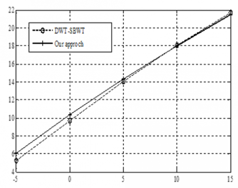
As that can be seen, results obtained from our approach report an enhancement of more than 1,5dB compared to the wavelet technique. However, reached results are very concurrent to those provided from DWT-SBWT as illustrated by Figure 3.
Figure 3. Quantitative comparison between proposed and DWT-SBWT [32] methods
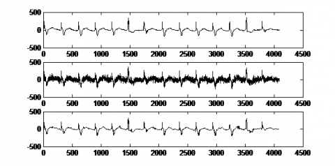
Additionally, to demonstrate the effectiveness of the proposed algorithm when applied on specific different ECG signals of different shapes, a quantitative evaluation is given in Table 2. As one can notice, the denoising process is done efficiently for different levels of input SNRs that are similarly done as the work in the study [32] and obtained results are very comparable of those of the DWT-SBWT. Additionally, a qualitative (visual) inspection is shown in Figure 4, Figure 5 Figure 6 and Figure 7.
For more convincing, results of the proposed approach are faced to those of the study [32] and those of the wavelet-based thresholding. The achieved comparison is summarized in Table 3.
From previous presented results, we can conclude that the suggested strategy is a valid concurrent of the state of the art algorithm of the study [32] and outperforms significantly the GS-WT [33] and the wavelet-based thresholding. Note the reported conclusion holds except for the case of input SNR=10dB where the improvement over the GS-WT is slightly lower than 1dB.
Table 1. Comparative results according to the average SNRout obtained from dataset1 records for different values of SNRin ranging from -5dB to 15 dB
|
SNRin (dB) |
-5 |
0 |
5 |
10 |
15 |
|
SNRout of our approach |
6.0703 |
10.3965 |
14.3076 |
17.9999 |
21.4464 |
|
SNRout of DWT-SBWT [32] |
5.2528 |
9.7188 |
14.0847 |
18.0943 |
21.6649 |
|
SNRout of wavelet- thresholding |
4,2664 |
8.4012 |
12.5424 |
16.0927 |
19.5915 |
|
ECG signal |
104.dat |
101.dat |
105.dat |
113.dat |
|
Specific Input SNR (dB) |
2.6969 |
7.8013 |
10.3621 |
15.7915 |
|
Proposed SNR out (dB) |
11.8099 |
16.7338 |
18.0777 |
23.0077 |
|
SNR out (dB) [32] |
11.0800 |
16.1197 |
18.5830 |
22.6416 |
Figure 4. 104.dat SNRI=2.6969 dB, Proposed SNR out=11.8099dB
Figure 5. 101.dat SNRI=7.8013dB, Proposed SNR out=16.7338dB
Figure 6. 105.dat SNRI=10.3621dB, Proposed SNR out=18.0777dB
Figure 7. 113.dat SNRI=15.7915dB, Proposed SNR out=23.0077dB
Table 3. Comparative results according to the average SNRout obtained from dataset2 records for different values of SNRin ranging from 0 dB to 15 dB
|
Input SNR (dB) |
0 |
5 |
10 |
15 |
|
SNRout of our approach |
9.9866 |
14.2769 |
17.5920 |
21.0737 |
|
SNRout of GS-WT [33] |
8.6300 |
12.5200 |
16.7300 |
19.6000 |
|
SNRout of wavelet-thresholding |
8.4012 |
12.5424 |
16.0927 |
19.5915 |
This work allowed us to re-explore the association of the wiener filter with the DONOHO’s wavelet thresholding applying the universal threshold. Obtained results are concurrent with those achieved in the most recent work of the study [32] and outperforms by more than 1.5dB the standard wavelet thresholding algorithm and the work of the study [33].
For the suggested association, the particularity highlighted by our work is that we obtain the best results by limiting the level of wavelet decomposition to 1-level unlike the most part of previous published works where fourth-level decomposition is frequently used for Arrythmia MIT-BIH database.
As one of future directions of research, one can improve the R-peaks and surrounding areas detection and incorporate the suggested technique in a wearable system.
[1] Nikolaev, N., Nikolov, Z., Gotchev, A., Egiazarian, K. (2000). Wavelet domain Wiener filtering for ECG denoising using improved signal estimate. In 2000 IEEE International Conference on Acoustics, Speech, and Signal Processing. Proceedings (Cat. No. 00CH37100), 6: 3578-3581. https://doi.org/10.1109/ICASSP.2000.860175
[2] Ghael, S., Sayeed, A.M., Baraniuk, R.G. (1997). Improved wavelet denoising via empirical Wiener filtering. In SPIE Technical Conference on Wavelet Applications in Signal Processing. 3169: 389-399. https://doi.org/10.1117/12.292799
[3] Smital, L., Vítek, M., Kozumplík, J., Provazník, I. (2013) Adaptive wavelet wiener filtering of ECG signals. IEEE Transactions on Biomedical Engineering, 60(2): 1-9 https://doi.org/10.1109/tbme.2012.2228482
[4] Chang, K.M., Liu, S.H. (2011). Gaussian noise filtering from ECG by Wiener filter and ensemble empirical mode decomposition. Journal of Signal Processing Systems, 64(2): 249-264. https://doi.org/10.1007/s11265-009-0447-z
[5] Tikkanen, P.E. (1999). Nonlinear wavelet and wavelet packet denoising of electrocardiogram signal. Biological Cybernetics, 80(4): 259-267. https://doi.org/10.1007/s004220050523
[6] Rakotomamonjy, A., Coast, D., Marche, P. (1999). Wavelet-based enhancement of signal-averaged electrocardiograms for late potential detection. Medical & Biological Engineering & Computing, 37(6): 750-759. https://doi.org/10.1007/BF02513378
[7] Abramovich, F., Benjamini, Y., Antoniadis, A., Oppenheim, G. (1995). Wavelets and Statistics. Springer-Verlag.
[8] Singh, B.N., Tiwari, A.K. (2006). Optimal selection of wavelet basis function applied to ECG signal denoising. Digital Signal Processing 16(3): 275-287. https://doi.org/ :10.1016/j.dsp.2005.12.003
[9] Sayadi, O., Shamsollahi, M.B. (2007). Multiadaptive bionic wavelet transform: Application to ECG denoising and baseline wandering reduction. EURASIP Journal on Advances in Signal Processing, (1): 1-11. https://doi.org/10.1155/2007/41274
[10] Poornachandra, S. (2008). Wavelet-based denoising using subband dependent threshold for ECG signal. Digital Signal Processing, 18(1): 49-55 https://doi.org/10.1016/j.dsp.2007.09.006
[11] Sharma, L.N., Dandapat, S., Mahanta, A. (2010). ECG signal denoising using higher order statistics in Wavelet subbands. Biomedical Signal Processing and Control, 5(3): 214-222. https://doi.org/10.1016/j.bspc.2010.03.003
[12] Zhang, D., Sui, W.T. (2010). Noise reduction of ECG signal based on morphological filtering and WT. Key Engineering Materials, 439-440: 12-16 https://doi.org/10;4028/www.scientific.net/KEM.439-440.12
[13] Joshi, S.S., Shrivastava, P. (2011). ECG beat detection using wavelet denoising. In Thinkquest~ 2010, pp. 8-11. https://doi.org/10.1007/978-81-8489-989-4_2
[14] Kabir, M.A., Shahnaz, C. (2012). Denoising of ECG signals based on noise reduction algorithms in EMD and wavelet domains. Biomedical Signal Processing and Control, 7(5): 481-489. https://doi.org/10.1016/j.bspc.2011.11.003
[15] Yadav, S.K., Sinha, R., Bora, P.K. (2015). Electrocardiogram signal denoising using non local wavelet transform domain filtering. IET Signal Processing, 9(1): 88-96. https://doi.org/10.1049/iet-spr.2014.0005
[16] Kumar, S., Panigrahy, D., Sahu, P.K. (2018). Denoising of Electrocardiogram (ECG) signal by using empirical mode decomposition (EMD) with non-local mean (NLM) technique. Biocybernetics and Biomedical Engineering, 38(2): 297-312. https://doi.org/10.1016/j.bbe.2018.01.005
[17] Wang, Z., Wan, F., Wong, C.M., Zhang, L. (2016). Adaptive Fourier decomposition based ECG denoising. Computers in Biology and Medicine, 77: 195-205. https://doi.org/10.1016/j.compbiomed.2016.08.013
[18] Zhu, J., Li, X. (2017). Electrocardiograph signal denoising based on sparse decomposition. Healthcare Technology Letters, 4(4): 134-137. https://doi.org/10.1049/htl.2016.0097
[19] Bayer, F.M., Kozakevicius, A.J., Cintra, R.J. (2019). An iterative wavelet threshold for signal denoising. Signal Processing, 162: 10-20. https://doi.org/10.1016/j.sigpro.2019.04.005
[20] Chatterjee, S., Thakur, R.S., Yadav, R.N., Gupta, L., Raghuvanshi, D.K. (2020). Review of noise removal techniques in ECG signals. IET Signal Processing, 14(9): 569-590. https://doi.org/10.1049/iet-spr.2020.0104
[21] Harkat, A., Benzid, R., Athamena, N. (2021). A multistage algorithm design for electrocardiogram signal denoising. Journal of Circuits, Systems and Computers, 30(4): 2150061-2150061. https://doi.org/10.1142/S0218126621500614
[22] Rakshit, M., Das, S. (2018). An efficient ECG denoising methodology using empirical mode decomposition and adaptive switching mean filter. Biomedical Signal Processing and Control, 40: 140-148. https://doi.org/10.1016/j.bspc.2017.09.020
[23] Bodile, R.M., Talari, V.K. (2021). Removal of power-line interference from ECG using decomposition methodologies and Kalman filter framework: a comparative study. Traitement du Signal, 38(3): 875-881. https://doi.org/10.18280/ts.380334
[24] Ouali, A.M., Ghanai, M., Chafaa, K. (2020). TLBO optimization algorithm based-Type2 fuzzy adaptive filter for ECG signals denoising. Traitement du Signal, 37(4): 541-553. https://doi.org/10.18280/ts.370401
[25] Lim, J.S. (1990). Two-dimensional signal and image processing. Englewood Cliffs.
[26] Mallat, S.G. (1989). A theory for multiresolution signal decomposition: the wavelet representation. IEEE Transactions on Pattern Analysis and Machine Intelligence, 11(7): 674-693. https://doi.org/10.1109/34.192463
[27] Donoho, D.L. (1995). De-noising by soft-thresholding. IEEE Transactions on Information Theory, 41(3): 613-627. https://doi.org/10.1109/18.382009
[28] Donoho, D.L., Johnstone, I.M. (1994). Threshold selection for wavelet shrinkage of noisy data. In Proceedings of 16th Annual International Conference of the IEEE Engineering in Medicine and Biology Society, 1: A24-A25. https://doi.org/10.1109/IEMBS.1994.412133
[29] Asma, T., Mouna, G., Assam, O.M., Kheireddine, C. (2019). Efficient filtering framework for electrocardiogram Denoising. International Journal Bioautomation, 23(4): 403-420. https://doi.org/10.7546/ijba.2019.23.4.000548
[30] Kohler, B.U., Hennig, C., Orglmeister, R. (2002). The principles of software QRS detection.IEEE Engineering in Medicine and biology Magazine, 21(1): 42-57. https://doi.org/ 10.1109/51.993193
[31] Moody, G.B., Mark, R.G. (2001). The impact of the MIT-BIH arrhythmia database. IEEE Engineering in Medicine and Biology Magazine. 20(3): 45-50. https://doi.org/ 10.1109/51.932724
[32] Talbi, M. (2020). New approach of ECG denoising based on 1-D double-density complex DWT and SBWT. Computer Methods in Biomechanics and Biomedical Engineering: Imaging & Visualization, 8(6): 608-620. https://doi.org/ 10.1080/21681163.2020.1763203
[33] Samann, F., Schanze, T. (2019). An efficient ECG denoising method using discrete wavelet with Savitzky-Golay filter. Current Directions in Biomedical Engineering, 5(1): 385-387. https://doi.org/10.1515/cdbme-2019-0097