Reham S. Saeed* | Bushra K. Oleiwi
© 2022 IIETA. This article is published by IIETA and is licensed under the CC BY 4.0 license (http://creativecommons.org/licenses/by/4.0/).
OPEN ACCESS
The outbreak of the new coronavirus (COVID-19) has created a disaster worldwide and it became a very severe and acute disease. COVID-19 prevalence is rapidly increasing around the world. Deep learning (DL) technology had become a hot topic in the context of computing and is widely applied in various medical applications. These techniques have proven to be one of the effective tools for clinicians in the automatic diagnoses of COVID-19. The goal of the present paper is to provide an overview of recently developed systems based on DL techniques that use various medical imaging modalities such as Computer Tomography (CT) and Chest X-Rays (CXR). This review focuses on systems that had been developed for the diagnosis of COVID-19 with the use of the DL methods, as well as the well-known datasets that are utilized for the training of those networks. Finally, the researcher reviewed 58 research papers based on different medical images. Overall, this article aims to assist experts (medical or otherwise) and technicians to understand how the DL approaches are utilized in this context and the way that they can potentially be expanded to combat COVID-19 outbreaks.
Chest X-Ray, CT-Scan, convolutional neural networks, COVID-19, deep learning, disease detection, medical applications, medical images
Since December 8, 2019, a set of the acute lung infections that have been referred to as Corona Virus Disease 2019 COVID-19 had taken place in Wuhan, Hubei, China. Patients who have been infected by COVID-19 have died as a result of severe pneumonia, pulmonary edema, or multiple organ failure. As of April 2, 2020, 82,735 COVID-19 cases had been confirmed in China, with 3,327 Chinese people dying due to the illness. Globally, COVID-19 has had an impact on over 200 countries and areas (a total of 858473 confirmed cases, and a cumulative death toll of 44064) [1]. The World Health Organization (WHO) declared in Mar. that COVID-19 outbreak might be considered a pandemic because this virus was spreading rapidly around the world [2]. The COVID-19 viruses, according to WHO data, had higher purpose death rates (about 3.30%) compared to the previous pandemics of influenza that had occurred in 1918 and 1957, and their spread rate has been even 40 times higher [3]. According to the WHO most recent report, the COVID-19 infected about 139 million people and died approximately 3 million people [4]. Typical clinical early signs include fevers, fatigue, headache, cough, dyspnea, chest tightness, and diarrhea [5].
The reverse transcription-polymerase chain reaction (RT-PCR) is currently considered as gold standard for diagnosing COVID-19 infection. None-the-less, sampling errors and low viral load can have an impact on RT-PCR results [6, 7]. As a result, these tests have a high rate of false negatives and may need to be repeated two or three times before the results are finally confirmed [8]. Many articles recommend chest imaging as one of the tools for the early COVID-19 screening. Many types of datasets as shown in Figure 1 are used for diagnosis and early detection with different accuracy and advantages.
Figure 1. Type of dataset
Hospital staff, nurses, doctors, and clinical facilities are critical in diagnosing this pandemic [9]. Many strategies were implemented so as to mitigate effects of COVID-19. Medical imaging is another technique for the analysis and forecasting of effects of covid-19 on human organism [10]. COVID-19 infected patients and healthy people may be analyzed concurrently using CT and CXR images. After diagnosis, infected patients of COVID-19 must be isolated, do suitable screening, and take sufficient precautions with prevention for the purpose of protecting the healthy individuals. A chain process occurs when contact with COVID-19 infected individuals causes the infection to spread from person to person [11].
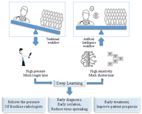
Since the coronavirus was discovered, it became a world-wide pandemic [12]. At the same time, managing the high number of cases has proved to be a challenging task for hospitals or healthcare staff. Thus, artificial intelligence (AI) plays an essential part in assisting the medical staff in the detection and diagnosis of COVID-19 as timely as possible, and most importantly, reducing the risk to the medical staff, saving effort and time, and providing high accuracy in diagnosis [13]. In medical science, DL is the most effective technique. It is an efficient and fast method for diagnosing and predicting diseases with a high accuracy for diagnosing and predicting diseases with high precision (Figure 2).
Figure 2. Using AI in medical
Despite the massive progress that comes with Artificial Intelligence development, challenges are yet, mainly when it involves human life. To get the best out of artificial intelligence, models must be trained using extensive amounts of input data. But differentiates AI technology from traditional technologies in healthcare is the ability to gather data, process it, and produce a well-defined output to the end-user. AI does this through machine learning algorithms and deep learning. These processes can recognize patterns in behavior and create their own logic. With COVID-19 spreading the most important benefit of using AI is to reduce the contact between medical staff and patient [14].
ML is a sub-set of AI [15] that seeks to uncover an unknown function, dependency, or structure between the input and output variables It is made up of various learning models, including Un-supervised Learning (UL), Supervised Learning (SL), and Reinforcement Learning (RL) [16]. There are three machine learning technique types, which are: Support Vector Machines (SVM) [17], Random Forests (RFs), and Neural Network Multilayer Perceptrons (MLPs) [18]. ML has demonstrated superior performance in a variety of image processing applications, including image analysis, classification, and segmentation [19, 20].
DL represents a sub-set of ML [21] that refers to a data exploration framework that utilizes hierarchical layers of linear and nonlinear transformations. DL-based algorithms, like the convolutional neural networks (CNN), recurrent neural networks (RNN), and long short-term memory (LSTM) were recently utilized by a variety of the researchers to combat this pandemic, which includes COVID-19 detection, classification, diagnosis, screening, and prediction [22-26].
CNN models are essentially feed-forward artificial neural networks (ANNs). Basically, CNNs are a group of neural networks with multiple convolutional layers that may be trained so as to analyze images and extract features for the computer vision [27, 28]. Designed to improve the use of spatial information through taking 2-D or 3-D images as input. CNNs are composed of the convolution layers that have been separated by pooling layers, succeeded by the fully connected layers, as in standard multi-layer NN [29]. (Figure 3). The purpose of the convolution layer is the detection of local features at a variety of the positions in input feature maps [30, 31]. The key benefit of the use of the CNNs instead of the conventional approaches is that it demands significantly less amount of the pre-processing [32] and reduce the number of parameters [33]. Recently, CNNs are commonly used for struggling with the COVID-19 disease. However, it is utilized for screening, diagnosing, classification, and predictions [34]. Because of the discriminative representations of the CNN features, object detection employs deep CNN in order to extract features [35].
Figure 3. General CNN
DL approaches are at the forefront of accurate COVID-19 diagnosis and prediction from medical imaging. It can be applied for the purpose of classifying the infected patients from the normal individuals or to segment infectious regions in CXR and CT images [36]. In this review, the research focused on deep learning-based approaches that used CXR or CT scan or both as a dataset in diagnosing COVID-19.
5.1 Diagnosis based on CXR images
For automatic diagnosis of COVID19, a DL model was proposed in Pelaez and Loayza [37] similar to the Visual Geometry Group-VGG block architecture based upon CNN with three incremental convolution blocks and a fully connected MLP with CXR images. And achieved 98.2% of accuracy. In Wang et al. [38] a deep CNN was designed for the detection of COVID-19 cases from the images of CXR called the COVID-Net. The model has tested on three cases no infections (i.e. normal), COVID-19 viral infection and non-COVID19 infections and was compared to other models and achieved good accuracy of 93.3%. Islam et al. [39] purposed a DL system based on a combination of LSTM and CNNs for automatically diagnosing COVID-19 from the images of CXR. CNN is utilized for the deep feature extraction in this system, and LSTM is utilized for the detections with the use of the obtained feature. The system achieved 99.4% accuracy. In Basu et al. [40], Domain extension transfer learning (DETL) is an approach that uses pre-trained deep CNN on a related large CXR data-set that is used to classify between normal, pneumonia, other illnesses, and COVID-19. The overall accuracy was 90.13%. In order to optimize hyper parameters for CNN training, Adam's optimization algorithm has been used. For COVID-19 detection in CXR images, a CNN-based Residual NN (ResNet50) has been proposed in Ref. [41]. The ResNet50 model, a 50-layer network, was used for classification of CXR images as COVID-19 and non-COVID categories, and it achieved 98% accuracy [41].
A new approach for detecting COVID-19 is proposed in Ref. [42] by combining a grey level co-occurrence matrix GLCM and a CNN approach to detect symptoms. Based on CXR images with light computer processing. The light version is constructed from GLCM simplicity and a single layer of CNN. The accuracy was 97.06%. CNN have been investigated by Tang et al. [43] For detecting COVID-19 on CXR images. The five CNN models that have been used are (DarkNet-19, ResNet101, SqueezeNet, VGG-16, and VGG19) with Natural images for pre-training models. Transfer learning was used for the modification of fully connected layer as well as the output layer for the binary classifications between normal and COVID-19 lung images. All five models had an accuracy above 90%. A Deep CNN model that had been referred to as the De-compose Transfer and Compose (DeTraC) is validated for classifying COVID-19 CXR images. Utilizing class de-composition mechanism, DeTraC can analyze any irregularity cases in an image data-set. Experimental results have shown the ability of DeTraC to detect cases of COVID-19 from a large image data-set that has been collected from hospitals worldwide, by achieving a 93.10% accuracy [44]. A conventional transfer learning approach with CXR images was initially used in Ref. [45], Utilizing five pre-trained DL models. The Xception model performed reasonably well, with a diagnostic accuracy of 96.75%. Using a mixture of deep features and ML classification, they developed a technique that further improves diagnostic accuracy. A 99.33% diagnostic accuracy is observed using Xception + SVM. As a result, the Xception model accuracy has been improved by 2.58% [45].
To develop COVID-CXNet, Haghanifar et al. [46] collected numerous CXR images from various sources utilizing the well-known CheXNet model. Five convolution layers that are succeeded by a flattening layer and three fully connected layers were used and this implementation stage does not employ batch normalization or pooling layers. While for high accuracy COVID-19 detection in the CXR images, several experiments were conducted by Khasawneh et al. [47], several groups were considered for the classification, including (COVID-19, Normal) (COVID-19, Pneumonia) and (COVID-19, Pneumonia, Normal). Different image dimensions and network architectures were employed. Using models of transfer learning, machine learning, and CNN were implemented and evaluated. The findings indicate that a CNN with minimized convolution and FC layers is having the ability to detect COVID-19 images in 2-class classifications, COVID-19/Normal and COVID-19/Pneumonia. To improve and test a ML model for COVID-19 diagnoses, and in order to present a tool for searching for similar patients based on X-rays scans, a classifier was built by Keidar et al. [48] utilizing pre-trained DL model (ReNet-50) and improved through the lung segmentation and data augmentation. The model had an accuracy of 89.7%. While in Yoo et al. [49] a DL-based decision-tree classifier was used so as to investigate the feasibility of COVID-19 detection from images of CXR. The suggested classifier is made up of 3 binary decision trees, every one of which has been trained with the use of a DL model with a CNN depending on PyTorch. The 1st decision tree detects whether the CXR images are normal or not, the 2nd one detects abnormal images with tuberculosis signs, while the 3rd one performs the same function for COVID-19. The 1st and 2nd have accuracies of 98% and 80%, while the 3rd decision tree has an average 95% accuracy [49].
Ozturk et al. [50] used raw CXR images with a novel model for automatic detection. The suggested model was designed in order to provide precise diagnostics for the binary classifications (COVID/ No-Findings) and multi classifications (COVID/No-Findings/Pneumonia). For the binary classes, the model accomplished a 98.08% accuracy, and for the multi class, it had an accuracy of 87.02%. DarkNet was used as a model. By using 17 convolution layers that have different filtering to every one of the layers. For detecting COVID-19 pneumonia infected patients, 3 different CNN based models (ResNet50, Inception-V3, and Inception-ResNet-V2) have been suggested by Narin et al. [51] with 98% accuracy, the ROC of pre-trained ResNet-50 provides the optimal classification efficiency. In addition to the other 2 suggested models (97% accuracy for InceptionV3 and 87% accuracy for the Inception-ResNetV2). From the PA's view of CXR scans with DL based on CNN models for classification into COVID-19 patients and healthy controls, three models were deployed (Inception V3, ResNeXt and Xception) and compared performance and accuracy. Xception model has the highest accuracy (97.97%) for detecting CXR images [52]. Elaziz et al. [53] present a new ML approach to classify CXR images to 2 groups: COVID-19 and non-COVID19. The features were obtained from CXR images with the use of the new Fractional Multi-channel Exponent Moments (FrMEMs). A parallel multicore computation framework is used for the acceleration of computation process. The most significant features have been then chosen with the use of a modified Manta-Ray Foraging Optimization based upon differential evolutions. There have been 2 COVID-19 x-ray data-sets utilized so as to assess the proposed method. For the first and second datasets, the proposed method achieved 96.09% and 98.09% of accuracy respectively.
By a thorough examination of eight pre-trained models. Those models had been trained, validated, and tested on 760 CXR images from five distinct classes. Pre-trained fine-tuned models on the ImageNet data-set have been computationally efficient and precise. DenseNet-121 model could achieve 98.69% accuracy for 4 classes of images, including healthy, COVID-19, bacterial pneumonia, and viral pneumonia images, and fine-tuned models had reached a better degree of the accuracy for 3 classes of images, including healthy, COVID-19, and SARS images [54].
Panahi et al. [55] had suggested a new method Fast COVID-19 Detector (FCOD) for rapid COVID-19 detection using CXR. The FCOD is DL model that is based upon the Inception architecture that detects COVID-19 by employing 17 depth-wise separable convolution layers. When compared to standard convolution layers, depth-wise separable convolutional layers reduce computation cost and time, and can help reduce the number of parameters. The results show that FCOD can classify COVID-19 with a 96% accuracy in 0.014 seconds for each case. In Das et al. [56], a Deep CNN based solution for detection of COVID-19 +ve patients was proposed using CXR images. To evaluate the efficacy of a solution, public available CXR images of coronavirus +ve and -ve cases have been used. In the test set-up, the solution had a classification accuracy of 95.7%. Also, a graphical user interface (GUI) application was developed for public uses. This application may be utilized with any device and by all types of the medical personnel for detecting of COVID patients by the utilization of the CXR images in a matter of seconds. A DL technique based upon the Pre-trained AlexNet Model was suggested by Ozsoz et al. for the classifications of the (normal CXR/COVID-19/ viral pneumonia/non-COVID-19/bacterial pneumonia). The proposed model had reached a 94.43% accuracy for non-COVID-19 viral pneumonia and normal, 91.43% accuracy for bacterial pneumonia and normal, 99.16% accuracy for COVID-19 pneumonia and normal CXR images, 99.62% accuracy in classifying COVID-19 pneumonia and non-COVID-19 viral pneumonia and for four classes, this model had achieved 93.42% accuracy, for multiclass datasets, the model could achieve an accuracy of 94% [57]. Table 1 summarizes diagnosis based upon CXR images. In this section, the author shows that using CXR as a dataset, in general, gives accepted accuracy (all above 80%). higher accuracy was found in Islam et al. [39] and Wang et al. [45] despite the number of the dataset, using CNN with a classifier (LSTM, SVM) as a mixture model gives better accuracy than using CNN to extract features and classify.
Table 1. Summary of COVID-19 diagnoses based upon the CXR images
|
Reference |
Dataset |
No. of images |
Model |
Accuracy |
|
[37] |
- COVID-19 X-ray image data-set - local hospital in Guayaquil - Different website’s data from Kaggle. |
838 |
Deep CNN with 3 convolutional blocks similar to VGG |
98.2% |
|
[38] |
COVIDx dataset |
13,975 |
COVID-Net |
93.3% |
|
[39] |
-Different website’s data from Kaggle and GitHub - Radiopaedia - NIH |
4575 |
Deep CNN-LSTM network |
99.4% |
|
[40] |
Italian Society of Medical Radiology and Interventional, Radiopaedia.org, J. Paul Cohen et al., hospital in Spain |
109656 |
Domain Extension Transfer Learning (DETL) |
90.13% |
|
[41] |
- Public dataset |
1824 |
ResNet50 |
98% |
|
[42] |
- Different website’s data from Kaggle and GitHub |
273 |
GLCM-CNN network |
97.06% |
|
[43] |
- COVIDx - University of Malaya (Biomedical department) |
691 |
Five models of CNN: (DarkNet-19, ResNet101, Squeeze Net, VGG-16, VGG-19) |
All above 90% |
|
[44] |
- Collected from several hospital around the world |
196 |
DeTraC |
93.1% |
|
[45] |
- Different website’s data from GitHub and Kaggle |
1102 |
Xception + SVM |
99.33% |
|
[46] |
- Pediatric CXR dataset - NIH CXR-14 dataset - Radiopaedia - Tuberculosis Chest X-ray Image Datasets |
N/M |
CheXNet |
N/M |
|
[47] |
- Different website’s data from Github - obtained by Kermany et al. |
6100 |
different network architectures, pre-trained networks, and machine learning models |
N/M |
|
[48] |
N/M |
2427 |
ReNet50 |
89.7% |
|
[49] |
N/M |
N/M |
DL-based three-level decision-tree classifier |
98% 80% 95% |
|
[50] |
- Different website’s data from GitHub - Chestx-ray8 |
N/M |
DarkNet |
98.08% 87.02% |
|
[51] |
- Different website’s data from Kaggle and GitHub |
100 |
Three models: (ResNet50, Inception V3, Inception-ResNetV2) |
98% 97% 87% |
|
[52] |
- Different website’s data from Kaggle. |
6432 |
Compare betweem 3 models: (Inception V3, Xception, ResNeXt) |
The best 97.97% |
|
[53] |
- Different website’s data from GitHub - collected by a team |
N/M |
Fractional Multichannel Exponent Moments (FrMEMS) |
96.09% 98.06% |
|
[54] |
- Collected from two different dataset |
N/M |
DenseNet121 |
98.69% |
|
[55] |
- Different website’s data from GitHub |
940 |
Fast COVID-19 Detector (FCOD) model based upon the Inception architecture |
96% |
|
[56] |
- Collected from different open sources |
1006 |
Deep CNN comprises of 3 pre-trained models (DenseNet, ResNet50-V2, Inception-V3) |
95.7% |
|
[57] |
- Different website’s data from Kaggle and GitHub - obtained by Kermany et al. |
11568 |
AlexNet for different classes |
All above 90% |
5.2 Diagnosis based on CT-scan images
Shah et al. [58] used different DL approaches for the differentiation of CT scans of COVID-19 and non COVID-19 CT. CTnet-10, a self-developed model with 82.1% accuracy, was designed for the diagnoses of COVID-19. DenseNet-169, VGG16, VGG-19, ResNet50, and inceptionV3 were also tested. When compared to all other DL models, the VGG-19 performed best, with a 94.52% accuracy. CTnet-10, a self-developed model, has been created with an 82.10% accuracy. Passing the CT scan image through several of the pre-existing models to improve accuracy. With an accuracy of 94.52%, the VGG-19 model was the best at classifying the images as COVID-19 +ve or –ve. Liang et al. [59] proposed a hybrid DL framework called CTNet, which combines CNN and transformer for detecting COVID-19 using 3-D images of CT scans. It includes a module of CNN feature extractor with SE attention that extracts enough features from the CTs and a transformer model that models the features of the 3-D CTs. The suggested CT-Net achieved a macro F-1 score of 88.21%. The ResNet-100 deep CNN along with LR classifier was used in Kavitha et al. [60] for the identification and diagnosis of the Coronavirus family rapidly by classifying the result as COVID-19, and normal. As a result, the accuracy was 99.15% with COVID-19 positive patients [60].
In Bhuyan et al. [61] a (full-resolution convolutional network) (FrCN) with a deep CNN is used to find a specific highly infectious area and classify patients to COVID-19 or non-COVID-19. the suggested model is tested for detection, segmentation, and classification. The classification accuracy performance is compared on test datasets validated with and without mass segmentation. A deep learning technique is presented by Verma et al. [62] that could be used for screening COVID-19 automatically. CT images are collected from multicenter case studies as the dataset for the model. According to the model, overall accuracy was around 79.39%. Two different methods with CT images were deployed by Cifci [63] for COVID-19 diagnosis at early stage. As a result of comparing methods, the Alex-Net was the best with an accuracy of 94.74% and the accuracy of Inception-V4 Net was 81.14%. To identify patients who have COVID-19, a DL-based system of CT diagnosis has been developed. Experimental results have indicated that the model could distinguish COVID-19 patients from bacterial pneumonia patients with 0.95 AUC. When 3 CT types were combined, the model realized a 0.93 recall for distinguishing COVID-19 patients from others. The DRENet was developed based on the pre-trained ResNet50, which is robust in detecting objects in images [64]. A DL model was developed by Gozes et al. [65] for detect, quantify and localize the intensity of COVID-19 through using the CTs. The algorithm is made up a series of image processing involving lung segmentation, 2-D slice, classification, and fine grain localization. As a result of classification for COVID vs. Non-COVID cases, the AUC was 0.948.
In JavadiMoghaddam and Gholamalinejad [66], a new DL framework was proposed and the pooling layer of the model has been made of pooling and Squeeze Excitation Block (SE-block) layer and to improve convergence time and efficiency of the COVID-19 diagnoses, Batch Normalization and Mish Function were used. Which is why, the suggested model compared with other popular deep NN and the achieved 99.03% accuracy. By Using DL techniques, Xu et al. [67] aimed to develop screening model in order to identify COVID-19 from Influenza-A, viral pneumonia and healthy samples utilizing the CTs of the lungs. The selection infection regions have been first segmented out of a lung CT image set with the use of a 3-dimensional DL model. Utilizing a location-attention classification model, those images have been classified to groups. As a result, the overall accuracy was 86.7%. For characterizing COVID-19, pneumonia, and normal CT lung images, a novel combination of DL of extracted features and Q-deformed entropy handcrafted features was used. This study employs the preprocessing so as to mitigate effects of the variations of intensity between the CT slices. The background of the CT lung scans is then isolated using histogram thresholding. CT lung scan is subjected to feature extraction using DL and a Q-deformed entropy algorithm. An LSTM NN classifier is used to classify the obtained features. The higher accuracy achieved 99.68% [68].
For COVID-19 diagnostic with CT images, a fully automatic DL system was proposed in Ref. [69] that consist of 3 parts, which are: the automatic segmentation of the lungs, suppression of non-lung areas, and COVID-19 diagnostic and prognostic analyses, with 2 models of DL (which are DenseNet-121-FPN for the segmentation of the lung area in the CT image, and suggested new COVID-19 Net for diagnostic and prognostic analyses of COVID-19). For detections of COVID-19 in an early phase, a new method was proposed in Ref. [70] for fusing and ranking deep features. A SVM is used as a classifier. With to subsets, the suggested approach had achieved a high performance on Subset-2 with an accuracy of 98.27%. Table 2 summarizes the diagnosis based on CT scan images. In this section, the author notes that using CT-scan as a dataset gives questionable accuracy that can’t be dependent as diagnosis result even with large number of data.
5.3 Diagnosis based on CXR and CT-scan images
By using different CNN with two types of datasets (CXR and CT scan) to compare the performances in the diagnosis of COVID-19. The Resnet-18, InceptionV3, and MobileNetV2 were tested on the two types and the result was all the best with CT scan type, The ResNet18 approach had the optimal overall sensitivity and precision of 98.60% and 98.5%, respectively, whereas InceptionV3 approach had the optimal specificity of 97.40%, and the MobileNet-V2 model has a very good sensitivity for the cases of COVID-19 [71]. El Asnaoui and Chawki [72] compare the coronavirus pneumonia detection and classification with the use of seven DL models, which include: VGG-16, VGG-19, DenseNet201, Inception ResNetV2, Inception V3, Resnet50, and MobileNetV2. The experiments were applied on CXR and CT datasets. Thus, the higher accuracy was 92%.
In Sedik et al. [73] a new detection system based on DL was proposed based on CNN and (ConvLSTM) with two datasets (CT images and CXR) and different cases (COVID-19/pneumonia/normal). X-rays and CTs were combined to test the proposed DL methods. This approach produced 100% accuracy. The primary goal of Yang et al. [74] was to investigate and compare several DL improved models for detecting COVID-19 in CXR and CT-scan medical images. Through the use of four robust pre-trained models of CNN for COVID-19 CTs for binary classification task: VGG16, DenseNet121, ResNet50, and ResNet152. The suggested Fast AI ResNet approach was developed to automatically determine the optimal architecture, preprocessing, and training parameters for the models. The diagnosis of COVID-19 utilizing CT images was over 96% accurate. Furthermore, in order to overcome data limitation and shorten training time, TL techniques were used. An improved VGG-16 deep transfer learning architecture had been used to complete binary and multi-class classification of the X-rays image tasks. With 99 percent accuracy, enhanced VGG16 detected X-rays from pneumonia and COVID-19. By utilizing the DL and TL algorithms in Ref. [75] to build a comprehensive dataset containing images of CXR, CT, and other datasets from multiple sources. Our technique is simple but effective in the detection of COVID-19. An X-ray and CT image analysis system, based on a CNN and a modified pre-trained Alex Net model were used. As a result, the utilized models achieved accuracy more than 98% by pre-trained network and 94.10% accuracy with the use of the modified CNN [75].
For diagnosing of the COVID-19, lung cancer, and pneumonia from a combination of CXR and CT scans, a multi-classification DL model was proposed by Ibrahim et al. [76]. This combination was chosen because a CXR is less effective in early disease stages, whereas a chest CT can be used even prior to when the symptoms appear, and a CT scan accurately detects the abnormal features that have been identified in the images. Utilizing those 2 types will result in increasing the data-set size as well, enhancing the accuracy of the classification. The efficiency of the 4 models (which are ResNet152 V2, VGG19-CNN, ResNet152 V2 + Gated Recurrent Unit (GRU), and ResNet152 V2 + Bi-directional GRU (BiGRU)) has been examined. Utilizing the public CT and CXR data-sets with 4 classes, a detailed evaluation of a variety of the DL models has been presented (Normal/ Pneumonia/ COVID-19/Lung cancer). According to results, VGG19 + CNN model performs better compared to the other 3 suggested models. Based on CXR and CT images, VGG19 and CNN model achieved accuracy level of 98.05%. Table 3 summarizes diagnosis based on the CXR as well as the CT scan images [76]. As a result, using a mixture of datasets needs more development, also the accuracy is questionable and needs to improve maybe with more layer of structure or using a classifier as well as CNN techniques.
Table 2. Summary of COVID-19 diagnosis based upon CT scan images
|
Reference |
Dataset |
No. of images |
Model |
Accuracy |
|
[58] |
N/M |
N/M |
CTnet-10 |
82.1% |
|
[59] |
- COV19-CT-DB |
+7323 |
CT-Net |
N/M |
|
[60] |
- Different website’s data from GitHub |
N/M |
RestNet-100 with LR |
99.15% |
|
[61] |
- Collected by Cohen et al. |
200 |
Deep CNN with FrCN |
+98% |
|
[62] |
- Collected online |
548 |
N/M |
79.39% |
|
[63] |
- Different website’s data from Kaggle |
5800 |
AlexNet and Inception-V4 |
94.74% 84.14% |
|
[64] |
- Collected from hospitals |
N/M |
DRE-Net |
86% |
|
[65] |
- Chainz - Zhejiang Province - El-Camino Hospital - University Hospitals of Geneva |
N/M |
ResNet-50 |
N/M |
|
[66] |
- Collected from two public hospitals |
19685 |
WCNN4 |
99.03% |
|
[67] |
- Collected from three hospitals in China |
618 |
N/M |
86.7% |
|
[68] |
- Radiopaedia and cancer imaging archive (TCIA) |
321 |
N/M |
99.68% |
|
[69] |
N/M |
5372 |
DenseNet121-FPN COVID-19Net |
N/M |
|
[70] |
N/M |
Subset1 6000 Subset2 6000 |
Pre-trained CNN with SVM |
95.6% 98.27% |
Table 3. Summary of COVID-19 diagnoses based upon CXR & CT scan images
|
Reference |
Dataset |
No. of images |
Model |
Accuracy |
|
[71] |
- HUST-19 - COVIDx |
24711 |
Three models of CNN: (ResNet-18, InceptionV3, MobileNetV2) |
N/M |
|
[72] |
- Two public image datasets |
6087 |
Seven models of CNN: have been used |
The best 92.18% |
|
[73] |
N/M |
N/M |
CNN and ConvLSTM |
100% |
|
[74] |
- several datasets from public repositories |
N/M |
four models of CNN: VGG16, DenseNet121, ResNet50, ResNet152 |
77% 83.7% 81% 80% |
|
[75] |
- Different website’s data from Kaggle and GitHub - BSTI - Radiopaedia |
526 |
Pre-trained CNN |
94.1% |
|
[76] |
- Different website’s data from GitHub - RSNA - Italian Society of the Medical and Interventional Radiology -Radiopaedia |
33,676 |
four models of CNN: VGG19-CNN, Res Net152V2, Res Net152V2 + (GRU), ResNet152V2 + (Bi-GRU) |
98.05% 95.31% 96.09% 93.36% |
The detection of COVID-19 from CXR and CT scans has been considered to be critical for doctors and patients in order to reduce cost and diagnostic time. DL and AI are both able to recognize images for tasks that have been taught to them. This research presents the most recent works for diagnoses of COVID-19 utilizing different DL approaches based on 2 types of medical images (X-ray and CT-scans). The researcher provides a list of all data sources used and models for easily understood by other researchers. This review can be further extended to cover other research directions such as using other types of medical images and CNN models.
There are many unique challenges for applying deep learning techniques and algorithms for the detection of COVID-19. Although deep learning-based COVID-19 detection from chest CT and X-ray images show promising results, its widespread adaptation still faces various societal and technical pushback. While deep learning techniques are highly automatable, it needs a large set of data to develop a robust system for diagnosis purposes. As COVID-19 is very new to research, the lack of standard data is a major challenge for diagnoses. On the other hand, the available imaging data for COVID-19 patients are incomplete, noisy, ambiguous, and inaccurately labeled in certain cases. To train a deep learning architecture with such massive and diverse data sets is very complex and a variety of problems must be resolved (e.g., data redundancy, sparsity, missing values). Almost all the reviewed systems used different data sets for the experiment. The developed systems collected data from internet sources, prepared it their way, and finally evaluated their systems using evaluation metrics. For this reason, it is quite difficult to conclude definitively which system yields the best result for COVID-19 detection.
In the future, efforts can be directed in more than one direction, firstly collecting the datasets of COVID-19 and other lung diseases from the local hospitals by the author. Secondly, optimization techniques like ant colony, gray wolf, or particle swarm optimization can be used. Finally, proposed an online and real-time approach diagnosis system to support physicians and avoid further spreading of the disease.
[1] Huang, Y., Zhao, N. (2021). Mental health burden for the public affected by the COVID-19 outbreak in China: Who will be the high-risk group? Psychology, Health & Medicine, 26(1): 23-34. https://doi.org/10.1080/13548506.2020.1754438
[2] Hadi, M.A., Ali, H.I. (2021). Control of COVID-19 system using a novel nonlinear robust control algorithm. Biomedical Signal Processing and Control, 64: 102317. https://doi.org/10.1016/j.bspc.2020.102317
[3] Hadi, M.A., Amean, Z.M. (2021). New strategy to control COVID-19 pandemic using lead/lag compensator. Biomedical Signal Processing and Control, 68: 102669. https://doi.org/10.1016/j.bspc.2021.102669
[4] Taha, B.A. (2021). Perspectives of photonics technology to diagnosis COVID–19 viruses: A short review. Journal of Applied Sciences and Nanotechnology, 1(1): 1-6. https://doi.org/10.53293/jasn.2021.11016
[5] Wan, S., Xiang, Y. I., Fang, W., et al. (2020). Clinical features and treatment of COVID‐19 patients in northeast Chongqing. Journal of Medical Virology, 92(7): 797-806. https://doi.org/10.1002/jmv.25783
[6] Abdulsahib, S.S. (2021). Identification of the respiratory tract infection due to methicillin-resistant by TaqMan real-time PCR. Medical Journal of Cell Biology, 9(2): 86-92. https://doi.org/10.2478/acb-2021-0012
[7] Xie, X., Zhong, Z., Zhao, W., Zheng, C., Wang, F., Liu, J. (2020). Chest CT for typical coronavirus disease 2019 (COVID-19) pneumonia: Relationship to negative RT-PCR testing. Radiology, 296(2): E41-E45. https://doi.org/10.1148/radiol.2020200343
[8] Corman, V.M., Landt, O., Kaiser, M., et al. (2020). Detection of 2019 novel coronavirus (2019-nCoV) by real-time RT-PCR. Eurosurveillance, 25(3): 2000045. https://doi.org/10.2807/1560-7917.ES.2020.25.3.2000045
[9] Abd Alsammed, S.M.Z. (2021). Implementation of Lung Cancer Diagnosis based on DNN in Healthcare System. Management.
[10] Hashim, A.T., Jabbar, A.K., Hassan, Q.F. (2021). Medical image encryption based on hybrid AES with chaotic map. In Journal of Physics: Conference Series, 1973(1): 012037.
[11] Yan, L., Zhang, H.T., Xiao, Y., et al. (2020). Prediction of criticality in patients with severe Covid-19 infection using three clinical features: A machine learning-based prognostic model with clinical data in Wuhan. MedRxiv, 27: 2020. https://doi.org/10.1101/2020.02.27.20028027
[12] Alwawi, B.K.O.C., Abood, L.H. (2021). Convolution neural network and histogram equalization for COVID-19 diagnosis system. Indonesian Journal of Electrical Engineering and Computer Science, 420-427.
[13] Thannoon, H.H., Ali, W.H., Hashim, I.A. (2019). A survey on deceptive detection systems and technologies. Engineering and Technology Journal, 37(3): 90-95. https://doi.org/10.30684/etj.37.3A.3
[14] Battineni, G., Chintalapudi, N., Amenta, F. (2020). AI chatbot design during an epidemic like the novel coronavirus. In Healthcare, 8(2): 154. https://doi.org/10.3390/healthcare8020154
[15] Abdulhussein, A.A., Raheem, F.A. (2020). Hand gesture recognition of static letters American sign language (ASL) using deep learning. Engineering and Technology Journal, 38(6): 926-937. https://doi.org/10.30684/etj.v38i6A.533
[16] Kwekha-Rashid, A.S., Abduljabbar, H.N., Alhayani, B. (2021). Coronavirus disease (COVID-19) cases analysis using machine-learning applications. Applied Nanoscience, pp. 1-13. https://doi.org/10.1007/s13204-021-01868-7
[17] Alwash, N.A., Kareem, H. (2021). Detection of COVID-19 based on chest medical imaging and artificial intelligence techniques. Engineering and Technology Journal, 39(10): 1588-1600. https://doi.org/10.30684/etj.v39i10.2200
[18] Mohammed, Z.A., Abdullah, M.N., Al Hussaini, I.H. (2021). Predicting incident duration based on machine learning methods. Iraqi Journal of Computers, Communications, Control and Systems Engineering, 21(1): 1-15.
[19] Mohammed, M.A., Abdulkareem, K.H., Al-Waisy, A.S., et al. (2020). Benchmarking methodology for selection of optimal COVID-19 diagnostic model based on entropy and TOPSIS methods. IEEE Access, 8: 99115-99131. https://doi.org/10.1109/ACCESS.2020.2995597
[20] Bashir, L.Z., Mahdi, N. (2013). Solving categorization problem using two models of machine learning. Iraqi Journal of Computers, Communication, Control & Systems Engineering, 13(3): 27-40. https://www.uotechnology.edu.iq/ijccce/issues/2013/vol13/no.03/full-text/04.pdf.
[21] Raheem, F.A., Hussain, A.W.A.A. (2020). Deep learning convolution neural networks analysis and comparative study for static alphabet asl hand gesture recognition. Journal of Xidian University, 14(4): 1871-1881. https://doi.org/10.37896/jxu14.4/212
[22] Zhou, Y., Wang, F., Tang, J., Nussinov, R., Cheng, F. (2020). Artificial intelligence in COVID-19 drug repurposing. The Lancet Digital Health, 2(12): e667-e676. https://doi.org/10.1016/S2589-7500(20)30192-8
[23] Bogu, G.K., Snyder, M.P. (2021). Deep learning-based detection of COVID-19 using wearables data. MedRxiv. https://doi.org/10.1101/2021.01.08.21249474
[24] He, X. (2020). Sample-efficient deep learning for COVID-19 diagnosis based on CT scans. IEEE Transactions on Medical Imaging. https://doi.org/10.1101/2020.04.13.20063941
[25] Hu, S., Gao, Y., Niu, Z., et al. (2020). Weakly supervised deep learning for covid-19 infection detection and classification from CT images. IEEE Access, 8: 118869-118883. https://doi.org/10.1109/ACCESS.2020.3005510
[26] Albayati, A.Q., Ameen, S.H. (2020). A method of deep learning tackles sentiment analysis problem in Arabic texts. Iraqi Journal of Computers, Communications, Control and Systems Engineering, 20(4): 9-20. https://doi.org/10.33103/uot.ijccce.20.4.2
[27] Abdullah, H.N., Abduljaleel, H.K. (2019). Deep CNN based skin lesion image denoising and segmentation using active contour method. Engineering and Technology Journal, 37(11): 464-469. https://doi.org/10.30684/etj.37.11A.3
[28] Uthaib, M.A., Croock, M.S. (2021). Multiclassification of license plate based on deep convolution neural networks. International Journal of Electrical & Computer Engineering (2088-8708), 11(6): 5266-5276. https://doi.org/10.11591/ijece.v11i6
[29] Ahmed, W.S. (2020). Motion classification using CNN based on image difference. In 2020 5th International Conference on Innovative Technologies in Intelligent Systems and Industrial Applications (CITISIA), pp. 1-6. https://doi.org/10.1109/CITISIA50690.2020.9371835
[30] Shen, D., Wu, G., Suk, H.I. (2017). Deep learning in medical image analysis. Annual review of biomedical engineering, 19: 221-248.
[31] Rahman, T., Chowdhury, M.E., Khandakar, A., et al. (2020). Transfer learning with deep convolutional neural network (CNN) for pneumonia detection using chest X-ray. Applied Sciences, 10(9): 3233. https://doi.org/10.3390/app10093233
[32] Khan, M., Mehran, M. T., Haq, Z.U., Ullah, Z., Naqvi, S.R., Ihsan, M., Abbass, H. (2021). Applications of artificial intelligence in COVID-19 pandemic: A comprehensive review. Expert Systems with Applications, 185: 115695. https://doi.org/10.1016/j.eswa.2021.115695
[33] Kodher, M., Saud, J.H., Hassan, H.S. (2021). Wheelchair Movement Based on Convolution Neural Network. Engineering and Technology Journal, 39(6): 1019-1030. https://doi.org/10.30684/etj.v39i6.1615
[34] Afifi, A., Hafsa, N.E., Ali, M.A., Alhumam, A., Alsalman, S. (2021). An ensemble of global and local-attention based convolutional neural networks for COVID-19 diagnosis on chest X-ray images. Symmetry, 13(1): 113. https://doi.org/10.3390/sym13010113
[35] Mahmoud, M.M., Nasser, A.R. (2021). Dual architecture deep learning based object detection system for autonomous driving. Iraqi Journal of Computers, Communication, Control & Systems Engineering, 21(2): 36-43. https://doi.org/10.33103/uot.ijccce.21.2.3
[36] Karthik, R., Menaka, R., Hariharan, M., Kathiresan, G.S. (2021). Ai for COVID-19 detection from radiographs: Incisive analysis of state of the art techniques, key challenges and future directions. IRBM. https://doi.org/10.1016/j.irbm.2021.07.002
[37] Pelaez, E., Loayza, F. (2020). A deep learning model to screen for Corona Virus Disease (COVID-19) from X-ray chest images. In 2020 Ieee Andescon, pp. 1-6. https://doi.org/10.1109/ANDESCON50619.2020.9272079
[38] Wang, L., Lin, Z.Q., Wong, A. (2020). Covid-net: A tailored deep convolutional neural network design for detection of COVID-19 cases from chest x-ray images. Scientific Reports, 10(1): 1-12. https://doi.org/10.1038/s41598-020-76550-z
[39] Islam, M.Z., Islam, M.M., Asraf, A. (2020). A combined deep CNN-LSTM network for the detection of novel coronavirus (COVID-19) using X-ray images. Informatics in Medicine Unlocked, 20: 100412. https://doi.org/10.1016/j.imu.2020.100412
[40] Basu, S., Mitra, S., Saha, N. (2020). Deep learning for screening covid-19 using chest x-ray images. In 2020 IEEE Symposium Series on Computational Intelligence (SSCI), pp. 2521-2527. https://doi.org/10.1109/SSCI47803.2020.9308571
[41] Rehman, A., Sadad, T., Saba, T., Hussain, A., Tariq, U. (2021). Real-time diagnosis system of COVID-19 using X-ray images and deep learning. It Professional, 23(4): 57-62. https://doi.org/10.1109/MITP.2020.3042379
[42] Sumari, P., Syed, S.J., Sheng, L.H. (2021). Light deep learning model architecture for chest X-ray based Covid-19 detection. In 2021 International Conference on Software Engineering & Computer Systems and 4th International Conference on Computational Science and Information Management (ICSECS-ICOCSIM), pp. 301-305. https://doi.org/10.1109/ICSECS52883.2021.00061
[43] Tang, G.S., Chow, L.S., Solihin, M.I., Ramli, N., Gowdh, N.F., Rahmat, K. (2021). Detection of COVID-19 Using Deep Convolutional Neural Network on Chest X-Ray (CXR) Images. In 2021 IEEE Canadian Conference on Electrical and Computer Engineering (CCECE), pp. 1-6. https://doi.org/10.1109/CCECE53047.2021.9569064
[44] Abbas, A., Abdelsamea, M.M., Gaber, M.M. (2021). Classification of COVID-19 in chest X-ray images using DeTraC deep convolutional neural network. Applied Intelligence, 51(2): 854-864. https://doi.org/10.1007/s10489-020-01829-7
[45] Wang, D., Mo, J., Zhou, G., Xu, L., Liu, Y. (2020). An efficient mixture of deep and machine learning models for COVID-19 diagnosis in chest X-ray images. PloS One, 15(11): e0242535. tps://doi.org/10.1371/journal.pone.0242535
[46] Haghanifar, A., Majdabadi, M.M., Choi, Y., Deivalakshmi, S., Ko, S. (2020). Covid-cxnet: Detecting covid-19 in frontal chest x-ray images using deep learning. arXiv preprint arXiv:2006.13807.
[47] Khasawneh, N., Fraiwan, M., Fraiwan, L., Khassawneh, B., Ibnian, A. (2021). Detection of COVID-19 from Chest X-ray images using deep convolutional neural networks. Sensors, 21(17): 5940. https://doi.org/10.3390/s21175940
[48] Keidar, D., Yaron, D., Goldstein, E., et al. (2021). COVID-19 classification of X-ray images using deep neural networks. European Radiology, 31(12): 9654-9663. https://doi.org/10.1007/s00330-021-08050-1
[49] Yoo, S.H., Geng, H., Chiu, T.L., et al. (2020). Deep learning-based decision-tree classifier for COVID-19 diagnosis from chest X-ray imaging. Frontiers in Medicine, 7: 427. https://doi.org/10.3389/fmed.2020.00427
[50] Ozturk, T., Talo, M., Yildirim, E.A., Baloglu, U.B., Yildirim, O., Acharya, U.R. (2020). Automated detection of COVID-19 cases using deep neural networks with X-ray images. Computers in Biology and Medicine, 121: 103792. https://doi.org/10.1016/j.compbiomed.2020.103792
[51] Narin, A., Kaya, C., Pamuk, Z. (2021). Automatic detection of coronavirus disease (covid-19) using x-ray images and deep convolutional neural networks. Pattern Analysis and Applications, 24(3): 1207-1220. https://doi.org/10.1007/s10044-021-00984-y
[52] Jain, R., Gupta, M., Taneja, S., Hemanth, D.J. (2021). Deep learning based detection and analysis of COVID-19 on chest X-ray images. Applied Intelligence, 51(3): 1690-1700. https://doi.org/10.1007/s10489-020-01902-1
[53] Elaziz, M.A., Hosny, K.M., Salah, A., Darwish, M.M., Lu, S., Sahlol, A.T. (2020). New machine learning method for image-based diagnosis of COVID-19. Plos One, 15(6): e0235187. https://doi.org/10.1371/journal.pone.0235187
[54] Kc, K., Yin, Z., Wu, M., Wu, Z. (2021). Evaluation of deep learning-based approaches for COVID-19 classification based on chest X-ray images. Signal, Image and Video Processing, 15(5): 959-966. https://doi.org/10.1007/s11760-020-01820-2
[55] Panahi, A.H., Rafiei, A., Rezaee, A. (2021). FCOD: Fast COVID-19 detector based on deep learning techniques. Informatics in Medicine Unlocked, 22: 100506. https://doi.org/10.1016/j.imu.2020.100506
[56] Das, A.K., Ghosh, S., Thunder, S., Dutta, R., Agarwal, S., Chakrabarti, A. (2021). Automatic COVID-19 detection from X-ray images using ensemble learning with convolutional neural network. Pattern Analysis and Applications, 24(3): 1111-1124. https://doi.org/10.1007/s10044-021-00970-4
[57] Ozsoz, M., Ibrahim, A.U., Serte, S., Al-Turjman, F., Yakoi, P.S. (2020). Viral and bacterial pneumonia detection using artificial intelligence in the era of COVID-19. Research Square. https://doi.org/10.21203/rs.3.rs-70158/v1
[58] Shah, V., Keniya, R., Shridharani, A., Punjabi, M., Shah, J., Mehendale, N. (2021). Diagnosis of COVID-19 using CT scan images and deep learning techniques. Emergency Radiology, 28(3): 497-505. https://doi.org/10.1007/s10140-020-01886-y
[59] Liang, S., Zhang, W., Gu, Y. (2021). A hybrid and fast deep learning framework for Covid-19 detection via 3D Chest CT Images. In Proceedings of the IEEE/CVF International Conference on Computer Vision, pp. 508-512.
[60] Kavitha, M., Jayasankar, T., Venkatesh, P.M., Mani, G., Bharatiraja, C., Twala, B. (2021). Covid-19 disease diagnosis using smart deep learning techniques. Journal of Applied Science and Engineering, 24(3): 271-277. https://doi.org/10.6180/jase.202106_24(3).0001
[61] Bhuyan, H.K., Chakraborty, C., Shelke, Y., Pani, S.K. (2021). COVID-19 diagnosis system by deep learning approaches. Expert Systems, pp. e12776. https://doi.org/10.1111/exsy.12776
[62] Verma, P., Dumka, A., Singh, R., et al. (2021). A deep learning based approach for patient pulmonary CT image screening to predict coronavirus (SARS-CoV-2) infection. Diagnostics, 11(9): 1735. https://doi.org/10.3390/diagnostics11091735
[63] Cifci, M.A. (2020). Deep learning model for diagnosis of corona virus disease from CT images. Int. J. Sci. Eng. Res, 11(4): 273-278. ISSN: 2229-5518.
[64] Song, Y., Zheng, S., Li, L., et al. (2021). Deep learning enables accurate diagnosis of novel coronavirus (COVID-19) with CT images. IEEE/ACM Transactions on Computational Biology and Bioinformatics, 18(6): 2775-2780. https://doi.org/10.1109/TCBB.2021.3065361
[65] Gozes, O., Frid-Adar, M., Sagie, N., Zhang, H., Ji, W., Greenspan, H. (2020). Coronavirus detection and analysis on chest CT with deep learning. arXiv preprint arXiv:2004.02640.
[66] JavadiMoghaddam, S., Gholamalinejad, H. (2021). A novel deep learning based method for COVID-19 detection from CT image. Biomedical Signal Processing and Control, 70, 102987. https://doi.org/10.1016/j.bspc.2021.102987
[67] Xu, X., Jiang, X., Ma, C., et al. (2020). A deep learning system to screen novel coronavirus disease 2019 pneumonia. Engineering, 6(10): 1122-1129. https://doi.org/10.1016/j.eng.2020.04.010
[68] Hasan, A.M., Al-Jawad, M.M., Jalab, H.A., Shaiba, H., Ibrahim, R.W., AL-Shamasneh, A.A.R. (2020). Classification of Covid-19 coronavirus, pneumonia and healthy lungs in CT scans using Q-deformed entropy and deep learning features. Entropy, 22(5): 517. https://doi.org/10.3390/e22050517
[69] Wang, S., Zha, Y., Li, W., et al. (2020). A fully automatic deep learning system for COVID-19 diagnostic and prognostic analysis. European Respiratory Journal, 56(2): 1-11. https://doi.org/10.1183/13993003.00775-2020
[70] Özkaya, U., Öztürk, Ş., Barstugan, M. (2020). Coronavirus (COVID-19) classification using deep features fusion and ranking technique. In Big Data Analytics and Artificial Intelligence Against COVID-19: Innovation Vision and Approach, pp. 281-295. https://doi.org/10.1007/978-3-030-55258-9_17
[71] Benmalek, E., Elmhamdi, J., Jilbab, A. (2021). Comparing CT scan and chest X-ray imaging for COVID-19 diagnosis. Biomedical Engineering Advances, 1: 100003. https://doi.org/10.1016/j.bea.2021.100003
[72] El Asnaoui, K., Chawki, Y. (2021). Using X-ray images and deep learning for automated detection of coronavirus disease. Journal of Biomolecular Structure and Dynamics, 39(10): 3615-3626. https://doi.org/10.1080/07391102.2020.1767212
[73] Sedik, A., Hammad, M., El-Samie, A., Fathi, E., Gupta, B.B., El-Latif, A., Ahmed, A. (2021). Efficient deep learning approach for augmented detection of Coronavirus disease. Neural Computing and Applications, pp. 1-18. https://doi.org/10.1007/s00521-020-05410-8
[74] Yang, D., Martinez, C., Visuña, L., Khandhar, H., Bhatt, C., Carretero, J. (2021). Detection and analysis of COVID-19 in medical images using deep learning techniques. Scientific Reports, 11(1): 1-13. https://doi.org/10.1038/s41598-021-99015-3
[75] Maghdid, H.S., Asaad, A.T., Ghafoor, K.Z., Sadiq, A.S., Mirjalili, S., Khan, M.K. (2021). Diagnosing COVID-19 pneumonia from X-ray and CT images using deep learning and transfer learning algorithms. In Multimodal Image Exploitation and Learning 2021, 11734: 117340. https://doi.org/10.1117/12.2588672
[76] Ibrahim, D.M., Elshennawy, N.M., Sarhan, A.M. (2021). Deep-chest: Multi-classification deep learning model for diagnosing COVID-19, pneumonia, and lung cancer chest diseases. Computers in Biology and Medicine, 132: 104348. https://doi.org/10.1016/j.compbiomed.2021.104348