Anisa C. Buchade![]() | MVV Prasad Kantipudi*
| MVV Prasad Kantipudi*![]()
© 2024 The authors. This article is published by IIETA and is licensed under the CC BY 4.0 license (http://creativecommons.org/licenses/by/4.0/).
OPEN ACCESS
The term "brain tumor" describes the unregulated increase in brain cells, which can have various adverse consequences. In the field of medical research, a variety of methods are employed to find brain tumor and the most reliable method still utilized by specialists is Magnetic Resonance Imaging (MRI). The noninvasive MRI method has developed into a primary emission brain tumor investigative tool. In order to accurately identify the extent of tumor, reliable, entirely an automatic segmentation method for the brain tumor and this is still being investigated. There is a higher possibility of success for the treatment when tumors are found early. Detecting brain tumor affected cells is tedious and time-consuming process. Identification and classification of brain tumors at the earliest is very essential for effective treatment. This article conducted an analysis of existing methodologies to apply various forms of deep learning techniques to MRI data. This review provides hybrid deep learning based brain tumor diagnosis approach which combines different deep learning methods like Convolutional Neural Networks (CNN), UNET Architecture, GoogLeNet and Gabor Filter for feature extraction. From extensive survey, this review concludes that deep learning approaches provide more accurate and efficient results than traditional machine learning algorithms. This survey highlights the current clinical challenges, potential future solutions and opens up the researcher's challenges to evolve systematic brain tumor detection system demonstrating clinically acceptable better accuracy which will assist the radiologists in diagnosis.
brain tumor detection, MRI, CT scan, deep neural networks, convolutional neural network, segmentation, classification
Cancer is a disorder caused by the unregulated growth of abnormal cells that may be fatal if left untreated. Cancer starts in a single cell and spreads throughout the body as it acquires characteristics that are now considered diagnostic of the disease [1, 2]. Cancer cells, in particular, can avoid normal regulating processes such as scheduled cell death and instead multiply indefinitely. Brain tumors are extremely dangerous; they account for a significant number of deaths in both infants and adults every year [3, 4].
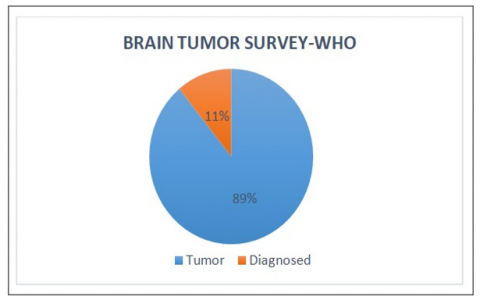
The most current World Health Organization (WHO) research estimates that 700,000 individuals worldwide have a brain tumor, with an additional 86,000 cases being identified from American population statistics sampled during the time period 2015-2019 and analyzed in 2022. Sixty-nine percent of those 700,000 individuals have normal tumors, while 31 percent have dangerous ones as shown in Figure 1 [5]. However, since 2019, 16,830 people have lost their lives to brain tumors, with only 35% making it through the disease. The large number of deaths has made this an increasingly popular subject of study in the realm of medical imaging. However, it's possible that human mortality rates from dangerous tumors could be increased with early discovery.
Figure 1. Brain tumor survey by WHO
The treatment plan will be determined based on the categorization of the tumor, which may range anywhere from 1 to 4. Grade 1 tumors are seen to be harmless, whereas grade 4 tumors are believed to be potentially life-threatening. Medical imaging plays an increasingly significant part, not just in normal practice but also in cutting-edge inquiry, as the quality of treatment that individuals get continues to improve. Because it provides such specific anatomical information about the human brain.
MRI is a useful tool for validating the presence of gliomas. Because of how common they are and how difficult they are to treat, research on brain tumors has emerged as a significant topic of focus in the field of medicine [6]. It is common practice to employ manipulated images of brain tumors in the diagnostic procedure.
These images have had their imaging data altered. There are a variety of diagnostic techniques that may be used, some of which includes MRI, X-ray, ultrasound, Computed Tomography (CT) etc. Despite the fact that these technologies improve the doctors' capacity to forecast how effectively treatment will work and how rapidly a tumor will grow [7], these methods are still unable to reveal all of the fine characteristics and locations of brain tumors. Using MRI to diagnose cancer has been proposed in a variety of different ways.
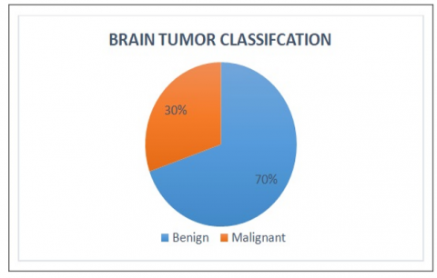
The typical human brain is composed of three different types of matter: cerebral fluid, white matter, and grey matter. White matter makes up the vast bulk of the material that makes up the brain. T1 and T2 are the two most frequent MRI sequences, and each one has its self-unique set of pros and cons in terms of the information and features it may disclose about various kinds of organs. T1-weighted magnetic resonance imaging, which is abbreviated as T1, is linked with contrast enhancement, while T2-weighted magnetic resonance imaging, which is abbreviated as T2, is related with fluid-attenuated inversion recovery [8]. A brain tumor is defined as any tumor that originates in the brain and develops as a result of uncontrolled cell division. One kind of cancer is known as malignant tumors, which are also known as hazardous tumors and the other type of tumor known as innocuous tumors as shown in Figure 2.
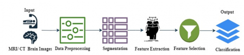
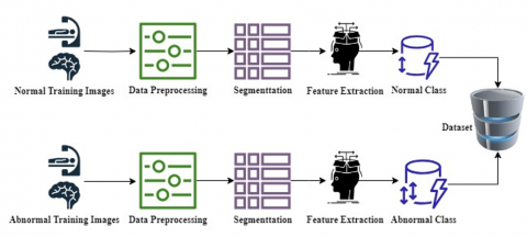
Procedures like CT or MRI scan provides complete structure of tumor if detected because it directs into the intracranial cavity generating lucid brain image of tumor. MRI scan scans by using powerful radio frequencies and magnetic fields to give thorough details of soft tissues. Whereas CT scan searches by transferring x ray beams. In the brain tumor detection preprocessing of MRI or CT brain images, Segmentation, feature extraction, feature selection and classification i.e. post processing of the images are involved as shown in Figure 3, which shows essential phases for any automatic brain tumor detection systems.
Figure 2. Tumor classes
1.1 Data preprocessing phase
It is major aspect of any image based system or application which is essential for following reasons:
1. Data preprocessing phase makes any images ready for the further level of processing like image or data segmentation, image feature extraction etc.
2. Preprocessing removes the labels or marks like name, date or other particulars from image which may impact classification of images.
3. To enhance the quality of images.
4. To eliminate noise of any type from the image.
1.2 Segmentation phase
Various regions and used to find out the interested area or definite area. Particularly it is used for dividing the components from images in order to recognize them as objects.
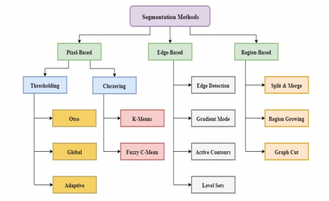
Liu et al. [9] partitioned image into three various groups of segmentations as shown in Figure 4 i.e. edge-based, pixel-based and region-based. Colecchia et al. [10] and Bhanothu et al. [11] also published various medical segmentation approaches of images. Some reviews are targeted segmentation of CT or MRI images [12, 13] which shows through comparisons of different approaches for segmentation of medical images.
The segmentation is the first most important method for classification of the brain tumor. It is found that most of the authors followed thresholding method by which proper segmentation results were not found. If segmentation and feature extraction performed with traditional filters such as Gaussian filter, median filter and even though using OTSU’s thresholding then accurate results were not found. Therefore, in order to increase accuracy, better methods for better segmentation needs to be used. Deep learning techniques can be used for better segmentation and better feature extraction in order to get better results [14].
1.3 Feature extraction and feature selection
Shahajad et al. [15] defined the goal of extracting features as reduction in original data sets depend upon computing of characteristics in particular which classifies as well as identifies various input patterns. Reduction in dimensionality is important factors of extraction of features phase which precisely locates the relevant image components as dense feature vector. This is helpful for the applications having immense image data sets, for which characteristic demonstrations have to minimize to facilitate quick tasks completion like image matching, retrieval of interested objects etc.
The feature extraction block diagram is demonstrated in Figure 5. Hollon et al. [16] demonstrated various well-known methods for feature extraction which are Grey Level Co-occurrence Matrix (GLCM), canny edge detection, local binary pattern (LBP) etc. Large numbers of researches have been carried out to compare the existing feature extraction features [17, 18].
Figure 5. Block diagram of feature extraction
Figure 6. Model of supervised learning algorithm
Feature selection technique is used to minimize unnecessary or redundant image features from the input CT or MRI images to select subgroups of the related matching features for building robust classifiers. Selection phase will most likely to increase construction precision and speed up of the ultimate classifier. As of theoretical perception, it can be stated that ultimate selection of features is used for the supervised methods of learning which requires broad investigation of all the credible subsections of the features. However, for substantial features conducting an entire inspection of all the features creating optimal set of features is unfeasible. Because of this reason supervised learning is designed to analyze suitable approximation of the greatest features set for precise classifier rather determination of optimal set.
1.4 Image classification
In machine learning and classification, researchers extended techniques to bring about a particular objective. Their work signifies development of various learning approaches for enhancing accomplishment of standard based upon use of data models or prior events [19-21]. Approval of training data sets in the supervised learning is depend upon patterns which bring labels to the output.
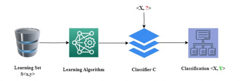
Figure 6 depicts model of supervised learning algorithm. Classification is when output classification values find outs diverse classes to relate the samples [22].
The factors of classifier illustrate training data one after another as classifiers collects from data model. Training data sent to the classification algorithm for building the classifier which has N number of data points which shown as, <xi, yi> N*n=1, with xi €Rd which is input paradigm for dimension d as well as yi€<−1, +1> corresponding label class for two classification tasks.
From the thorough observations or review, this paper is structured in the overall background information regarding approaches to develop an efficient system for brain tumor detection. Also this review discussed and analysed about fundamentals of the MRI. This review is organized as follows, brief introduction to types of tumors and essential phases for any automatic brain tumor detection systems such as image segmentation, feature extraction, classification, selection etc. in section 1. Discovery of literature review, various deep learning techniques with their state-of-the-art evaluations, limitations of conventional and manual approaches, gaps found during thorough examination of existing literature, brief discussion on numerous hybrid deep learning models for feature extraction, selection and classification techniques for helping researchers to build more efficient and more accurate system in section 2. Survey of various open datasets in section 3, overall comparative analysis with hybrid methodologies in analyzing brain tumor detection in section 4 and conclusion and future scope in brain tumor detection is explained in section 5.
In this paper, we conduct a systematic review of the literature on the topic of medical imaging assessment, focusing on studies that employ deep learning models and CNN. Image segmentation is technique of breaking image to discrete parts that cannot be recombined with any other aspects of the picture. Image segmentation is also known as image partitioning. It is the most crucial part of any medical imaging operation, and it may help with the identification, visualization as well as characterization of interested regions.
In spite of the fact that several studies have been conducted on the topic, segmentation continues to be a challenging task for a variety of reasons. Some of these reasons include the variety of information included within the image, the presence of packed objects, occlusion, noise reduction, and non-uniform material texture [23]. There is now a computer-assisted method for interpreting MRI scans and locating anomalies in a manner that is both accurate and expedient. Recent developments in pattern recognition and rapid processing have made it possible to implement this method. In the course of the last few years, the discipline of medical image analysis has seen a shift towards a greater emphasis on picture segmentation as a topic for study.
The computer-aided method of diagnosing brain abnormalities enables quicker illness categorization, which in turn enables treatment to begin at an earlier stage. Because it exposes minute structural changes that are difficult to detect with computed tomography (CT) guided imaging, MRI is now the most frequent non-invasive scanning approach used by radiologists. This is due to the fact that it was developed in the 1970s [24]. On the other hand, determining the sort of tumor a patient has a laborious process that is taken into account during the classification step of AI-powered solutions. As a direct result of this, a gap is a convenient solution for intraoperative brain surgery problems that arise while the procedure is being performed. The solution to this issue was found by doing research on an Intelligent Deep Learning (DL) framework.
This article gives a summary of previously published research on the use of MRI imaging to the classification of brain tumors. We conducted an in-depth analysis of the primary segmentation methods described in every research journals. This survey provides researchers with a comprehensive analysis of problem as well as new insights into the ways in which a variety of image segmentation as well as machine learning (ML) methods utilized to find brain tumor. When compared to systems considered being at the cutting edge of technology as well as those considered to be state-of-the-art, it has been established that deep learning algorithms are more effective at segmenting tumors from brain MRI images [25]. Noise, artifacts, and brightness non-uniformity make an already challenging process in MRI much more difficult. The task involves classifying brain tumor MRIs. The manual segmentation of an MRI picture is a labor-intensive, time-consuming process that is highly reliant on the individual user. In order to achieve accurate and automated brain tumor segmentation, the goal of this study is to conduct the research that has been offered here in order to provide an overview of well-known methods that can be used to overcome the challenges that have been outlined above.
The following are some of the limitations of conventional and manual approaches for diagnosing tumors:
1. The diagnosis is often dependent on the expertise of the physician and calls for intensive patient monitoring.
2. Although it is often accurate as the ailment worsens, it does not seem to be as effective as semi-automatic therapies in the earlier stages of the issue.
3. As a last point, the method is labor-intensive, which encourages the exploration of other approaches to the problem. In certain instances, the boundary of the cancer is not precisely identified, and hence, the tumor is not entirely drained, which results in the tumor regrouping. Radiologists who operate in clinics often choose semi-automatic options over manual treatments due to the former's greater time commitment.
Table 1. Brain tumor detection using deep learning techniques
|
Reference |
Methodology Used |
Evaluation |
|
[15] |
Shallow CNN |
Accuracy97.77% |
|
[20] |
Deep Neural Network (DNN) |
Accuracy 98.87% |
|
[21] |
DNN |
Accuracy 98.00% |
|
[22] |
ResNet50 Network |
Accuracy 92.34% |
|
[23] |
CNN |
Accuracy 99.00 % |
|
[24] |
CNN |
Accuracy 98.96% |
|
[25] |
Principal component analysis (PCA) using template-based K-means. |
Accuracy= 95.00%, Sensitivity= 97.36%, Specificity = 100.00% |
|
[26] |
CNNbased on deep neural networks. |
Specificity = 99.00% Accuracy= 98.80%. |
|
[27] |
Deep wavelet AE |
Accuracy= 99.30% |
|
[28] |
Segmentation using Otsu |
Accuracy= 90.00% |
|
[29] |
CNN that has been pretrained using VGGNET and RESNET |
Specificity= 98.00% Accuracy= 96.80%, Sensitivity= 96.00%, Accuracy =99.00% |
|
[30] |
Supervised SVM and AE |
Accuracy = 99.91% |
|
[31] |
Several ML |
Accuracy= 97.10% |
|
[32] |
SVM |
Accuracy 92.00 % |
|
[33] |
DNN |
71.00 % |
|
[34] |
CNN |
94.60% |
|
[35] |
3D CNN and a U-Net. |
Enhancing tumor: 75.00% Tumor core: 84.60% Whole Tumor: 90.60% |
|
[36] |
DCNN and RCNN classifier |
Accuracy = 97.30% |
|
[37] |
Region Proposal Network (RPN) |
Accuracy = 77.60% |
|
[38] |
CNN, ResNet |
Accuracy = 95.50 |
|
[39] |
DCNN |
Accuracy = 95.00% |
|
[40] |
DCNN |
Accuracy = 70.90% |
|
[41] |
SVM, RCNN, DNN with Support Value |
Accuracy=97.21% Precision =97.90% Recall=97.01% |
|
[42] |
SVM |
Accuracy= 97.10% |
|
[43] |
Region Proposal Network (RPN) |
average precision of 77.60% |
|
[44] |
modified CNN, ResNet, DenseNet |
Accuracy = 95.49%. |
|
[45] |
DCNN |
Accuracy = 86.20% |
Various ML techniques with competitor findings regarding valuations of performance and accuracy were analyzed and discussed, as shown in Table 1. Following are the limitations or gaps found during thorough examination of existing literature, which can be useful for the researchers to study further and overcome these limitations.
This review is carrying out on numerous feature extraction, selection and classification techniques with below hybrid deep learning methods.
2.1 Convolutional Neural Network (CNN)
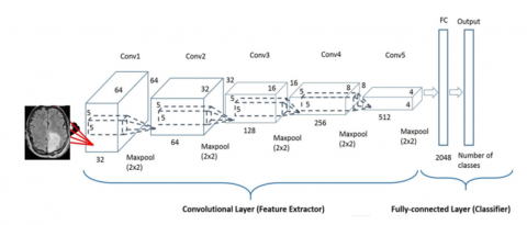
CNN is a neural network which employs a unique layer known as convolution layer. Convolution layer, sub sampling or pooling layer, fully connected layer and nonlinearity layer are some of the layers that make up CNN. Each convolution layer has a filter that is shared by many neurons within that layer. These filters are smaller in size than the picture. The features from the input picture are extracted using the filters. The receptive field is the region of the picture from which the filter extracts the features, and the extracted feature is termed a feature map, implying that the filter will dot product with the preceding layer. Figure 7 shows a logic execution perspective of a suggested CNN for detection of anomaly in images.
The next layer is sub sampling layer also called as pooling layer. In this the feature map gets generated in preceding convolution layer is used by each neuron in the pooling layer. Pooling layer is primarily used for reducing amount of inputs. There are two pooling types i.e. average pooling and maximum pooling. In max pooling, the greatest value from the feature map is discovered and those pixels can be replaced with the single pixel with the highest value. Average pooling finds the average value from the feature map and replaces those pixels with a single pixel with the average value [36, 37]. This can receive the reduced version of the preceding image at the conclusion of this layer. The procedure will be repeated with this reduced picture as an input to the next convolution layer. The number of levels in the network determines how often the operation is repeated. The number of layers is not set and will change depending on the situation. The last layer is completely linked, each neuron in completely connected layer and is linked to each neuron, as illustrated in Figure 7.
The convolutional layer, pooling layer and output layer are three layers which builds basic convolutional neural network. In some of the cases, pooling layer is not required. As shown in Figure 7, standard convolutional neural network design with three convolutional layers is ideally suited for classification. It has an input layer, several hidden layers (convolutional, normalization, and pooling repeats), fully linked and output layer. Layer of output neurons in one layer communicate with neurons in the next layer, allowing for easier scaling of higher-resolution pictures [28]. The pooling or sub-sampling operations may be employed to decrease the input dimensions. The input picture is seen as a collection of tiny sub-regions known as "receptive fields" in a CNN model. On the input layer, a convolutional mathematical process is used to simulate the response to the next layer.
Figure 7. Logical process of CNN for disease detection
2.2 UNET architecture
In order to achieve exact localization in biomedical picture segmentation, U-Net, a deep neural network, uses data augmentation. A breakthrough in deep learning for medical picture segmentation was made by the U-Net study, which was published in 2015. The concept of U-Net is actually not new because it was already covered in the U-Net publication, which has received more than 2000 citations as of the time this thesis was written, serves as the best baseline for the majority of medical picture segmentation tasks [46].
Numerous up sampling layers, a skip connection which focuses feature maps and learnable weight filters are the entire components of the U-Net design. The suggested approach is depending upon the U-Net architecture, which allows for demonstration of good performance with little number of training data sets. The suggested technique expands U-Net architecture targeting to include tumor component detection in MRI images. The suggested network comprises of two strategies, the concatenation and the pyramid pooling approaches. Pyramid pooling enables network to improve level of pixels categorization depend upon context data provided, while the proposed approach of the concatenation, permits a profound network. The fundamental concept behind the architecture is about how network’s traditional vision for ideal local sparse structure can be estimated and enclosed by readily accessible dense components.
2.3 GoogLeNet
GoogLeNet is deep CNN which developed at Google by researchers. In 2014, it was introduced and won ImageNet Large Scale Visual Recognition Challenge in same year with 6.67% top five error rate.
GoogLeNet is very useful for inception module which contains number of parallel convolutional layers along with various filter sizes subsequently by pooling layer. This design benefits network to study features at various resolutions and numerous scales when keeping manageable computational cost [47]. Network furthermore includes auxiliary classifier at middle layers which benefit network to prevent overfitting and to learn more discriminative features.
GoogLeNet develops on ideas of previous CNNs such as LeNet. LeNet was one of successful applications of deep learning. However it is more complex and deeper as compared to LeNet.
2.4 Sparsed Gabor filter (SGF)
Gabor filter can be applied for Multidimensional texture extraction.
Properties of SGF:
i] SGF has a compact Gabor filter bank.
ii] SGF reduces computational complexity of texture feature extraction.
iii] SGF produces low dimensional feature representation with improved sample-to-feature ratio and hence it improves the performance of texture classification
From this extensive literature survey, it is concluded that hybrid deep learning approaches provides more accurate and more efficient results than traditional machine learning algorithms.
3.1 DICOM image sample sets
Digital Imaging and Communications in Medicine (DICOM) datasets are only used for academic research and instruction [48, 49]. Each and every one of these DICOM files has been compressed using the JPEG 2000 transfer syntax. Even though DICOM datasets are internationally accepted for medical imaging as standard format, these have not been usually used for ultimate identification of series. These datasets are greatly flexible, which in turn results in imperative data changeability. DICOM dataset entities have numerous attributes which includes such as patient demographics, pixel data, the image acquisition modality, technical parameters and the clinical site.
3.2 BRATS database
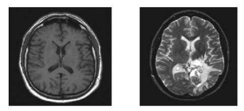
The BraTS dataset was developed as a standard for assessing and designing algorithms for segmentation and diagnosis of brain tumor. The brain tumor MRI scans are part of BraTS dataset collection which was collected from several institutes. Research organizations, hospitals and colleges are few of the institutions which contributed to the BraTs dataset. The patients gave their informed agreement whose MRI scans were used to create the BraTs dataset to have their medical information used for research. The images were taken from the challenge hosted by B. Menze et al. MICCAI 2012 [50]. These photos were used in the study by the aforementioned researchers. Figure 8 shows the MR images of normal and abnormal brain tissues. The competition was a direct outcome of the research conducted by these academics. The image database for the challenge now includes content from scholarly organizations. The MICCAI 2012 Competition on Multimodal Brain Tumor Segmentation is the name of the competition. This held a competition called Multimodal Brain Tumor Segmentation (BRATS) competition in connection with MICCAI 2012 conference. The conferences as well as tournament are being held at the same venue. The goal of this competition is to compare and contrast the various approaches being studied for automated brain tumor segmentation. Thirty distinct patients with a diagnosis of glioma have had their multi contrast MR images used as the training set.
(a) (b)
Figure 8. (a) MRI of normal brain (b) MRI of abnormal brain
There is a wide spectrum of severity among those who have had or been treated for surgical resection. Expert opinions on matters like "active tumor" and "edoema" are also included. All patients have access to magnetic resonance (MR) scans in T1, T2, and FLAIR formats, in addition to post-gallium T1 imaging. Data was extrapolated to a resolution of 1 mm in each direction after the skull was removed using linear co-registration of all volumes with the T1 contrast image. This was done to guarantee the highest level of realism in the final product. This was done so that the final product would be as accurate to life as possible. Not a single effort was made to compile the many happenings into a central hub from which they could be studied and understood more readily.
3.3 Simulated brain database: BrainWeb
As the use of computer-aided data analysis of medical image data becomes more widespread, there is correspondingly an increased need for the validation of the processes.
Unfortunately, evaluating in vivo data does not have a “ground truth” or “gold standard” that can be used. An alternate method of dealing with the verification problem is provided by the simulated brain database (SBD). The SBD contains collection of real MRI image data volumes which is created by MRI simulator. To evaluate performance of different image analysis methods, these kinds of data can be used by neuroimaging community [51].
In this paper, a comparison of the latest machine learning techniques performed where various ML techniques having competitor findings were analyzed and discussed regarding valuations of performance and accuracy as shown in Table 1. The important frames of surveying and analyzing CT and MRI brain images was the classification and segmentation as shown in Table 1. Various available open datasets for either normal or abnormal brain images or their versions are briefly analyzed as shown in Table 2 which is significant for training the model.
Techniques, Evaluations, Highlights described the use of performance parameter of various literatures to give general idea of each study. For brain tumor detection, comparative accuracies of various techniques used by various authors are shown in Figure 9. Also various hybrid methodologies for analyzing brain tumor with their observations are shown in Table 3. The detection of brain tumor is still very much demanding because of variable size, structure and shape of tumor appearance. From extensive research review it’s observed that when various kinds of deep learning approaches such as CNN, UNET, GoogLeNet, Gabor filter etc combined to form hybrid architecture then it gives better performance and accuracy than traditional machine learning approaches to segment, classify and extract the features of brain tumor.
Figure 9. Comparison of evaluation of accuracy
Table 2. Datasets for brain tumor detection and their versions
|
Sr. No. |
Dataset |
Version |
|
1 |
MICCAI BRATS |
2012, 2013, 2014, 2015, 2016, 2017, 2018 |
|
2 |
Harvard |
- |
|
3 |
RIDER |
- |
|
4 |
ISBR |
- |
|
5 |
BrainWeb |
- |
|
6 |
ISLES |
2015, 2016, 2017 |
4.1 The loss function
The loss function of discriminator is the sum of two cross-entropy function H
$\operatorname{Loss}(D)=H\left(\mathrm{real}_{e b}, 1\right)+H\left(e b_1, 0\right)=[-1 \times$ $\log D\left(\right.$ real $\left.\left._{e b}\right)-(1-1) \log \left(1-D\left(e b_{\text {real }}\right)\right)\right]+$$$\begin{gathered}{\left[-0 \times \log D\left(e b_g\right)-(1-0) \log \left(1-D\left(e b_g\right)\right)\right]} =-\log D\left(e b_{\text {real }}\right)-\log \left(1-D\left(e b_g\right)\right.\end{gathered}$$ (1)
where, $e b_g \sim p$data $(e b)$ i.e. $e b_{\text {real }}$ is image region taken from the training dataset and $e b_g$ is image region from testing dataset.
4.2 The time complexity
The sum of total of time that is spent by the proposed system processing each convolution layer,
$O\left(\sum_{l=1}^d n_{l-1} \cdot s_1^2 \cdot n_1 \cdot m_1^2\right)$ (2)
In this specific instance, the index of the convolutional layer has been assigned the value 1 and the variable d has been assigned the role of representing total number of convolution layers present in network. Additionally being known as the various input channels that are used in the lth layer, the total number of filters that are utilized in the lth layer is n. This number is also referred to as the filtering capacity of the layer [52]. Another name for this number is number of filters that is employed in lth layer. S Spatial size of the completed feature map turns out to be m, although the filter seems to have a size of s in this respect. On the other hand, the filter looks to have a size of s. It’s cost time with fully connected layer and pooling layers are between 5% and 10% of the entire amount of time spent calculating. This time cost was not included in the composition that was supplied before. The evaluation of the five approaches, which were evaluated according to the following standards of performance, formed the basis for the comparison that was carried out.
True Positive $(\mathrm{TP})=$ No of resulted images having brain tumo
True Negative $(\mathrm{TN})=$ No of images that haven't tumor
False Positive(FP) $=$No of images that haven't tumor and detected positive
False Negative $(\mathrm{FN})=$No of images have tumor and not detected
Precision $=\mathrm{TP}(\mathrm{TP}+\mathrm{FP})$
Recall $=\mathrm{TP}(\mathrm{TP}+\mathrm{FN})$
Accuracy $=(\mathrm{TP}+\mathrm{TN}) /(\mathrm{TP}+\mathrm{TN}+\mathrm{FP}+\mathrm{FN})$
Table 3. Hybrid methodologies in analyzing brain tumor
|
Methodologies |
References |
Purpose |
Observations |
|
Genetic algorithm (GA), Wavelet transforms (WT) and supervised learning methods. |
[53] |
Brain tissues classification in MRI images |
This methodology is accurate, non-invasive, inexpensive and easy to operate. |
|
Morphological operations, Sobel edge detection and k-mean. |
[54] |
Brain Lesions segmentation in CT scan and MRI images |
With manual delineation achieves high accuracy of 96.8% |
|
Support vector machine (SVM), Fuzzy C Means (FCM) |
[55] |
Brain tumor detection using MRI Images |
In minimum execution time, provides more effective and more accurate results for classification of brain tumor MRI images |
|
SVM, Non sub Sampled Contourlet Transform (NSCT) and k means. |
[56] |
Classification of MRI images having Brain Tumor. |
Achieves higher accuracy for classification. |
|
Artificial Neural Network (ANN), Gabor feature extraction and Fuzzy Clustering. |
[57] |
Classification of Brain tumor using MRI and CT images |
Achieves higher accuracy for classification of 98% and classifiers output helps radiologist to make better decisions. |
|
Kernel Support Vector Machine (KSVM), K-Means, Berkeley Wavelet Transform (BWT), Gray Level Co-occurrence Matrix (GLCM) and Principal Component Analysis (PCA) |
[58] |
Detection of Brain tumor using MRI and CT images |
Achieves higher accuracy, higher performance and proposed technique is used for screening for clinical purpose and for the radiologists. |
In modern societies, one of the main concerns is saving human lives from known diseases such as brain tumors. With the recent advancements in technologies, medical imaging has been stimulated by artificial intelligence techniques such as deep learning. Finding brain tumors from CT or MR images is rapid technological development and it is challenging task in medical research field to find brain tumor accurately and efficiently. Some notable architectures such as UNet and GoogLeNet based models have shown significant potential for enhancing the state-of-the-art with careful pre-processing, advanced training schemes and weight initialization. To improve efficiency, this review proposed several hybrid approaches to identify and locate brain tumor from MRI images. This review discovered comparison for various hybrid technologies and its evaluation of accomplished percentages in specificity, accuracy and sensitivity for identified approaches of brain tumor from MRI and CT images.
The detection of brain tumor is still very much demanding because of variable size, structure and shape of tumor appearance. Even though tumor segmentation techniques have shown higher potential in detecting and analyzing tumors in MRI or CT images, there are still much improvements required to efficiently classify, segment, estimate area and volume of tumor region. Existing research work has challenges and limitations for identifying regions of tumor as well as classification of unhealthy and healthy images. In short, this brain tumor survey covers latest work done so far with their challenges & limitations and all important aspects.
This will be really helpful to develop understanding and to perform new research in little time with correct direction for the researchers. The various deep learning techniques contributed significantly to identify brain tumor than traditional machine learning approaches. These techniques deliver better output results when testing and training are performed on similar acquisition features such as resolution, intensity range etc. Whereas, a little variation in the testing or training MR images directly impacts the robustness of methods. Therefore hybrid deep learning techniques can be used for better feature extraction, segmentation and classification extraction in order to get significant and accurate results in detecting brain tumor.
[1] Fernando, T., Gammulle, H., Denman, S., Sridharan, S., Fookes, C. (2021). Deep learning for medical anomaly detection–a survey. ACM Computing Surveys (CSUR), 54(7): 1-37. https://doi.org/10.1145/3464423
[2] Almadhoun, H.R., Abu-Naser, S.S. (2022). Detection of brain tumor using deep learning. International Journal of Academic Engineering Research (IJAER), 6(3): 29-47.
[3] Islam, M.K., Ali, M.S., Miah, M.S., Rahman, M.M., Alam, M.S., Hossain, M.A. (2021). Brain tumor detection in MR image using superpixels, principal component analysis and template based K-means clustering algorithm. Machine Learning with Applications,5:100044. https://doi.org/10.1016/j.mlwa.2021.100044
[4] Naseer, A., Yasir, T., Azhar, A., Shakeel, T., Zafar, K. (2021). Computer-aided brain tumor diagnosis: Performance evaluation of deep learner CNN using augmented brain MRI. International Journal of Biomedical Imaging, 2021: 5513500. https://doi.org/10.1155/2021/5513500
[5] NBTC. (2019). Quick brain tumor facts. Retrieved from https://braintumor.org/brain-tumor-information/brain-tumor-facts/.
[6] Abd El Kader, I., Xu, G., Shuai, Z., Saminu, S., Javaid, I., Ahmad, I.S., Kamhi, S. (2021). Brain tumor detection and classification on MR images by a deep wavelet auto-encoder model. Diagnostics, 11(9): 1589. https://doi.org/10.3390/diagnostics11091589
[7] Gesperger, J., Lichtenegger, A., Roetzer, T., Salas, M., Eugui, P., Harper, D.J., Merkle, C.W., Augustin, M., Kiesel, B., Mercea, P.A., Widhalm, G., Baumann, B., Woehrer, A. (2020). Improved diagnostic imaging of brain tumors by multimodal microscopy and deep learning. Cancers, 12(7): 1806. https://doi.org/10.3390/cancers12071806
[8] Truong, A.H., Sharmanska, V., Limbӓck-Stanic, C., Grech-Sollars, M. (2020). Optimization of deep learning methods for visualization of tumor heterogeneity and brain tumor grading through digital pathology. Neuro-Oncology Advances, 2(1): vdaa110. https://doi.org/10.1093/noajnl/vdaa110
[9] Liu, D., Zhang, D., Song, Y., Zhang, F., O’Donnell, L.J., Cai, W. (2018). 3D large kernel anisotropic network for brain tumor segmentation. In: Cheng, L., Leung, A., Ozawa, S. (eds) Neural Information Processing. ICONIP 2018. Lecture Notes in Computer Science(), vol 11307. Springer, Cham. https://doi.org/10.1007/978-3-030-04239-4_40
[10] Colecchia, F., Ruffle, J.K., Pombo, G.C., Gray, R., Hyare, H., Nachev, P. (2020). Knowledge-driven deep neural network models for brain tumour segmentation. Journal of Physics: Conference Series, 1662(1): 012010. https://doi.org/10.1088/1742-6596/1662/1/012010
[11] Bhanothu, Y., Kamalakannan, A., Rajamanickam, G. (2020). Detection and classification of brain tumor in MRI images using deep convolutional network. In 2020 6th international conference on advanced computing and communication systems (ICACCS), Coimbatore, India, pp. 248-252. https://doi.org/10.1109/ICACCS48705.2020.9074375
[12] Sheeba, L., Mitra, A., Chaudhuri, S., Sarkar, S.D. (2021). Detection of exact location of brain tumor from MRI data using big data analytics. Systematic Rev. Pharmacy, 12(5): 378-381.
[13] Amin, J., Sharif, M., Haldorai, A., Yasmin, M., Nayak, R.S. (2022). Brain tumor detection and classification using machine learning: A comprehensive survey. Complex & Intelligent Systems, 8(4): 3161-3183. https://doi.org/10.1007/s40747-021-00563-y
[14] Díaz-Pernas, F.J., Martínez-Zarzuela, M., Antón-Rodríguez, M., González-Ortega, D. (2021). A deep learning approach for brain tumor classification and segmentation using a multiscale convolutional neural network. Healthcare, 9(2): 153. https://doi.org/10.3390/healthcare9020153
[15] Shahajad, M., Gambhir, D., Gandhi, R. (2021). Features extraction for classification of brain tumor MRI images using support vector machine. In 2021 11th International Conference on Cloud Computing, Data Science & Engineering (Confluence), Noida, India, pp. 767-772. https://doi.org/10.1109/Confluence51648.2021.9377111
[16] Hollon, T.C., Pandian, B., Adapa, A.R., et al. (2020). Near real-time intraoperative brain tumor diagnosis using stimulated Raman histology and deep neural networks. Nature Medicine, 26(1): 52-58. https://doi.org/10.1038/s41591-019-0715-9
[17] Ali, M., Gilani, S.O., Waris, A., Zafar, K., Jamil, M. (2020). Brain tumour image segmentation using deep networks. IEEE Access, 8: 153589-153598. https://doi.org/10.1109/ACCESS.2020.3018160
[18] Nadeem, M.W., Ghamdi, M.A.A., Hussain, M., Khan, M.A., Khan, K.M., Almotiri, S.H., Butt, S.A. (2020). Brain tumor analysis empowered with deep learning: A review, taxonomy, and future challenges. Brain Sciences, 10(2): 118. https://doi.org/10.3390/brainsci10020118
[19] Huang, Z., Du, X., Chen, L., Li, Y., Liu, M., Chou, Y., Jin, L. (2020). Convolutional neural network based on complex networks for brain tumor image classification with a modified activation function. IEEE Access, 8: 89281-89290. https://doi.org/10.1109/ACCESS.2020.2993618
[20] Banerjee, S., Mitra, S., Masulli, F., Rovetta, S. (2020). Deep radiomics for brain tumor detection and classification from multi-sequence MRI. SN Computer Science, 1(4): 209. https://doi.org/10.1007/s42979-020-00214-y
[21] Gajja, M. (2020). Brain tumor detection using mask R-CNN. Journal of Advanced Research in Dynamical and Control Systems, 12(SP8): 101-108. https://doi.org/10.5373/jardcs/v12sp8/20202506
[22] Benjamin, B. (2020). Improved detection of fmri activation in brain tumor patients through MP-PCA denoising. https://doi.org/10.26226/morressier.5e8335ba7cb08a046ef7c728
[23] Dutta, S., Bandyopadhyay, S. (2020). Cross-Validated AdaBoost Classifier Used for Brain Tumor Detection. https://doi.org/10.20944/preprints202006.0351.v1
[24] Akil, M., Saouli, R., Kachouri, R. (2020). Fully automatic brain tumor segmentation with deep learning-based selective attention using overlapping patches and multi-class weighted cross-entropy. Medical Image Analysis, 63: 101692. https://doi.org/10.1016/j.media.2020.101692
[25] Sun, L., Zhang, S., Chen, H., Luo, L. (2019). Brain tumor segmentation and survival prediction using multimodal MRI scans with deep learning. Frontiers in Neuroscience, 13: 810. https://doi.org/10.3389/fnins.2019.00810
[26] Lundervold, A.S., Lundervold, A. (2019). An overview of deep learning in medical imaging focusing on MRI. Zeitschrift für Medizinische Physik, 29(2): 102-127. https://doi.org/10.1016/j.zemedi.2018.11.002
[27] Fabelo, H., Halicek, M., Ortega, S., Shahedi, M., Szolna, A., Piñeiro, J.F., Sosa, C., O’Shanahan, A.J., Bisshopp, S., Espino, C., Márquez, M., Hernández, M., Carrera, D., Morera, J., Callico, G.M., Sarmiento, R., Fei, B. (2019). Deep learning-based framework for in vivo identification of glioblastoma tumor using hyperspectral images of human brain. Sensors, 19(4): 920. https://doi.org/10.3390/s19040920
[28] Kumar, S., Negi, A., Singh, J.N. (2019). Semantic segmentation using deep learning for brain tumor MRI via fully convolution neural networks. In: Satapathy, S., Joshi, A. (eds) Information and Communication Technology for Intelligent Systems . Smart Innovation, Systems and Technologies, vol 106. Springer, Singapore. https://doi.org/10.1007/978-981-13-1742-2_2
[29] Deepak, S., Ameer, P.M. (2019). Brain tumor classification using deep CNN features via transfer learning. Computers in Biology and Medicine, 111: 103345. https://doi.org/10.1016/j.compbiomed.2019.103345
[30] Nandeesh, M.D., Meenakshi, M. (2019). Tumor detection using enhanced FCM for multimodal brain images. 2019 2nd International Conference on Intelligent Computing, Instrumentation and Control Technologies (ICICICT). https://doi.org/10.1109/icicict46008.2019.8993400
[31] Siar, M., Teshnehlab, M. (2019). Brain tumor detection using deep neural network and machine learning algorithm. In 2019 9th International Conference on Computer and Knowledge Engineering (ICCKE), Mashhad, Iran, pp. 363-368. https://doi.org/10.1109/ICCKE48569.2019.8964846
[32] Bakas, S., Reyes, M., Jakab, A., et al. (2018). Identifying the best machine learning algorithms for brain tumor segmentation, progression assessment, and overall survival prediction in the BRATS challenge. arXiv preprint arXiv:1811.02629. https://doi.org/10.48550/arXiv.1811.02629
[33] Jiang, Y., Hou, J., Xiao, X., Deng, H. (2018). A brain tumor segmentation new method based on statistical thresholding and multiscale CNN. In: Huang, DS., Gromiha, M., Han, K., Hussain, A. (eds) Intelligent Computing Methodologies. ICIC 2018. Lecture Notes in Computer Science(), vol 10956. Springer, Cham. https://doi.org/10.1007/978-3-319-95957-3_26
[34] Abiwinanda, N., Hanif, M., Hesaputra, S.T., Handayani, A., Mengko, T.R. (2019). Brain tumor classification using convolutional neural network. In: Lhotska, L., Sukupova, L., Lacković, I., Ibbott, G.S. (eds) World Congress on Medical Physics and Biomedical Engineering 2018. IFMBE Proceedings, vol 68/1. Springer, Singapore. https://doi.org/10.1007/978-981-10-9035-6_33
[35] Mohsen, H., El-Dahshan, E.S.A., El-Horbaty, E.S.M., Salem, A.B.M. (2018). Classification using deep learning neural networks for brain tumors. Future Computing and Informatics Journal, 3(1): 68-71. https://doi.org/10.1016/j.fcij.2017.12.001
[36] Ismael, M.R. (2018). Hybrid model-statistical features and deep neural network for brain tumor classification in MRI images. Dissertations. 3291. Western Michigan University: Kalamazoo, MI, USA.
[37] Ladefoged, C.N., Marner, L., Hindsholm, A., Law, I., Højgaard, L., Andersen, F.L. (2019). Deep learning based attenuation correction of PET/MRI in pediatric brain tumor patients: evaluation in a clinical setting. Frontiers in Neuroscience, 12: 1005. https://doi.org/10.3389/fnins.2018.01005
[38] Suter, Y., Jungo, A., Rebsamen, M., Knecht, U., Herrmann, E., Wiest, R., Reyes, M. (2019). Deep learning versus classical regression for brain tumor patient survival prediction. In: Crimi, A., Bakas, S., Kuijf, H., Keyvan, F., Reyes, M., van Walsum, T. (eds) Brainlesion: Glioma, Multiple Sclerosis, Stroke and Traumatic Brain Injuries. BrainLes 2018. Lecture Notes in Computer Science(), vol 11384. Springer, Cham. https://doi.org/10.1007/978-3-030-11726-9_38
[39] Virupakshappa, Amarapur, B. (2020). Computer-aided diagnosis applied to MRI images of brain tumor using cognition based modified level set and optimized ANN classifier. Multimedia Tools and Applications, 79(5-6): 3571-3599. https://doi.org/10.1007/s11042-018-6176-1
[40] Benson, E., Pound, M.P., French, A.P., Jackson, A.S., Pridmore, T.P. (2019). Deep hourglass for brain tumor segmentation. In: Crimi, A., Bakas, S., Kuijf, H., Keyvan, F., Reyes, M., van Walsum, T. (eds) Brainlesion: Glioma, Multiple Sclerosis, Stroke and Traumatic Brain Injuries. BrainLes 2018. Lecture Notes in Computer Science(), vol 11384. Springer, Cham. https://doi.org/10.1007/978-3-030-11726-9_37
[41] Afshar, P., Mohammadi, A., Plataniotis, K.N. (2018). Brain tumor type classification via capsule networks. In 2018 25th IEEE International Conference on Image Processing (ICIP), Athens, Greece, pp. 3129-3133. https://doi.org/10.1109/ICIP.2018.8451379
[42] Narayana, T.L., Reddy, T.S. (2018). An efficient optimization technique to detect brain tumor from MRI images. In 2018 International Conference on Smart Systems and Inventive Technology (ICSSIT), Tirunelveli, India, pp. 168-171. https://doi.org/10.1109/ICSSIT.2018.8748288
[43] Polly, F.P., Shil, S.K., Hossain, M.A., Ayman, A., Jang, Y.M. (2018). Detection and classification of HGG and LGG brain tumor using machine learning. In 2018 International Conference on Information Networking (ICOIN), Chiang Mai, Thailand, pp. 813-817. https://doi.org/10.1109/ICOIN.2018.8343231
[44] Jagan, A. (2018). A new approach for segmentation and detection of brain tumor in 3D brain mr imaging. 2018 Second International Conference on Electronics, Communication and Aerospace Technology (ICECA). https://doi.org/10.1109/iceca.2018.8474874
[45] Dubey, Y. K., Mushrif, M. M., Pisar, K. (2018). Brain tumor type detection using texture features in Mr Images. 2018 IEEE Region 10 Humanitarian Technology Conference (R10-HTC). https://doi.org/10.1109/r10-htc.2018.8629800
[46] Hasan, A.M., Jalab, H.A., Meziane, F., Kahtan, H., Al-Ahmad, A.S. (2019). Combining deep and handcrafted image features for MRI brain scan classification. IEEE Access, 7: 79959-79967. https://doi.org/10.1109/ACCESS.2019.2922691
[47] Biswas, B., Soroardi, H.S., Islam, M.J. (2018). Brain tumor detection with tumor region analysis using adaptive thresholding and morphological operation. 2018 4th International Conference on Electrical Engineering and Information & Communication Technology (iCEEiCT). Dhaka, Bangladesh, https://doi.org/10.1109/CEEICT.2018.8628107
[48] http://www.osirix-viewer.com/resources/dicom-image-library/.
[49] https://www.kaggle.com/datasets/navoneel/brain-mri-images-for-brain-tumor-detection.
[50] http://www2.imm.dtu.dk/projects/BRATS2012/data.html.
[51] https://brainweb.bic.mni.mcgill.ca/.
[52] Zaw, H.T., Maneerat, N., Win, K.Y. (2019). Brain tumor detection based on Naïve Bayes classification. In 2019 5th International Conference on engineering, applied sciences and technology (ICEAST), Luang Prabang, Laos, pp. 1-4. https://doi.org/10.1109/ICEAST.2019.8802562
[53] Sert, E., Özyurt, F., Doğantekin, A. (2019). A new approach for brain tumor diagnosis system: Single image super resolution based maximum fuzzy entropy segmentation and convolutional neural network. Medical Hypotheses, 133: 109413. https://doi.org/10.1016/j.mehy.2019.109413
[54] Kalaiselvi, T., Kumarashankar, P., Sriramakrishnan, P., Karthigaiselvi, S. (2019). Brain tumor detection from multimodal MRI brain images using pseudo coloring processes. Procedia Computer Science, 165: 173-181. https://doi.org/10.1016/j.procs.2020.01.094
[55] Deepa, A.R., Sam Emmanuel, W.R. (2019). An efficient detection of brain tumor using fused feature adaptive firefly backpropagation neural network. Multimedia Tools and Applications, 78: 11799-11814. https://doi.org/10.1007/s11042-018-6731-9
[56] Natarajan, A., Kumarasamy, S. (2019). Efficient segmentation of brain tumor using FL-SNM with a metaheuristic approach to optimization. Journal of Medical Systems, 43(2): 25. https://doi.org/10.1007/s10916-018-1135-y
[57] Ari, A., Hanbay, D. (2018). Deep learning based brain tumor classification and detection system. Turkish Journal of Electrical Engineering and Computer Sciences, 26(5): 2275-2286. https://doi.org/10.3906/elk-1801-8
[58] Zhou, C., Chen, S., Ding, C., Tao, D. (2019). Learning contextual and attentive information for brain tumor segmentation. In: Crimi, A., Bakas, S., Kuijf, H., Keyvan, F., Reyes, M., van Walsum, T. (eds) Brainlesion: Glioma, Multiple Sclerosis, Stroke and Traumatic Brain Injuries. BrainLes 2018. Lecture Notes in Computer Science(), vol 11384. Springer, Cham. https://doi.org/10.1007/978-3-030-11726-9_44