E. Arulkumar | S. Thanikaikarasan* | Tansir Ahamad | Saad M Alshehri
© 2023 IIETA. This article is published by IIETA and is licensed under the CC BY 4.0 license (http://creativecommons.org/licenses/by/4.0/).
OPEN ACCESS
In this study, cupric oxide nanoparticles was synthesized by chemical precipitation method. Relevant unique structural, surface morphology with chemical composition and optical properties are investigated. The improvement in their crystallinity and purification can be further attained by post calcinations process. The most two prominent intense peaks (-111) and (111), were found, indicated a crystalline form of CuO. The SEM images showed that the distribution of crystalline particles which are agglomeration with cluster shape. The EDX analysis of chemical composition percentage found to be the copper is completely oxidized with containing of dual element (Cu and O).
copper oxide, nanoparticle, wet chemical precipitation, X-ray diffraction
The major development of nanoscale to microscale of transition metal oxide nanoparticles have showing their excessive field of catalysis, sensing, supercapacitors due to their combination formation of nano to bulk creation. The growth and uses of metal oxide nanoparticles in consumer products and the release of these metal oxide nanoparticle into the environment is inevitable [1]. Therefore, there is a high demand for sensitive and rapid systematic methods for the detection of metal oxide nanoparticles. Due to the electroactivity of most of the metal oxide nanoparticles, electrochemistry represents a facile and suitable method that could be used for this purpose [2]. The electrochemistry of metal oxide nanoparticles for direct determination of particle in solution with in the electrode surface and interface as well as detecting and measuring redox potential value of the nanoparticles [3]. There is a nanoparticle based electrochemical capacitors are improved the higher capacitive value which has leads to improve the properties of longer life cyclic, very much power density storage, charge-discharge properties compared normal capacitors and batteries. Although the supercapacitors make ultracapacitor and it involves for energy storage device due to its huge range of application in various fields, such as electric vehicles, electric utilities, load cranes, factory power backup and forklifts [4,5]. Therefore, the growth of energy storage equipment is the significant to the effective use of energy while improving its structure and function. The lithium-ion batteries and zinc-ion batteries are a current market place for energy storage because of their increasing lightness, portability, and columbic efficiency of cyclic life[6.7]. The aqueous metal ion batteries are still in initial stage and mainly focused on the development of cathode materials. The Al, Zn, Cu, Ni and Co metals are deliveries a high volumetric energy density and has outstanding theoretical capacitance of Ni 3750 (F/g) [8] and Co have 3560 (F/g) [9]. The copper electrode layers used to be a highcapacity for Li-ion batteries due to the lower surface potential barrier for diffusion of electron-hole [10] as well as the copper oxide enhances the viscosity and thermal conductivity of nanofluids. The challenge with copper oxide is that it is a more desirable material to synthesis, has controllable size and shape, and is effective and affordable.
The CuO nanomaterials was synthesized through chemical co-precipitation method [11,12]. The 50 ml of deionized water is used to dissolve 0.02 M cupric acetate monohydrate Cu(CH3.COO)2.H2O) and the solution was continuously heated upto 80℃ and stirred at the temperature for 30 mins until its pH adjusted upto 8 ± 0.2. After cooling down the solution aged for 24 hours to settled precipitation of CuO. The precipitate was filtered and the obtained yield was washed with anhydrous ethanol and deionized water more than three times to remove the excess of impurities. Afterward the obtained powder was dry air at 24 hours and annealed at 500℃ at furnace for 4 hours. The formation Copper Oxide is takes place according to the following Eq's (1) and (2):
$\mathrm{Cu}(\mathrm{OAC})_2+\mathrm{NH}_4 \mathrm{OH} \rightarrow \mathrm{Cu}(\mathrm{OH})_2+2 \mathrm{NH}^{+}+\mathrm{H}_2 \mathrm{O}$ (1)
$\mathrm{Cu}(\mathrm{OH})_2$ rapdily converted to $\mathrm{CuO}$ on heating:
$\mathrm{Cu}(\mathrm{OH})_2 \rightarrow \mathrm{CuO}+\mathrm{H}_2 \mathrm{O}$ (2)
X-ray diffraction pattern was carried out to analyze the phase and crystallite natue of the Copper Oxide nanoparticle. The surface morphology along with composition n was attained using scanning electron microscopy. Ultraviolet Visible NIR spectroscopic measurements was carried out to estimate the percentage of absorbance and transmittance for the prepared samples.
3.1 Structural properties
X-ray diffraction pattern was carried out to find out the structural features with crystalline nature of syntheszed $\mathrm{CuO}$ nanoparticles. There is no peak response to elemental $\mathrm{Cu}$ and $\mathrm{O}$ present in the sample. The peaks for the diffraction angles that correspond to the Miller indices of the monoclinic (110), $(-111),(111),(-202),(020),(202),(-113),(-311),(220)$, (311) and (-222) planes,respectively [11-13]. The two most intense peaks corresponds to (-111) and (111), indicated a crystalline form of $\mathrm{CuO}$ with $2 \theta$ values of 35.53 and 38.75 . As a result of this method, the calculated lattice parameters for the monoclinic $\mathrm{CuO}$ space group C2/c are (a=5.5005Å and b=c=4.9656Å). The peaks of FWHM indicating that the $\mathrm{CuO}$ is a very small crystal and the average particle size of prepared $\mathrm{CuO}$ nanoparticles is calculated with the help of Debye-Scherrer formula.
$D=\frac{0.9 \lambda}{\beta \cos \theta}$ (3)
where $\lambda$ is the wavelength of X-rays used (CuK $\alpha$=1.5406Å), $\beta$ is the full width half maximum value in terms of radians, $\theta$ refers to diffraction angle, the calculated particle size was $22.65 \mathrm{~nm}$.
The dislocation density is calculated as following equation:
$\delta=\frac{1}{D^2}$ (4)
Figure 1. X ray diffraction pattern of CuO nano particles synthesized by Co-preciptitation method
The particle size (D), dislocation density (δ) and strain (ε) of the nanoparticles is evident that the micro strain values decrease with increase in particle size. The calculated dislocation density indicates intermediate crystallization of the CuO nanoparticle. The definition of strain is the restoring force that can be applied to a material surface to prevent growth of crystallites along the surface. The following equation is used to determine the amount of strain in the produced CuO.
$\varepsilon=\frac{\beta \cos \theta}{4}$ (5)
The calculated value of crystallite size, strain and dislocation density was found to be $22.65 \mathrm{~nm}, 0.1084 \times 10^{-3}$ line ${ }^{-2}$ metre $^{-4}, 2.5 \times 10^{14} \mathrm{~cm}^{-2}$
3.2 Morphology and composition
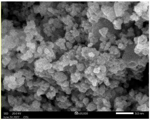
The surface morphological view with composition of synthesized CuO nanoparticles was analyzed using Energy dispersive X-ray analysis attached with scanning electron microscopy. The clustering of small grains by coprecipitations approach is shown in Figure 2. Similar beaviour of surface morphology CuO nanoparticles nanoparticles synthesized using chemical co-precipitation reported by Rajeshwari Sivaraj et al [14].The sizes of the grains is found to be in the range 500 nm. EDX analysis attached with scanning electron microscopy used to find out the elements present in the sample. The atomic percentage in the EDX results demonstrated that the sample's phase belongs to CuO, and the results are consistent with X-ray diffraction analysis with phase corresponds to CuO.
Figure 2. Scanning electron microscopic image of CuO nanoparticles sythesized by Co-precipitation method
It confirms that the produced CuO nanoparticles contain all of their constituent elements, including copper (Cu) and oxygen (O) which is given in Table 1. The presence atomic percentage in the synthesized sample confirmed the toichiometry.The strong peaks of the EDX data showed that the produced CuO nanoparticles had an excellent crystalline phase and that the EDX peak positions were consistent with CuO. The observation CuO diffraction with strong intensity and narrow width in the EDX spectrum, show that the resultant products have crystalline in nature. As a result, we can draw the conclusion that co-precipitation significantly influenced particle size. These results are very consistent with previous research.
Figure 3. Typical EDX spectra of Copper Oxide nanoparticle synthesized by Co-Precipitation method
Table 1 Atomic percentage of CuO nanoparticle prepared by Co-
|
Element |
line |
Mass % |
Atom % |
|
O |
K |
18.84 ± 0.16 |
47.97 ± 0.42 |
|
Cu |
K |
81.16 ± 0.61 |
52.03 ± 0.39 |
|
|
|
100.00 |
100.00 |
|
Spc_004 Fitting ratio 0.2219 |
|||
3.3 Optical properties
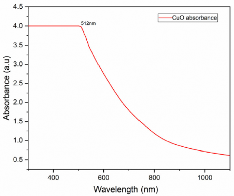
The UV-Visible absorption spectra of CuO nanoparticles shows maximum absorption peak in UV region at 549 nm is shown if Figure 4.
Figure 4. Optical absorbance spectra of CuO nanoparticles sythesized by Co-Precipitation method
The Tauc plot helps us to calculate the band gap energy in a crystalline semiconductor. The band gap of the CuO nanoparticles was obtained by plotting absorptivity (αhν)2 as a function of energy (hν). Extrapolating the linear portion of the curve to absorption axis gives the band gap energies of CuO nanoparticles which were 2.1 eV. This band gap energy value for CuO nanoparticles is slightly higher than theoretical bandgap value 1.8 eV. Generally, the band gap increases with decreasing particle size due to quantum confinement [15]. But the band gap does not follow the same trend always, and it can be affected by other parameters, also like defects present in the oxides. In the present study, defects prompted effects are dominating over particle size effect. As a result, a slightly higher band gap is obtained for as-synthesized CuO nanoparticles [16]. For direct transition, their optical absorption near the band edge follows the equation.
$(\alpha h v)=\mathrm{A}\left(h v-E_g\right)^{1 / 2}$ (5)
where α, h, v, Eg, and A are the absorption coefficient, Plank constant, light frequency, band gap energy, and constant a respectively. The band gap energy was obtained by extrapolating the linear portion of the (α h v)n curve versus hν to zero. Figures 5 illustrate the UV–Visible diffuse spectra as well as the Tauc plot of the CuO where the bandgap is found to be 1.4 eV [17].
Figure 5. Plot of (hν) versus (αhν)2 for CuO nanoparticles synthesized by Co precipitation method
Synthesis and characterization of nano-sized copper oxide powders via co precipitation method using Cupric acetate precursors was reported. X-ray diffraction with Prominent peaks around $2 \theta$ values 35.53 and 38.75 which are combinations of (-111) and (111) respectively, have been identified as the characteristics of monoclinic CuO. Furthermore, the crystallite size calculated from XRD data through Debye-Scherrer method produced average size of 22.65 nm for the nanoparticles. The distribution of CuO crystalline particles was visible in SEM pictures, which were agglomerations of cluster. Optical properties showed an absorption band at 549 nm, which is the characteristic band for Cu completely oxidized and the energy gap value around 2.1 eV with slightly higher than theoretical band gap valuedue to the formation of crystal defect.
The authors extend their sincere appreciation to the Researchers Supporting Project Number (RSP2023R6), King Saud University, Riyadh, Saudi Arabia for funding this research.
[1] Mahendra Singh Yadav, A.K. Sinha, M.N. Singh, Ashish Kumar (2021). Electrochemical study of copper oxide and activated charcoal based nanocomposite electrode for supercapacitor. Materials Today: Proceedings. 46, 5722-5729. (https://doi.org/10.1016/j.matpr.2021.02.134)
[2] Wei Zhe Teo, Adriano Ambrosi, Martin Pumera (2013). Direct electrochemistry of copper oxide nanoparticles in alkaline media. Electrochem commun. 28, 51–53. (https://doi.org/10.1016/j.elecom.2012.12.006)
[3] Jinlei Meng, Zhanhong Yang, Linlin Chen, Haigang Qin, Fan Cui, Yinan Jiang, Xiao Zeng. (2020). Energy storage performance of CuO as a cathode material for aqueous zinc ion battery. Mater. Today Energy. 15, 100370. (DOI: 10.1016/j.mtener.2019.100370Zhu H, Yang Q, Liu D, Du Y, Yan S, Gu M, Zou Z. (2021), Direct Electrochemical Protonation of Metal Oxide Particles. J Am Chem Soc. 143 (24) 9236-9243. (doi: 10.1021/jacs.1c04631.)
[4] Guofa Cai, Xu Wang, Mengqi Cui, Peter Darmawan, Jiangxin Wang, Alice Lee-Sie Eh, Pooi See Lee (2015). Electrochromo-supercapacitor based on direct growth of NiO nanoparticles, Nano Energy, 12, 258–267. (https://doi.org/10.1016/j.nanoen.2014.12.031)
[5] Achintya Dutta, Ramakrishna Nayak, M. Selvakumar, Dheeraj Devadiga, P. Selvaraj, S. Senthil Kumar. (2021). Graphite/copper nanoparticle-based high-performance micro supercapacitor with porous wet paper-based PVAPVP blend polymer electrolyte, Mater. Lett., 295, 129849. (https://doi.org/10.1016/j.matlet.2021.129849)
[6] Da Deng. (2015). Li-ion batteries: basics, progress, and challenges, Energy Sci. Eng., (https://doi.org/10.1002/ese3.95)
[7] Xu, W., Wang, Y. (2019). Recent Progress on Zinc-Ion Rechargeable Batteries. Nano-Micro Lett., 11, 90. (https://doi.org/10.1007/s40820-019-0322-9)
[8] Sung-Wook Kim, Ik-Hee Kim, Sun-I Kim, and Ji-Hyun Jang. (2017). Nickel hydroxide supercapacitor with a theoretical capacitance and highrate capability based on hollow dendritic 3D-nickel current collectors, Chem. Asian. J., 12, 1291-1296. (https://doi.org/10.1002/asia.201700454)
[9] Srikant Sahoo, Ashis Kumar Satpati. (2017). Electrochemical capacitance properties of cobalt oxide entangled over MWCNT and cobalt oxide AC composites, J. Electroanal. Chem., 801, 416–424. (https://doi.org/10.1016/j.jelechem.2017.08.022)
[10] Langner, T., Sieber, T. & Acker, J. (2021). Studies on the deposition of copper in lithium-ion batteries during the deep discharge process. Sci. Rep. 11, 6316. (https://doi.org/10.1038/s41598-021-85575-x)
[11] K. Jhansi, S. Chandralingam, N. Manohar Reddy, Padma Suvarna, Ch. Ashok, K. Venkateswara Rao. (2017). CuO nanoparticles Synthesis and Characterization for Humidity Sensor-Application, J. mater. sci. nanotechnol., 04, 27. (https://doi.org/10.15436/2377-1372.16.020)
[12] Phiwdang, Sineenart Suphankij, Wanichaya Mekprasart and Wisanu Pecharapa. (2013). Synthesis of CuO Nanoparticles by Precipitation Method Using Different Precursors, Energy Procedia, 34, 740 – 745. (https://doi.org/10.1016/j.egypro.2013.06.808)
[13] Hafsa Siddiqui, Mohammad Ramzan Parra, M. S. Qureshi, M. M. Malik, and Fozia Z. Haque. (2018). Studies of structural, optical, and electrical properties associated with defects in sodium-doped copper oxide (CuO/Na) nanostructures, J mater Sci, 53, 8826-8843. (https://doi.org/10.1007/s10853-018-2179-6)
[14] Rajeshwari Sivaraj, Pattanathu K.S.M. Rahman, P. Rajiv, Hasna Abdul Salam, R. Venckatesh. (2014). Biogenic copper oxide nanoparticles synthesis using Tabernaemontana divaricate leaf extract and its antibacterial activity against urinary tract pathogen, Spectrochimica Acta Part A: Molecular and Biomolecular Spectroscopy, 133, 178-181. (https://doi.org/10.1016/j.saa.2014.05.048)
[15] Elizabeth C. Pastrana, Steveen J. Loarte, Carlos D. Gonzales-Lorenzo, Roxana Y.P. Alta, Hugo A. Alarcon. (2021). Fabrication and characterization of copper (II) oxide/iron (III) oxide thin film heterostructures for trace arsenic (III) removal in water, Thin Solid Films, 717, 138440 (https://doi.org/10.1016/j.tsf.2020.138440)
[16] PriyambadaSahoo, M.J.Sneha, B.P.Mandal, AmbeshDixit. (2022). Strain induced bandgap engineering in multiferroic CuO nanoparticles: Competing micro-strain and geometrical size in nanometer scales, Mater. Lett., 324, 132747. (https://doi.org/10.1016/j.matlet.2022.132747)
[17] Carmen Gherasim, Petronela Pascairu, Mihai Asandulesa, Marius Dobromir, Florica Doroftei, Nicusor Fifere, Andrei Dascalu, Anton Airinei, (2022). Copper Oxide Nanostructures: Preparation, Structural, Dielectric and Catalytic Properties, Ceramics Internationals, 48, 25556-25568, (https://doi.org/10.1016/j.ceramint.2022.05.235)
[18] Singh, P.K., Kumar, P., Hussain. (2016). Synthesis and characterization of CuO nanoparticles using strong base electrolyte through electrochemical discharge process. Bull Mater Sci, 39, 469–478. (https://doi.org/10.1007/s12034-016-1159-1)