Ahmed Ehsan Jassem*![]() | Mohamed Hamzah Al-Maamori | Ahmed Fadhil Hamzah
| Mohamed Hamzah Al-Maamori | Ahmed Fadhil Hamzah![]()
© 2024 The authors. This article is published by IIETA and is licensed under the CC BY 4.0 license (http://creativecommons.org/licenses/by/4.0/).
OPEN ACCESS
Nano lead was utilized to manufacture shielding aprons, serving as a practical substitute for the commonly employed sheet lead aprons in providing protection against medical radiation. In addition, metallic nano lead and chlorophyll were evaluated as alternative materials for these aprons, The metallic lead nanoparticles were dispersed using ultrasound. An investigation was carried out to assess the efficacy of four different anti-radiation shielding sheets made of metallic nano lead and chlorophyll at different concentrations (10, 20, and 30 wt% and 35 wt%, respectively) using bio silicon as the foundation material The selection of concentrations was predicated upon prior scholarly investigations. The radiation dosages emitted by these sheets were compared to those emitted by a lead standard. An experiment was carried out to determine the tensile strength of radiation shielding sheets throughout the production process, specifically focusing on the appropriate mixing procedure. The assessment of transmission dosage was performed utilising the lead equivalent test technique (IEC 61331-3:2014) on X-ray protective equipment, in compliance with the European Industrial Standard. The experiment entailed quantifying the levels of radiation transmission using a lead standard of varied thicknesses (0.05, 0.1, 0.15, 0.2, 0.25, 0.3, and 0.35 mm) and five distinct types of radiation shielding sheets. The data were recorded at tube voltages of 30, 60, 90, 120, 150, and 180 kilovolts peak (kVp). According to the results, using a composite made up of 20 wt% metallic nano lead and 35 wt% chlorophyll effectively assessed a dose equivalent to 0.3 mm of lead. Compared to conventional lead plates, the suggested shield exhibits a transmission dose of 0.115 mR. Conversely, traditional shield plates with a thickness of 0.3 mm demonstrate a transmission dose of 0.119 mR. Therefore, it has the capability to be used instead of lead sheets, making it a viable sheet for medical radiation shielding and demonstrating favorable economic feasibility.
shield anti X-ray, medical radiation, chlorophyll, nano lead, radiation shielding materials, bio-silicon composites
Medical radiation shielding plays a crucial role in safeguarding individuals against the detrimental consequences of radiation exposure, including cancer, genetic abnormalities, and tissue harm. Medical radiation shielding is employed in diverse applications, including diagnostic imaging, radiation therapy, and radio pharmacy. Shielding materials are employed in various applications to safeguard patients, medical personnel, and the environment from avoidable radiation exposure. Lead aprons are utilized by both patients and medical personnel to protect their organs from the dispersion of radiation during X-ray procedures. Lead containers are utilized for the purpose of storing and transporting radioactive materials that are employed in the field of nuclear medicine. Concrete walls are employed for the purpose of enclosing radiation therapy rooms in order to avert radiation leakage [1-3].
The efficacy of medical radiation shielding relies on various aspects, including the radiation type and energy, shielding material thickness and density, and the exposure duration and distance. The effectiveness of a material in blocking radiation can be quantified using the linear attenuation coefficient, which represents the likelihood of radiation interacting with the material over a given distance. Greater linear attenuation coefficients correspond to superior shielding performance. The linear attenuation coefficient is influenced by the atomic number, density, and energy of the material. In general, materials that have higher atomic number and density have larger linear attenuation coefficients, resulting in a greater ability to attenuate radiation. Nevertheless, the energy of the radiation also impacts the effectiveness of the shielding. Low-energy gamma rays exhibit a greater tendency to be absorbed by materials with low atomic numbers, such as water, fat, and bone, as opposed to those with high atomic numbers, such as lead. High-energy gamma rays exhibit a greater propensity for absorption in materials with higher atomic numbers compared to materials with lower atomic numbers [4].
Aral et al. [5] conducted a study to examine the effectiveness of tungsten, bismuth, and barium sulphate powders as possible alternatives to lead for shielding against X-ray radiation. The X-ray attenuation ratios were tested at tube voltages of 80, 100, and 150 kV, in accordance with medical safety norms. The study's results demonstrate that a coating containing bismuth, with a thickness of 1.55mm, has the ability to reduce 90% of X-ray radiation at 100 kV. Similarly, a tungsten protective coating of 1.73mm in thickness delivers an equivalent amount of attenuation [5].
Burlington Medical, LLC utilises several materials, including the bilayer PM3-b and the monolayer PM6-m, to manufacture anti-X-ray shielding. The findings indicate that these materials possess the capacity to endure radiation energy at tube voltages within the 50-60 kVp range, surpassing the performance of lead-free alternatives [6].
Kim et al. [7] utilised barium sulphate as a primary material for protection against tungsten, molybdenum, rubber, and silicon. This approach is being evaluated at various tube voltages, ranging from 50 to 150 kVp. The results indicate that combining barium, tungsten, molybdenum, and silicon produces an equal amount of lead, measured at 0.3 mm. Therefore, it has the capability to function as a replacement for lead sheets, making it a viable material for medical radiation shielding [7].
Abdolahzadeh et al. [8] conducted a study on the production of shielding materials against gamma and X-ray radiation. They focused on high-density polyethylene (HDPE)-based nanocomposites containing tungsten oxide, barium sulphate, and bismuth trioxide at different concentrations. The results indicate that as the number of nanoparticles increases, the transmission factor and HVL values drop, while the linear attenuation coefficient dramatically increases for tube voltages of 50, 60, 80, and 120 kVp [8].
Aral et al. [9] conducted a study on the X-ray attenuation and flex resistance qualities of shielding materials that are free of lead and made from textiles. Tungsten and barium sulphate powders were utilised as radiopaque additions in textiles. The X-ray attenuation ratios were determined at tube voltages of 80kV, 100kV, and 150kV, in compliance with medical protection regulations. The findings indicated that the inclusion of tungsten compounds in the silicone rubber coating resulted in superior attenuation ratios compared to the samples coated with barium sulphate-silicone rubber at identical thicknesses. Tungsten-silicone rubber coated textiles exhibited excellent resistance to bending, but minor fractures were noticed on the barium sulphate silicone rubber covering [9].
A polyvinyl chloride (PVC) film tinted with malachite green was utilized to undertake the measurement of high radiation exposures. The experimental findings indicate that the dye demonstrates light absorption at a distinct wavelength of 628 nm within the energy spectrum of 0 to 125 kGy, operating at a temperature lower than 60℃. This is regarded as a radiation detector at this specific wavelength [10].
Significant advancements have been made in the field of X-ray shielding technology. Nevertheless, there are several issues with these shields. These include the emergence of micro-cracks over time as a result of usage, the substantial weight of the shields leading to weariness among workers during extended periods of work, and the manufacturing expenses associated with producing these shields. The present work employed metal nanolead containing chlorophyll as an alternative to conventional lead sheets to fabricate a radiation shielding sheet that is more environmentally sustainable due to the green dye possesses the capacity to selectively absorb a portion of the radiation that is directed towards it. Additionally, various components were incorporated into the nanolead-chlorophyll mixture to enhance its limited miscibility. The objectives of this study were to devise a composition and methodology aimed at improving the rate of shielding as well as assess its efficacy. Furthermore, the optimal mixing composition was ascertained by quantifying the radiation doses at 30-180 kVp to evaluate the shielding efficacy of recently produced sheets made from lead.
2.1 Materials used
The shield under consideration will incorporate silicone rubber MM928 (RTV 754) obtained from CHT Germany GmbH as the binder or foundation material. Additionally, it will contain metal nano-lead powder with a particle size of 90 nm, and chlorophyll derived from plants abundant in chlorophyll, such as celery.
2.2 Preparation samples
The production process involved the combination of PEG 400, chlorophyll, rubber silicon, and nano-lead to create four distinct varieties of radiation shielding sheets. Chlorophyll extraction from plants abundant in this pigment, such as celery and other specimens, involves the use of a xylene solvent. (Due to its solubility in silicone rubber, the xylene solvent possesses the capacity to undergo volatilization subsequent to the desiccation of the fabricated shield at a temperature of 50℃). The process entails immersing the celery in xylene for a duration of twenty-four hours, followed by filtration to remove impurities. Subsequently, the extract is subjected to drying procedures to yield chlorophyll powder.
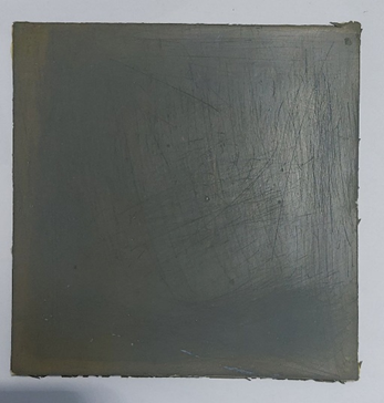
The chlorophyll is quantified to determine its concentration, yielding different value of (5, 15, 25, 35 and 45) wt%. Subsequently, varying amounts (10, 20, and 30 wt%) of nano lead are introduced to the chlorophyll. The mixture of nano lead and chlorophyll is achieved by employing a small quantity of xylene and subjecting it to ultrasound waves for a duration of 45 minutes. Additionally, hydrophilicity of the silicone rubber is enhanced by incorporating 5%wt of PEG 400 (PEG 400 is utilized to convert silicone rubber from hydrophobic to hydrophilic, thereby promoting the adhesion and uniformity of chlorophyll within the silicone rubber, this, in turn, enhances the efficacy of the shielding) [9, 10]. The suspension is incorporated into the silicone compound and subjected to magnetic mixing for a duration of eight hours. Subsequently, the hardener is introduced to the silicone and mixed again using the magnetic mixer for a period of three minutes. The mixture is poured into a glass mold measuring 15 × 15 cm. A vacuum is applied for 15 minutes to eliminate any bubbles present in the shield. Subsequently, the mold is transferred to an oven and subjected to a temperature of 50℃ and a pressure of 5 bar for a period of one hour. Following this, the mold is removed from the oven and allowed to solidify in order to obtain a silicone shield (Figure 1). The homogeneous distribution of metal nano-lead in the fabricated shield can be verified using a scanning electron microscopy (SEM) investigation, as depicted in Figure 2.
Figure 1. A radiation shielding sheet composed of silicon, which serves as a finished product
The dimensions of sample are (150 × 150 × 2) mm
2.3 Measurements
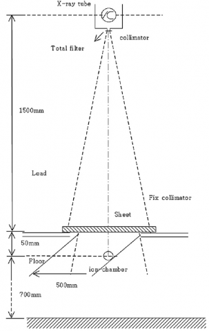
In order to measure the effectiveness of the protective barrier, the researchers utilized a method for testing lead equivalence as outlined in the European Industrial Standard for X-ray protective supplies (IEC 61331-3:2014) [11, 12]. This method was chosen to evaluate the efficacy of the sheet. The assessment of the X-ray generator's energy efficiency (DefiniumTM 6000, GE Healthcare, USA) was also performed. Figure 2 illustrates the geometric factors that are essential for evaluating the effective energy.
Figure 2. A diagram is utilized for the purpose of quantifying the half-value layer, which serves to provide insights into the effective energy [7, 13]
The investigation employed a tube current of 200 mA and an exposure time of 0.1 s. The X-ray tube was outfitted with intrinsic filtering options measuring 0.1 mm of beryllium (Be) and 4.0 mm of beryllium (Be), respectively. The collimator possessed a constant filtration of 1.5 mm of aluminium (Al), along with an extra filtration of 1.5 mm of aluminium (Al). The tube voltage was modified to several kilovolt peak (kVp) values, especially 30, 60, 90, 120, 150, and 180 kVp, which are within the range commonly employed in clinical settings. The determination of the half-value layer involved modifying the dimensions of the aluminium absorber. The assessment of the dosage of exposure was conducted by (QUART didoNEO R model). At present the temperature is maintained at 22℃ and the pressure is at 1 atmosphere.
To calculate the half-value layer, derive the linear absorption coefficient (µ) from calculating the slope of graph of y = -ax. The logarithm was utilized to convert the equation to exponential (I = I0e-µx) [14]. When I = 1/2 Io in half value layer. The equation expressed as (half-value layer = 0.693/µ), where l is the parameter being measured. To calculate energy efficiency. The mass absorption coefficient from Hubbell's table was used to get the energy efficiency of the corresponding half-value layer [14, 15].
Figure 3. The present configuration conforms to the lead equivalent testing methodology as stipulated in the European Industrial Standard for X-ray protective equipment [16, 17]
Figure 3 depicts the geometric factors that were taken into account during the performance evaluation of the manufactured shielding sheets. The experiment showcased the effectiveness of the shielding by doing a direct comparison of the transmission doses with those of lead equivalents. The accomplishment was attained through the measurement of radiation doses subsequent to the placement of a shield between the X-ray beam and the ionization dosimeter. The experiment was performed on ten times, and the average value was employed for the purpose of analysis. To assess the effectiveness of shielding, a series of experiments were undertaken to analyze the transmission doses of lead standards with varying thicknesses. The lead standards used in the studies had thicknesses of 0.05, 0.1, 0.15, 0.2, 0.25, 0.3, and 0.35 mm [18].
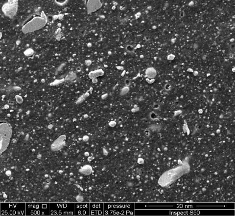
The protective sheets that were made for this study are 2 mm thick. The tensile strength (kg/cm3) and density (g/cm3) are shown in Table 1. Table 2 shows the important details about the characteristic and effectiveness of the X-ray that was used to check the rate of protection. The verification of nano-lead dispersion was conducted using scanning electron microscopy (SEM) analysis, as depicted in Figure 4. It is observed that the dispersion rate of metallic lead particles approaches 90%, hence ensuring the safety of the production process employed. Furthermore, the fabricated shields successfully underwent a toxicity test, as evidenced by the presence of chlorophyll pigment blooms on the surface and the volatilization of Xylene solvent. It was tested how well the nano lead with chlorophyll acted as a shield at a tube voltage of 60 kilovolts peak (kVp). We found that the linear absorption coefficient was 0.1946 /mm and the half-value layer was 2.40 mmAl. These values were found without any other filter. Based on the mass absorption coefficient table of Hubbell [19, 20], at 30.58 keV. Because of this, the useful energy can be raised by raising the tube voltage and adding stronger add filtering, since these things affect the quality of the X-rays. The sheets' ability to block radiation was judged by the quality of the X-rays that were taken.
Figure 4. The SEM image for shield content 20wt% of Pb and 35wt% chlorophyll
Table 1. Properties of the sheet products
|
Sheets |
Density [g/cm3] of Chlorophyll |
Density [g/cm3] of Chlorophyll with Nano Lead |
Tensile Strength [MPa] of Chlorophyll |
Density [g/cm3] of Chlorophyll with Nano Lead |
|
Non |
1.45 |
1.45 |
85 |
85 |
|
B |
1.45 |
1.51 |
83 |
140 |
|
C |
1.36 |
1.59 |
75 |
200 |
|
D |
1.3 |
1.68 |
69 |
270 |
|
E |
1.25 |
|
64 |
356 |
|
F |
1.19 |
|
52 |
320 |
Table 2. Tube voltage and effective energy
|
Tube Voltage [kVp] |
Inherent Filter |
Added Filter |
Half Value [mmAl] |
Effective Energy [keV] |
|
30 |
1.0 mmBe |
|
0.36 |
19.9 |
|
60 |
4.0 mmBe + 1.5 mmAl |
|
2.4 |
30.6 |
|
90 |
|
4.07 mmAl +0.31 mmCu |
4.91 |
48.3 |
|
120 |
|
4.07 mmAl +0.31 mmCu |
4.419 |
66 |
|
150 |
|
4.07 mmAl +0.31 mmCu |
10.26 |
76.7 |
|
180 |
|
4.07 mmAl +0.31 mmCu |
12.312 |
92.03 |
Table 3. The standard lead transmission doses
|
Dose (kVp) |
mmPb |
Transmission Doses with Thickness |
||||||
|
|
NON |
Thickness 0.05 (mm) |
Thickness 0.1 (mm) |
Thickness 0.15 (mm) |
Thickness 0.2 (mm) |
Thickness 0.25 (mm) |
Thickness 0.3 (mm) |
Thickness 0.35 (mm) |
|
30 |
6.3612 |
1.266 |
0.424 |
0.148 |
0.08 |
0.031 |
0.013 |
0.004 |
|
60 |
18.735 |
5.574 |
2.802 |
1.465 |
1.119 |
0.668 |
0.481 |
0.305 |
|
90 |
44.435 |
18.4 |
11.195 |
6.7 |
5.61 |
3.7 |
2.8 |
1.915 |
|
120 |
75.9 |
36.17 |
25.46 |
16.3 |
14.1 |
9.6 |
7.5 |
5.3 |
|
150 |
112.21 |
59.01 |
41.23 |
28.14 |
25.15 |
17.5 |
13.8 |
9.8 |
|
180 |
134.652 |
70.812 |
49.476 |
33.768 |
30.180 |
21.000 |
16.560 |
11.760 |
Note: The unit of measurement is mR.
For the comparison, the shielding effectiveness of both the lead standard and the new shielding sheets had to be tested. Using the same method as before to measure the buffering capacity of the lead standard was enough to do this. In Table 3, it can see the transfer doses of regular lead. A positive relationship was found between the shielding ability and the thickness of the lead. This means when increase the thickness lead had better shielding qualities. So, this study shows the average transmission rates and lead equivalent values of the protecting sheets that were made so that we can see how well they work at blocking compared to regular lead.
The shields containing the chlorophyll pigment showed the ability to absorb X-rays, as the percentage of the chlorophyll formula increased, the ability to absorb it increased. The best radiation absorption at 35 wt%, as shown in Table 4. The shielding efficacy was enhanced by the incorporation of nano lead with chlorophyll. This was achieved by combining nano-lead with 35% chlorophyll and 5% polyethylene glycol in silicone rubber, therefore reducing the inter-particle porosity. Furthermore, the process of vacuum defoaming was employed in a sequential manner prior to the hardening stage in order to enhance the packing of particles and improve the miscibility inside the silicon resin. The transmission dosages are presented in Table 4, which categorizes them based on the composition of the shielding sheets used and the thickness of the lead standard. The sheet with a composition of 20 wt% nano lead with 35 wt% chlorophyll exhibited identical shielding effectiveness to a 0.3 mm lead equivalent at 150 kVp and 180 kVp.
Table 4. A comparison between radiation shielding sheeting and transmission doses, dose (kVp)
|
Dose |
Samples |
Non |
5% |
15% |
25% |
35% |
45% |
|
30 |
chlorophyll |
6.361 |
5.618 |
3.587 |
1.598 |
0.981 |
0.925 |
|
|
10%Pb+35% chlo |
|
|
|
|
0.105 |
|
|
|
20%Pb+35% chlo |
|
|
|
|
0.001 |
|
|
|
30%Pb+35% chlo |
|
|
|
|
0.015 |
|
|
|
mmPb |
0.000 |
0.039 |
0.063 |
0.095 |
0.134 |
0.236 |
|
60 |
chlorophyll |
18.735 |
18.036 |
9.872 |
3.589 |
2.875 |
2.597 |
|
|
10%Pb+35% chlo |
|
|
|
|
0.158 |
|
|
|
20%Pb+35% chlo |
|
|
|
|
0.015 |
|
|
|
30%Pb+35% chlo |
|
|
|
|
0.098 |
|
|
|
mmPb |
0.000 |
0.036 |
0.059 |
0.092 |
0.121 |
0.221 |
|
90 |
chlorophyll |
44.435 |
43.581 |
38.258 |
24.528 |
14.587 |
14.254 |
|
|
10%Pb+35% chlo |
|
|
|
|
0.215 |
|
|
|
20%Pb+35% chlo |
|
|
|
|
0.01 |
|
|
|
30%Pb+35% chlo |
|
|
|
|
0.115 |
|
|
|
mmPb |
0.000 |
0.033 |
0.056 |
0.089 |
0.120 |
0.219 |
|
120 |
chlorophyll |
75.9 |
75.015 |
72.532 |
68.925 |
65.032 |
64.987 |
|
|
10%Pb+35% chlo |
|
|
|
|
0.287 |
|
|
|
20%Pb+35% chlo |
|
|
|
|
0.108 |
|
|
|
30%Pb+35% chlo |
|
|
|
|
0.187 |
|
|
|
mmPb |
0.000 |
0.033 |
0.052 |
0.084 |
0.117 |
0.215 |
|
150 |
chlorophyll |
112.21 |
112.058 |
110.132 |
106.932 |
102.687 |
102.655 |
|
|
10%Pb+35% chlo |
|
|
|
|
0.308 |
|
|
|
20%Pb+35% chlo |
|
|
|
|
0.115 |
|
|
|
30%Pb+35% chlo |
|
|
|
|
0.223 |
|
|
|
mmPb |
0.000 |
0.032 |
0.051 |
0.086 |
0.119 |
0.217 |
|
180 |
chlorophyll |
134.652 |
134.622 |
134.098 |
133.187 |
131.532 |
131.51 |
|
|
10%Pb+35% chlo |
|
|
|
|
0.315 |
|
|
|
20%Pb+35% chlo |
|
|
|
|
0.119 |
|
|
|
30%Pb+35% chlo |
|
|
|
|
0.259 |
|
|
|
mmPb |
0.000 |
0.032 |
0.053 |
0.086 |
0.118 |
0.218 |
Note: The unit of measurement is mR.
The use of radiation in medical technology and equipment has significantly contributed to the diagnosis and treatment of several ailments. The application of radiation in invasive medical procedures, such as angiography, has witnessed significant growth, resulting in an escalation of radiation exposure for both patients and healthcare personnel. Consequently, ensuring individual radiation protection has emerged as a crucial concern.
Within contemporary medical discourse, the primary constituent of shielding materials is lead. Consequently, aprons crafted from nano-lead and chlorophyll have gained prominence as a cost-effective, easily manufacturable, and pleasant alternative. This is attributed to the inherent flexibility of the sheet, which not only enhances comfort but also mitigates toxicity concerns. Furthermore, the process of mass manufacture and dissemination poses significant challenges.
The lead equivalence of current lead aprons ranges from 0.25 to 0.5 mm. A lead apron with a thickness of 0.5 mmPb has a shielding effectiveness of over 95% against direct radiation at 100 kVp. Hence, it is imperative to have a minimum shielding thickness of 0.3 mmPb in order to account for the reduction in shielding efficacy. this study has demonstrated that nano-lead, when combined with chlorophyll at concentrations of 20 wt% and 35 wt% respectively, exhibits a remarkable capacity to protect against various forms of medical radiation. This finding is visually represented in Figure 5. One of the challenges associated with this particular shielding method is to the intricate dispersion of nano-lead, a process that necessitates a significant amount of time for adequate preparation prior to commencing mass production. Furthermore, achieving optimal shielding and efficiency necessitates the reduction of lead particle size.
Figure 5. A comparison of the radiation shielding sheet, focusing on the shielding capacity of micro lead, chlorophyll, and lead in the 40-80 keV X-ray energy region for diagnostic purposes (The absorption of nano lead with chlorophyll was found to be greater than that of lead)
The present investigation resulted in the development of a protective barrier against X-ray radiation. Within the realm of medicine, the comparative analysis is conducted between bio-silicon, possessing a thickness of 2 mm and exhibiting a low weight, and the existing lead sheet shields with a thickness of 0.3 mm that are presently employed in healthcare facilities. The researchers considered the effectiveness of the shield in environments with high levels of radiation, and determined that a plate thickness of 1cm is sufficient to provide protection against low radiation fields.
A medical radiation shielding sheet was produced by incorporating varying concentrations (10, 20 and 30 wt%) of nano lead and chlorophyll (at varying concentrations of 35 wt%) into bio silicon. This approach proved to be more cost-effective compared to previously suggested shielding methods, while still achieving a comparable shielding effect to that of lead.
The radiation attenuation properties of shielding sheets, with a thickness of 2 mm, were evaluated at tube voltages of 30, 60, 90, 120, 150, and 180 kilovolts peak (kVp). The use of a composite consisting of metallic nano lead at a concentration of 20 wt% in conjunction with chlorophyll at a concentration of 35 wt% shown notable efficacy in accurately measuring a dosage at 150 Kvp. Compared to conventional lead plates, the suggested shield exhibits a transmission dose of 0.115 mR. Conversely, traditional shield plates with a thickness of 0.3 mm demonstrate a transmission dose of 0.119 mR. The concentration of lead exhibited a remarkably high tensile strength.
The preparation of shielding sheets involved the classification of materials and the subsequent assessment of their respective shielding abilities. This evaluation aimed to identify the composition that would yield the most effective shielding performance. The shielding efficacy of the nano lead composite, consisting of 20 wt% lead and 35 wt% chlorophyll, was shown to be comparable to that of a 0.3 mm lead equivalent.
[1] Sarihan, M. (2022). Simulation of gamma-ray shielding properties for materials of medical interest. Open Chemistry, 20(1): 81-87. https://doi.org/10.1515/chem-2021-0118
[2] Massalha, S., Almufleh, A., Small, G., Marvin, B., Keidar, Z., Israel, O., Kennedy, J.A. (2019). Strategies for minimizing occupational radiation exposure in cardiac imaging. Current Cardiology Reports, 21: 1-6. https://doi.org/10.1007/s11886-019-1157-1
[3] Vano, E. (2015). Occupational radiation protection of health workers in imaging. Radiation Protection Dosimetry, 164(1-2): 126-129. https://doi.org/10.1093/rpd/ncu354
[4] Chang, Q., Guo, S., Zhang, X. (2023). Radiation shielding polymer composites: Ray-interaction mechanism, structural design, manufacture and biomedical applications. Materials & Design, 233: 112253. https://doi.org/10.1016/j.matdes.2023.112253
[5] Aral, N., Banu Nergis, F., Candan, C. (2016). An alternative X-ray shielding material based on coated textiles. Textile Research Journal, 86(8): 803-811. https://doi.org/10.1177/0040517515590409
[6] Trajkovska Petkoska, A. (2023). Assessment of the attenuation properties of commercial lead-free radiation-shielding composite materials against medical X-rays. Journal of Composites Science, 7(10): 424. https://doi.org/10.3390/jcs7100424
[7] Kim, S.C., Dong, K.R., Chung, W.K. (2012). Medical radiation shielding effect by composition of barium compounds. Annals of Nuclear Energy, 47: 1-5. https://doi.org/10.1016/j.anucene.2012.04.014
[8] Abdolahzadeh, T., Morshedian, J., Ahmadi, S. (2023). Novel polyethylene/tungsten oxide/bismuth trioxide/barium sulfate/graphene oxide nanocomposites for shielding against X-ray radiations. International Journal of Radiation Research, 21(1): 79-87. https://doi.org/10.52547/ijrr.21.1.11
[9] Aral, N., Nergis, F.B., Candan, C. (2016). Investigation of X-ray attenuation and the flex resistance properties of fabrics coated with tungsten and barium sulphate additives. Journal of Textile & Apparel/Tekstil ve Konfeksiyon, 26(2): 166.
[10] Kattan, M., Daher, Y., Alkassiri, H. (2007). A high-dose dosimeter-based polyvinyl chloride dyed with malachite green. Radiation Physics and Chemistry, 76(7): 1195-1199. https://doi.org/10.1016/j.radphyschem.2006.12.004
[11] Schmid, E., Panzer, W., Schlattl, H., Eder, H. (2012). Emission of fluorescent x-radiation from non-lead based shielding materials of protective clothing: A radiobiological problem? Journal of Radiological Protection, 32(3): N129. https://doi.org/10.1088/0952-4746/32/3/N129
[12] Büermann, L. (2016). Determination of lead equivalent values according to IEC 61331-1: 2014—Report and short guidelines for testing laboratories. Journal of Instrumentation, 11(09): T09002. https://doi.org/10.1088/1748-0221/11/09/T09002
[13] Alsaab, A.H., Zeghib, S. (2023). Analysis of X-ray and gamma ray shielding performance of prepared polymer micro-composites. Journal of Radiation Research and Applied Sciences, 16(4): 100708. https://doi.org/10.1016/j.jrras.2023.100708
[14] Al-Akhras, M.A.H., Aljarrah, K., Al-Omari, A., Al-Khateeb, H.M., Albiss, B.A., Azez, K., Makhadmeh, G. (2008). Influence of extremely low energy radiation on artificial tissue: Effects on image quality and superficial dose. Spectroscopy, 22(5): 419-428. https://doi.org/10.3233/SPE-2008-0357
[15] Suleiman, I.K. (2018). Determination of linear absorption coefficient for different materials absorbers of varying lengths first colliminator radioactive source sodium iodide detector (NaI) ratemeter (Decade Scaler Counter). International Journal of Modern Research in Engineering and Technology (IJMRET), 3(8): 28-38.
[16] Schöpf, T., Pichler, T. (2016). Radiation protection clothing in X-Ray diagnostics–Influence of the different methods of measurement on the lead equivalent and the required mass. Fortschr Röntgenstr, 188(8): 768 - 775. 10.1055/s-0042-106651
[17] Wargo, R.R., Aljabal, A.F., Lin, P.J.P. (2020). Evaluation and verification of a simplified lead equivalency measurement method. Journal of Applied Clinical Medical Physics, 21(2): 152-156. https://doi.org/10.1002/acm2.12810
[18] Çetin, H., Yurt, A., Yüksel, S.H. (2017). The absorption properties of lead-free garments for use in radiation protection. Radiation Protection Dosimetry, 173(4): 345-350. https://doi.org/10.1093/rpd/ncw004
[19] McCaffrey, J.P., Shen, H., Downton, B., Mainegra‐Hing, E. (2007). Radiation attenuation by lead and nonlead materials used in radiation shielding garments. Medical Physics, 34(2): 530-537. https://doi.org/10.1118/1.2426404
[20] Akça, B., Erzeneoğlu, S.Z. (2014). The mass attenuation coefficients, electronic, atomic, and molecular cross sections, effective atomic numbers, and electron densities for compounds of some biomedically important elements at 59.5 KeV. Science and Technology of Nuclear Installations, 2014: 901465. https://doi.org/10.1155/2014/901465