Acute Toxicity of Aluminum, Cobalt, and Lithium Chlorides in Albino wistar Mice: Determination of Lethal Doses

Wasan Abdulmunem Taha*![]() | Sura Samir Mohammed
| Sura Samir Mohammed![]() | Lubna Samir Mohammed
| Lubna Samir Mohammed![]()
© 2024 The authors. This article is published by IIETA and is licensed under the CC BY 4.0 license (http://creativecommons.org/licenses/by/4.0/).
OPEN ACCESS
Chlorides are considered one of the dangerous pollutants that pose a threat to human and animal health. The study aims to determine the acute lethal doses (LD50) of aluminum, cobalt, and lithium chlorides in Albino wistar mice. Albino wistar mice were divided into four groups with the mice were randomly divided into 4 groups, 8 mice for experimental group and 6 mice for control group. Short tests were conducted to determine the acute lethal dose of the metals under study. Over 168 hours, mice were exposed to four different concentrations of (AlCl2) metal in concentrations of 40, 50, 60 and 70 mg/kg per liter of water, where 1 liter of water was placed in each bottle, while the second group was exposed to cobalt chloride (CoCl2) in concentrations 75, 100, 125 and 150 mg/kg per liter of water, where 1 liter of water was placed in each bottle. As well as, the third group, it was exposed to (LiCl2) at concentrations 75, 100, 125 and 150 mg/kg per liter of water, while the fourth group included the control group. 1 liter of water was placed in each bottle and it was given orally. LC50 was calculated by use Finney's probit analysis method where LC50 it is abbreviation "lethal concentration 50%" or "intermediate lethal concentration". It is the concentration of a material (in the water) which it killed a half the members of populations after a specific during of exposed to toxic metal. This value is so importance in toxicology because it gives an indicated of materials poisoning. The LC50 is inverse proportionate to toxicity. The current study concluded the dose that killed 50% of the Albino wistar mice was 50mg/kg of AlCl2, 100mg/kg of CoCl2, while it was 125mg/kg of lithium chloride. Long-term use of greater doses of chlorides can have varying degrees of negative impact on various organs in mice. This study provides context for a more thorough investigation of the toxicity of chlorides compounds on humans, which may be significant for public health.
Albino wistar mice, acute lethal dose, heavy metals, metal chloride toxicity
Both humans and animals regularly come into contact with their environment, leading to exposure to diverse chemicals and heavy metals. These substances have the potential to bioaccumulation in the body, accumulating in tissues with limited excretion capabilities [1].
Exposure to heavy metals is inevitable because they are widely distributed in the environment, food, and water. High concentrations of heavy metals are measured in a variety of human, animal, and aquatic fish organs. Even though many metals are necessary trace elements, prolonged exposure to them and high doses of their salts can be harmful to both people and animals [2]. Chlorides are significant ions that occur naturally and are present in all natural environments. In the majority of aqueous environments, chlorine behaves as a conservative ion, which means that the interactions of water with soils, sediments, and rocks do not slow down chloride's flow [2]. As a result, it might be utilized as a sign of more contamination. An "advance warning" of the existence of additional, more dangerous pollutants can be provided by abnormally high concentrations. According to Davis et al. [3] the concentration of Cl- in natural waters can range from less than 1 mg/L in rainfall and some freshwater aquifers to more than 100,000 mg/L in old ground waters.
Cl-concentricity in precipitation in midcontinent regions is often less than 1 mg/L, and always less than 0.1 mg/L [4]. Upon contacting earth roof, Cl concentricity in water raise as a result of interacts with soils, rocks, and biota (waste products), as well as the impacts of evaporation [5]. Chlorides are the most superabundant ions in freshwater, with a concentricity super than 860ppm [6]. Highly levels of Cl- in roof water are mostly due to significance evaporation. Chloride is non-toxicity to humans, but high levels make water unbootable because the salty relish [6].
Chloride is corrosive to steel, thus it can erode pipes in water treatment and industrial plants. Elevated Cl- levels in drinking water supplies can raise treatment costs since it is corrosive and gives water a salty relish. At concentrations as low as 210 mg/L, elevated Cl- in surface water has been associated to harm to aquatic animals, terrestrial plants, and both [7].
Daily interactions between humans and animals and their surroundings expose them to a variety of toxins and Heavy metals have the ability to undergo bioaccumulation in the body, leading to their accumulation in tissues with limited excretion capacity [8]. Aluminum (Al) is the third most prevalent metallic element in the crust of the Earth [9]. Al is utilized in many common daily applications, including water treatment, wood preservation, shampoos, vitamins, food additives, packaging materials, antiperspirants, toothpaste, pharmaceuticals, and their usage as fillers in plastics contribute to widespread exposure, making it difficult to avoid contact with these substances [10]. Al frequently enters humans through ambient air and tainted food and drink [11]. Additionally, cement companies' wastewater and industrial particulate matter include large amounts of aluminum, which exposes workers to quantities of aluminum beyond the permissible limit [12]. Al is often consumed in doses of between 70 and 140 mg per week, and although having a low gastrointestinal absorption capacity (less than 1%), Al may accumulate over time in vital organs like the kidney, liver, and brain, where it may cause apparent neurotoxicity and cytotoxicity [13]. Al is thus listed among the hazardous materials with the highest priority as per the records of the Agency for Toxic Substances and Disease Registry (ATSDR) [8]. According to growing evidence, the pathophysiology of neurodegenerative diseases like Alzheimer's disease, Parkinsonism, dementia, and amyotrophic lateral sclerosis may be influenced by aluminum accumulation [2]. Aluminum exposure has been demonstrated to cause abnormalities in memory development in animal studies, which are comparable to those of dementia and Alzheimer's disease [14]. According to a prior studies aluminum changes cholinergic and noradrenergic neurotransmission by damaging neurons, interfering with glucose metabolism, interrupting signal transduction, and producing reactive oxygen species (ROS) [2].
Aluminum chloride has been linked to negative effects on behavioral parameters in wistar rats, negative aluminum treatment in wistar rats resulted in increased anxiety levels, negatively impacting anxiety-related behavior. Additionally, at higher doses, aluminum demonstrated neurodegenerative properties, leading to adverse effects on the histology of the cerebral cortex in adult wistar rats [14]. Moreover, the brain, particularly the grey and white matter, was found to be a favored site for aluminum accumulation [15].
Cobalt (Co) is an essential hint metal to human life [16]. Co, an indispensable ferromagnetic transition metal, plays a vital role in supporting mammalian well-being [17], that plays an important role in the vitamin B12 synthesis [14]. Co is frequently used as a dietary supplement, preservative, in beverages, cosmetics, and medical equipment as well as a medicinal agent to treat various disorders [18]. The main sources of Co include meat, vegetables, and drinking water. According to studies on laboratory animals exposed to metal ions over an extended period of time, these ions build up in organs such the kidney, liver, spleen, heart, stomach, intestines, muscle, brain, and testes. Additionally, whole blood, serum, and urine all have higher concentrations of Co [19]. Although absorption varies depending on the species, water-soluble cobalt compounds absorb more than non-water-soluble forms [20].
The toxicity effects of the initial documentation of Co occurred in 1966, when individuals who consumed beer experienced the development of cardiomyopathy, which was marked by pericardial effusion, elevated hemoglobin levels, and congestive heart failure. This condition was attributed this outcome can be attributed to the presence of Co sulfate, which was added to the beer as a stabilize [14]. The stated median fatal dosage for mammals ranges from 150 mg kg-1 to 500 mg kg-1 where high quantities of soluble cobalt salt exposure is toxic [21]. As well as, the cardiovascular, hematological, neurological, endocrine, and reproductive systems are severely adversely affected by oxygen radicals produced due to cobalt (II) complexes in biological systems [22, 23]. Earlier research has the findings revealed that Co modification mitochondrial permeability by triggering the opening of transmutation holes, resulting in mitochondrial bulges and a decline in the electrical membrane potential. Additionally, Co has a high affinity for sulfhydryl groups, which inhibits key enzymes involved in the mitochondrial respiratory chain. Additionally, it has been shown that Co inhibits synaptic transmission by blocking calcium (Ca2+) channels presynaptically, which reduces neurotransmitter-induced postsynaptic responses. Additionally, it has been demonstrated that Co treatment in rats causes the reduction in neurotransmitters such as dopamine, norepinephrine, and serotonin suggest the potential of Co to adversely affect memory function [9]. Cobalt chloride (CoCl2) exposure over an extended period of time may result in a series of metabolic reactions that alter signal transmission, protein production, and other biological processes, which may, on the one hand, contribute to the toxicity of cobalt ions and, on the other, impair animal development [24]. Lithium is naturally coexisting with many other minerals such as magnesium and boron and takes only a small proportion in most of its deposits, as the lightest metal element, is widely used in various fields such as lithium electrolytes for secondary batteries, lithium additives for functional materials, lithium-based lubricating greases and medical additives [25]. (Li) exerts its effects on a wide extent of cellular activities meanwhile inhibits inositol producing, affects protein kinase C signals and suppress glycogen synthesize kinase-3β (GSK-3β). GSK-3, a serine/threonine protein kinase initially identify as the key regulators of glycogen metabolism, contains twice extremely homologous isoforms (α, β) of human. A β shape of GSK-3, GSK-3β, plays very paramount functions in metabolism, cells subsistence, diffusion, and differentiation [26]. The ability contribution lipid GSK-3β overthrow is investigation previously by treating yellow croaker fishes by (LiCl), especially, gene express and fat acid synthesis activities (FAS) and liver lipoprotein lipase (LPL) are inhibition by (LiCl) along with cholesterol (C) plasma levels, triglycerides (TG), liver and plasma non-esterified fat acids (NEFA) [26, 27]. Hui et al. [27] exhibited that LiCl selective inhibition the phosphorylation and activity of p38 MAPK in respond to IL-1b thus prevents the stimulation of MMP-1 and MMP-13 and the results cartilage degeneration that followed. Other researches LiCl is showed to inducing apoptosis and to obstruct the sulphation of glycosaminoglycans which is probable to impacts on proteoglycan hydration and tissues mechanisms [27].
All mice were purchased from Iraqi Center for Cancer Research and Medical Genetics (Baghdad- Iraq). Totally 40 mice (4 weeks old) were housed in the laboratories of the General Company for Pharmaceutical Industry - Baghdad, at 25℃, 50.55% humidity, 12 h light/12 h dark cycle, with free access to water and food. The mice were divided into one control group and three experimental groups, with 8 mice in each group. Short tests were conducted to determine the acute dose of the metals under study. Over 168 hours, mice were exposed to four different concentrations of (AlCl2) metal in concentrations of 40, 50, 60 and 70 mg/kg per liter of water, where 1 liter of water was placed in each bottle, while the second group was exposed to cobalt chloride (CoCl2) in concentrations 75, 100, 125 and 150 mg/kg per liter of water, where 1 liter of water was placed in each bottle. As well as, the third group, it was exposed to (LiCl2) at concentrations 75, 100, 125 and 150 mg/kg per liter of water, where 1 liter of water were placed in each bottle. All these doses were the total daily doses. During this period, the bottles water were changed every two days with continuous ventilation and fed twice daily with recording of all the changes observed during the trial period.
The data are presented as the mean ± standard error of the mean. The results include the F ratio, the number of degrees of freedom, the outcome, and the significance value. Statistical analyses were conducted using SPSS v20.0 software (IBM, Armonk, NY, USA). An unpaired Student's t‑test and repeated analysis of variance (ANOVA), followed by the Bonferroni post hoc test, were employed when suitable. A significance level of P<0.05 was considered indicative of a statistically significant difference.
In environmental studies, lethal dosage levels often refer to a chemical's concentration in water. A conventional experiment involves exposing groups of animals to a concentration for a predetermined amount of time, as stated in the (Organization for Economic Cooperation and Development) (OECD) Guidelines for the Testing of Chemicals. Up to 7 days of clinical observation are given to the fish. The LC50 value was the amount of the chemical present in the water that, throughout the course of the observation time, killed 50% of the test animal [28].
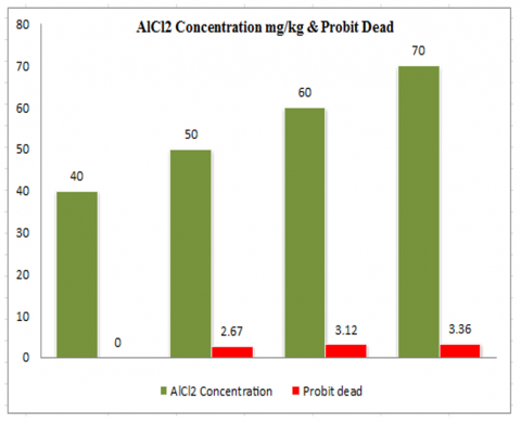
Mice were exposed to doses of 40, 50, 60 and 70 mg/kg of AlCl2. Table 1 displays LC50 value of AlCl2 which determined after subjecting the samples to a 168-hour exposure period, Finney's probit analysis method was utilized in conjunction with SPSS Statistical Software for data analysis. Probit analysis is used to analyze many kinds of dose-response or binomial response experiments in a variety of fields; Probit Analysis is commonly used in toxicology to determine the relative toxicity of chemicals to living organisms. In this pattern an expecting dose corresponding to every observed dose measuring experimentally may be determines from the observation mortality, and the deduced dose, so obtained, were called ‘‘normal equivalent deviations” or ‘‘N.E.D.”. For avoid these difficulties, a modern chart of statistics unit called “probits” is devise (]) in which the zero of the ordinary statistics tables of diverge has equate to the number 5, and the diverge of the regular curve, in terms of U, adding algebraically to gain the probit identical for all percentages kill. Probits to transforms the dose-mortality curves to an upright line. In the table was gave the probit corresponds for all percentages mortality listed along the left border to probit table (as shown in Table A).
Table 1. LC50 values of mice exposed to various AlCl2 concentrations for 168 hours
|
Concentration (AlCl2 mg/L) |
Log10 (Concentration) |
Dead |
Probit Kill |
|
40 |
1.602059991 |
0 |
0 |
|
50 |
1.698970004 |
1 |
2.67 |
|
60 |
1.77815125 |
3 |
3.12 |
|
70 |
1.84509804 |
5 |
3.36 |
Figure 1. Concentration of AlCl2 mg/kg and probit dead
Figure 1 displays the measured percentage of mice mortality for AlCl2 in static tests conducted continuously for various hours and concentrations. Show the line graph depicting the probit analysis of the toxicity data for AlCl2. Using Finney's probit analysis approach, probit kill was discovered at a concentration of 50 mg/kg.
Al compounds are recognized to impede gastrointestinal motility and lead to delayed stomach emptying in both humans and rats, which might be the underlying reason for this observed mortality. Al exerts its effects by disrupting the serotonin and dopamine synthesis pathways, contributing to these effects, which are directly engaged in the regulation of digestive and nutritional behaviour as well as the management of satiety; therefore death is likely caused by this action [15]. According to the study by Yousef et al. [12] exposure to Al compounds results in liver necrosis, significant cellular membrane degradation, the release of intracellular enzymes and bilirubin into the blood circulation, as well as morphological kidney lesions [29]. AlCl2 is then entered into the blood-brain barrier (BBB) and accumulated in the brain, primarily in the hippocampus, prolonged accumulation of Al causes neurotoxicity through the development of neurofibrillary tangles and amyloid aggregates [30]. Other study reveals that AlCl2 administrate was a damaging effect on hippocampal constructing. This was in the form of neuronal cell damage, shrunken cell bodies with pericellular haloes. Moreover, previous studies reported that reactive oxygen species (ROS) production and oxidative stress are linked to the use of AlCl2 [31]. They added that brain tissue is very exposure to the hazardous effect of ROS because their elevated rates of oxygen consume, present of profuse polyunsaturated fatty acids in the cell and organs’ membranes and little antioxidant enzymes [32]. In addition, AlCl2 can induced lipid peroxidation by interacts with plasma membrane lipids. Previous researchers reported that membrane lipids' peroxidation can result in rise in membrane leakage, mitochondrial dysfunctional, DNA, lipids and protein damages, and results in cellular degradation and finally cells dying [33].
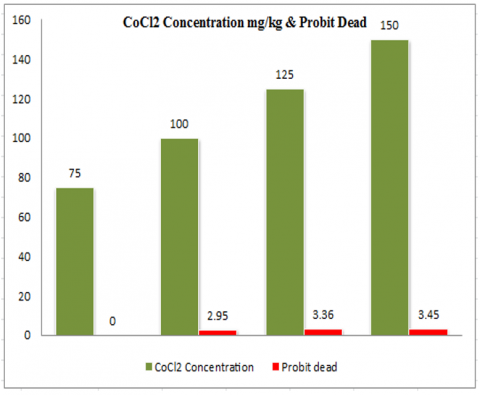
Mice were exposed to doses of 75, 100, 125 and 150 mg/kg of CoCl2. Table 2 displays LC50 value of CoCl2 which determined after subjecting the samples to a 168-hour exposure period, Finney's probit analysis method was utilized in conjunction with SPSS Statistical Software for data analysis.
Table 2. LC50 values of mice exposed to various CoCl2 concentrations for 168 hours
|
Concentration (CoCl2 mg/L) |
Log10 (Concentration) |
Dead |
Probit Kill |
|
75 |
1.875061263 |
0 |
0 |
|
100 |
2 |
2 |
2.95 |
|
125 |
2.096910013 |
6 |
3.36 |
|
150 |
2.176091259 |
5 |
3.45 |
Figure 2 displays the measured percentage of mice mortality for CoCl2 in static tests conducted continuously for various hours and concentrations. It shows the probit line graph of the CoCl2 toxicity data. Using Finney's probit analysis approach, probit kill was discovered at a concentration of 75 mg/kg.
Figure 2. Concentration of CoCl2 mg/kg and probit dead
Table 3. LC50 values of mice exposed to various LiCl2 concentrations for 168 hours
|
Concentration (LiCl2 mg/L) |
Log10 (Concentration) |
Dead |
Probit Kill |
|
75 |
1.875061263 |
0 |
0 |
|
100 |
2 |
0 |
0 |
|
125 |
2.096910013 |
4 |
3.25 |
|
150 |
2.176091259 |
7 |
3.52 |
Mice were exposed to doses of 75, 100, 125 and 150 mg/kg of CoCl2. Table 3 displays LC50 value of CoCl2 which determined after subjecting the samples to a 168-hour exposure period, Finney's probit analysis method was utilized in conjunction with SPSS Statistical Software for data analysis.
Numerous researchers have reported on the interactions between oxidative stress, inflammation, and apoptosis in physiological derangements brought on by toxicants [34]. According to reports, the hypoxic reaction brought on CoCl2 is linked to an increase in the generation of reactive oxygen species (ROS), which can cause cellular damage and death in biological systems [34]. Co (II) combines with H2O2 directly in a Fenton-type reaction to form ROS, which when produced in excess can damage cell survival by irreversibly altering the DNA; CoCl2 operates as an oxidative stress-inducing agent [21].
Figure 3 displays the measured percentage of mice mortality for LiCl2 in static tests conducted continuously for various hours and concentrations. Show the probit line graph of the LiCl2 toxicity data. Using Finney's probit analysis approach, probit kill was discovered at a concentration of 75 mg/kg.
Figure 3. Concentration of LiCl2 mg/kg and probit dead
Mice were exposed to doses of 75, 100, 125 and 150 mg/kg of LiCl2. Table 3 displays LC50 value of LiCl2 which determined after subjecting the samples to a 168-hour exposure period, Finney's probit analysis method was utilized in conjunction with SPSS Statistical Software for data analysis.
Lithium's negative side effects can be classified according to the organs or systems they affect, including the cardiovascular, gastrointestinal, thyroid, neurological, thyroid, metabolic, cognitive, dermatological, and sexual systems. However, a number of negative side effects may manifest at any point throughout the use of lithium [35].
In the present study, the LC50 values for AlCl2, CoCl2, and LiCl2 after 168 hours were found to be 40 mg/kg, 75 mg/kg, and 75 mg/kg respectively. The detailed results are presented in Tables 1, 2, and 3.
This version clarifies the units for the LC50 values and adds clarity to the reference to the tables. The findings distinctly demonstrate that as the concentration of the toxicant increases, the mortality rate rises for a specific fixed time. Similarly, for a particular concentration, an increase in exposure time also leads to an elevation in mortality. The toxicant's consistent mode of action involves accumulation up to dangerous levels, ultimately resulting in fatalities [36]. Furthermore, the damaging effects of these metals on the kidney, brain, and liver may also contribute to the observed deaths [12]. In serum the dose of (Li) ranging between 0.8 to 1.2 mM were recommendation and highly uses, but they were association by a highly rate of side effects includes gastro intestinal, renal, neurological and endocrine disorderliness. Li show to reduction of neuronal inositol level by inhibitions of inositol monophosphatase (IMPase), that converting myo-inositol monophosphates to myo-inositol, to reconstitute the membrane phospholipids [37]. The poisonous effect impact of LiCl on fetal developing was exploring on different type organism, such as squid, Xenopus, zebrafish and sea urchin. The result shows that LiCl prevents the growth straight the animal vegetal axis and anterior midline of the squid fetal [37]. In Xenopus laevis it alters the body axial patterning, produced fetal with decreased posterior but exaggerated anterior texture and embryos with truncation of anterior structures. LiCl also stimulates significance phenotypic abnormalities in zebrafish growth, including pericardial and yolk sac oedema, dispersed pigment cells [37-39].
In the current investigation, mice exposed to different concentrations of AlCl2, CoCl2 and LiCl2 mg/ kg have displayed a high mortality rate when exposed to high concentrations of these metals, where the LC50 of AlCl2 was 50 mg/ kg, while the LC50 of CoCl2 and LiCl2 was 75 mg/ kg. Therefore, the current study recommends conducting many studies in order to know the effect of these metals on other body functions such as endocrine pathway including sexual hormone such as Follicle stimulating hormone (FSH), estrogen, progesterone, testosterone etc.
The authors thank the Iraqi Center for Cancer Research and Medical Genetics (Iraq-Baghdad) for their help in providing experimental animals. We would also like to thank the Deanship of the College of Applied Sciences for providing the laboratories to complete the practical part of the manuscript.
Table A. Finney's table for transformation of percentage of mortality to probit values [39]
|
% |
0 |
1 |
2 |
3 |
4 |
5 |
6 |
7 |
8 |
9 |
|
0 |
- |
2.67 |
2.95 |
3.12 |
3.25 |
3.36 |
3.45 |
3.52 |
3.59 |
3.66 |
|
10 |
3.72 |
3.77 |
3.82 |
3.87 |
3.92 |
3.96 |
4.01 |
4.05 |
4.08 |
4.12 |
|
20 |
4.16 |
4.19 |
4.23 |
4.26 |
4.29 |
4.33 |
4.36 |
4.39 |
4.42 |
4.45 |
|
30 |
4.48 |
4.50 |
4.53 |
4.56 |
4.59 |
4.61 |
4.64 |
4.67 |
4.69 |
4.72 |
|
40 |
4.75 |
4.77 |
4.80 |
4.82 |
4.85 |
4.87 |
4.90 |
4.92 |
4.95 |
4.97 |
|
50 |
5.00 |
5.03 |
5.05 |
5.08 |
5.10 |
5.13 |
5.15 |
5.18 |
5.20 |
5.23 |
|
60 |
5.25 |
5.28 |
5.31 |
5.33 |
5.36 |
5.39 |
5.41 |
5.44 |
5.47 |
5.50 |
|
70 |
5.52 |
5.55 |
5.58 |
5.61 |
5.64 |
5.67 |
5.71 |
5.74 |
5.77 |
5.81 |
|
80 |
5.84 |
5.88 |
5.92 |
5.95 |
5.99 |
6.04 |
6.08 |
6.13 |
6.18 |
6.23 |
|
90 |
6.28 |
6.34 |
6.41 |
6.48 |
6.55 |
6.64 |
6.75 |
6.88 |
7.05 |
7.33 |
|
- |
0 |
0.1 |
0.2 |
0.3 |
0.4 |
0.5 |
0.6 |
0.7 |
0.8 |
0.9 |
|
99 |
7.33 |
7.37 |
7.41 |
7.46 |
7.51 |
7.58 |
7.65 |
7.75 |
7.88 |
8.09 |
[1] Al Dera, H. S. (2016). Protective effect of resveratrol against aluminum chloride induced nephrotoxicity in rats. Saudi Medical Journal, 37(4): 369-378. https://doi.org/10.15537%2Fsmj.2016.4.13611
[2] Gluhcheva, Y., Atanasov, V., Zhorova, R., Madzharova, M., Ivanova, J., Mitewa, M. (2010). Cobalt bioaccumulation in mouse blood plasma and liver. Biotechnology & Biotechnological Equipment, 24(sup1): 311-314. https://doi.org/10.1080/13102818.2010.10817853
[3] Davis, S.N., Whittemore, D.O., Fabryka-Martin, J. (2005). Uses of chloride/bromide ration in studies of potable water. Groundwater Monitoring & Remediation, 36(2): 338-350. https://doi.org/10.1111/j.1745-6584.1998.tb01099.x
[4] Stirpe, C.R., Cunningham, M.A., Menking, K.M. (2017). How will climate change affect road salt export from watersheds? Water, Air, & Soil Pollution, 228: 362. https://doi.org/10.1007/s11270-017-3455-9
[5] An, N., Tang, C.S., Xu, S.K., Gong, X.P., Shi, B., Inyang, H.I. (2018). Effects of soil characteristics on moisture evaporation. Engineering Geology, 239: 126-135. https://doi.org/10.1016/j.enggeo.2018.03.028
[6] Hunt, M., Herron, E., Green, L. (2012). Chlorides in fresh water. The University of Rhode Island.
[7] Kaushal, S.S., Groffman, P.M., Likens, G.E., Belt, K.T., Stack, W.P., Kelly, V.R., Band, L.E., Fisher, G.T. (2005). Increased salinization of fresh water in the northeastern United States. In Proceedings of the National Academy of Sciences, 102(38): 13517-13520. https://doi.org/10.1073/pnas.0506414102
[8] Othman, M.S., Fareid, M.A., Abdel Hameed, R.S., Abdel Moneim, A.E. (2020). The protective effects of melatonin on aluminum-induced hepatotoxicity and nephrotoxicity in rats. Oxidative Medicine and Cellular Longevity, 2020: 7375136. https://doi.org/10.1155/2020/7375136
[9] Al-Olayan, E.M., El-Khadragy, M.F., Abdel Moneim, A.E. (2015). The protective properties of melatonin against aluminium-induced neuronal injury. International Journal of Experimental Pathology, 96(3): 196-202. https://doi.org/10.1111/iep.12122
[10] Abdel Moneim, A.E., Othman, M.S., Mohmoud, S.M., El-Deib, K.M. (2013). Pomegranate peel attenuates aluminum-induced hepatorenal toxicity. Toxicology Mechanisms and Methods, 23(8): 624-633. https://doi.org/10.3109/15376516.2013.823634
[11] Bulan, Ö.K., Bayrak, B.B., Sarikaya-Ünal, G., Yanardağ, R. (2019). The influence of melatonin supplementation against aluminum-induced toxicity in brains of male rats. Journal of Research in Pharmacy, 23(2): 275-283. https://doi.org/10.12991/jrp.2019.134
[12] Yousef, M.I., Mutar, T.F., Kamel, M.A.E.N. (2019). Hepato-renal toxicity of oral sub-chronic exposure to aluminum oxide and/or zinc oxide nanoparticles in rats. Toxicology Reports, 6: 336-346. https://doi.org/10.1016/j.toxrep.2019.04.003
[13] Tietz, T., Lenzner, A., Kolbaum, A.E., Zellmer, S., Riebeling, C., Gürtler, R., et al. (2019). Aggregated aluminium exposure: Risk assessment for the general population. Archives of Toxicology, 93: 3503-3521. https://doi.org/10.1007/s00204-019-02599-z
[14] Alexa, T., Luca, A., Dondas, A., Bohotin, C.R. (2015). Preconditioning with cobalt chloride modifies pain perception in mice. Experimental and Therapeutic Medicine, 9(4): 1465-1469. https://doi.org/10.3892/etm.2015.2235
[15] Bekhedda, H., Menadi, N., Demmouche, A., Ghani, A., Mai, H. (2020). Histological study of the effects of aluminum chloride exposure on the brain of wistar rats female. Journal of Drug Delivery and Therapeutics, 10(3-s): 37-42. https://doi.org/10.22270/jddt.v10i3-s.4152
[16] Simonsen, L.O., Harbak, H., Bennekou, P. (2012). Cobalt metabolism and toxicology—A brief update. Science of the Total Environment, 432: 210-215. https://doi.org/10.1016/j.scitotenv.2012.06.009
[17] Gluhcheva, Y., Pavlova, E., Petrova, E., Tinkov, A.A., Ajsuvakova, O.P., Skalnaya, M.G., Vladov, I., Skalny, A.V. (2020). The impact of perinatal cobalt chloride exposure on extramedullary erythropoiesis, tissue iron levels, and transferrin receptor expression in mice. Biological Trace Element Research, 194: 423-431. https://doi.org/10.1007/s12011-019-01790-8
[18] Rasgele, P.G., Kekecoglu, M., Gokalp, F.D.M. (2013). Induction of micronuclei in mice bone marrow cells by cobalt and copper chlorides. Archives of Environmental Protection, 39(1): 75-82. https://doi.org/10.2478/aep-2013-0007
[19] Gluhcheva, Y.G., Atanasov, V.N., Ivanova, J.M., Pavlova, E.H. (2014). Chronic exposure to cobalt compounds—An in vivo study. Central European Journal of Biology, 9: 973-981. https://doi.org/10.2478/s11535-014-0334-x
[20] Tanaka, T., Matsumoto, M., Inagi, R., Miyata, T., Kojima, I., Ohse, T., Fujita, T., Nangaku, M. (2005). Induction of protective genes by cobalt ameliorates tubulointerstitial injury in the progressive Thy1 nephritis. Kidney International, 68(6): 2714-2725. https://doi.org/10.1111/j.1523-1755.2005.00742.x
[21] Oyagbemi, A.A., Omobowale, T.O., Awoyomi, O.V., Ajibade, T.O., Falayi, O.O., Ogunpolu, B.S., Okotie, U.J., Asenuga, E.R., Adejumobi, O.A., Hassan, F.O., Ola-Davies, O.E., Saba, A.B., Adedapo, A.A., Yakubu, M.A. (2019). Cobalt chloride toxicity elicited hypertension and cardiac complication via induction of oxidative stress and upregulation of COX-2/Bax signaling pathway. Human & Experimental Toxicology, 38(5): 519-532. https://doi.org/10.1177/0960327118812158
[22] Finley, B.L., Monnot, A.D., Paustenbach, D.J., Gaffney, S.H. (2012). Derivation of a chronic oral reference dose for cobalt. Regulatory Toxicology and Pharmacology, 64(3): 491-503. https://doi.org/10.1016/j.yrtph.2012.08.022
[23] Permenter, M.G., Dennis, W.E., Sutto, T.E., Jackson, D.A., Lewis, J.A., Stallings, J.D. (2013). Exposure to cobalt causes transcriptomic and proteomic changes in two rat liver derived cell lines. PloS One, 8(12): e83751. https://doi.org/10.1371/journal.pone.0083751
[24] Gluhcheva, Y., Pavlova, E., Atanasov, V., Vladov, I. (2014). Cobalt chloride treatment and iron metabolism in immature mice. Ecological Engineering and Environment Protection, 2014(1): 18-23.
[25] Wen, X., Ma, P., Zhu, C., He, Q., Deng, X. (2006). Preliminary study on recovering lithium chloride from lithium-containing waters by nanofiltration. Separation and Purification Technology, 49(3): 230-236. https://doi.org/10.1016/j.seppur.2005.10.004
[26] Lee, Y., Kim, S.M., Jung, E.H., Park, J., Lee, J.W., Han, I.O. (2020). Lithium chloride promotes lipid accumulation through increased reactive oxygen species generation. Biochimica et Biophysica Acta (BBA)-Molecular and Cell Biology of Lipids, 1865(2): 158552. https://doi.org/10.1016/j.bbalip.2019.158552
[27] Hui, W., Litherland, G.J., Jefferson, M., Barter, M.J., Elias, M.S., Cawston, T.E., Rowan, A.D., Young, D.A. (2010). Lithium protects cartilage from cytokine-mediated degradation by reducing collagen-degrading MMP production via inhibition of the P38 mitogen-activated protein kinase pathway. Rheumatology, 49(11): 2043-2053. https://doi.org/10.1093/rheumatology/keq217
[28] Burden, N., Benstead, R., Benyon, K., Clook, M., Green, C., Handley, J., et al. (2020). Key opportunities to replace, reduce, and refine regulatory fish acute toxicity tests. Environmental Toxicology and Chemistry, 39(10): 2076-2089. https://doi.org/10.1002/etc.4824
[29] Morsy, G.M., Abou El-Ala, K.S., Ali, A.A. (2016). Studies on fate and toxicity of nanoalumina in male albino rats: Oxidative stress in the brain, liver and kidney. Toxicology and Industrial Health, 32(2): 200-214. https://doi.org/10.1177/0748233713498462
[30] Di, S., Yu, M., Guan, H.B., Zhou, Y.Y. (2021). Neuroprotective effect of Betalain against AlCl3-induced Alzheimer’s disease in Sprague Dawley Rats via putative modulation of oxidative stress and nuclear factor kappa B (NF- κ B) signaling pathway. Biomedicine & Pharmacotherapy, 137: 111369. https://doi.org/10.1016/j.biopha.2021.111369
[31] Abdel Dayem, D.A.M., Ahmed, A.F., Abdel Hafez, S.M.N., Gamal El-Tahawy, N.F., Abdel-Aleem, S. (2023). Possible effects of aluminum chloride induced toxicity on hippocampus of adult male albino rats. Minia Journal of Medical Research, 34(3): 74-80. https://doi.org/10.21608/mjmr.2023.215259.1408
[32] Al-Otaibi, S.S., Arafah, M.M., Sharma, B., Alhomida, A.S., Siddiqi, N.J. (2018). Synergistic Effect of Quercetin and α-Lipoic Acid on Aluminium Chloride Induced Neurotoxicity in Rats. Journal of Toxicology, 2018: 2817036. https://doi.org/10.1155/2018/2817036
[33] Singh, A., Kukreti, R., Saso, L., Kukreti, S. (2019). Oxidative stress: A key modulator in neurodegenerative diseases. Molecules, 24(8): 1583. https://doi.org/10.3390/molecules24081583
[34] Noeman, S.A., Hamooda, H.E., Baalash, A.A. (2011). Biochemical study of oxidative stress markers in the liver, kidney and heart of high fat diet induced obesity in rats. Diabetology & Metabolic Syndrome, 3: 1-8. https://doi.org/10.1186/1758-5996-3-17
[35] Buraimoh, A.A., Ojo, S.A. (2013). Effects of aluminium chloride exposure on the histology of lungs of wistar rats. Journal of Applied Pharmaceutical Science, 3(1): 108-112. https://doi.org/10.7324/JAPS.2013.30121
[36] Spurgeon, D., Lahive, E., Robinson, A., Short, S., Kille, P. (2020). Species sensitivity to toxic substances: Evolution, ecology and applications. Frontiers in Environmental Science, 8: 588380. https://doi.org/10.3389/fenvs.2020.588380
[37] Ruocco, N., Costantini, M., Santella, L. (2016). New insights into negative effects of lithium on sea urchin Paracentrotus lividus embryos. Scientific Reports, 6(1): 32157. https://doi.org/10.1038/srep32157
[38] Liu, D., Mai, K., Zhang, Y., Xu, W., Ai, Q. (2016). GSK-3β participates in the regulation of hepatic lipid deposition in large yellow croaker (Larmichthys crocea). Fish Physiology and Biochemistry, 42: 379-388. https://doi.org/10.1007/s10695-015-0145-7
[39] Hamidi, M.R., Jovanova, B., Panovska, T.K. (2014). Toxic оlogical evaluation of the plant products using Brine Shrimp (Artemia salina L.) model. Macedonian Pharmaceutical Bulletin, 60(1): 9-18. https://doi.org/10.33320/maced.pharm.bull.2014.60.01.002