Aulia Chintia Ambarita![]() | Sri Mulyati*
| Sri Mulyati*![]() | Nasrul Arahman
| Nasrul Arahman![]() | Muhammad Roil Bilad
| Muhammad Roil Bilad![]()
© 2024 The authors. This article is published by IIETA and is licensed under the CC BY 4.0 license (http://creativecommons.org/licenses/by/4.0/).
OPEN ACCESS
Hydrophobic membranes often have fouling problems; thus, surface modification is required to increase membrane hydrophilicity. This can be achieved by the addition of materials containing hydrophilic groups, such as dragon blood resin (DBR). This study aims to identify the properties of DBR that are critical for the modification of hydrophobic membranes. The DBR used is a grade A resin from the species Daemonorops draco Blume. DBR was characterized by analyzing its functionalities, morphology, elemental composition, surface charge and antimicrobial activity. The results show the presence of hydroxyl groups, including O-H, C-O and C=O functions, along with the elemental composition of oxygen, nitrogen and silica, as well as a negative charge (-44.42 mV). These are expected to contribute positively to the improved hydrophilicity of the membrane. However, DBR does not exhibit antibacterial activity against E. coli, therefore the addition of DBR is limited to enhance antifouling properties of membranes. The results are useful as a primary knowledge for the modification of hydrophobic membranes based on biomaterial.
dragon blood resin, hydrophobic membrane, modifying agent, antifouling, antibacterial
Dragon blood resin (DBR) is a traditional medicine derived from various plant species. DBR comes from various endemic plant taxa in different regions around the world, namely Dracaena (Agavaceae), which originates from Central Asia and North Africa; Daemonorops (Palmae) from Southeast Asia, Malaysia, and Indonesia; Croton (Euphorbiaceae) in the Amazon basin region of South America; and Pterocarpus (Leguminosae) in India [1-4]. It has been used in traditional medicine systems worldwide for over 1000 years, including Arabic medicine, African medicine, and traditional Chinese medicine [5]. The resin is known for its medicinal properties, such as wound healing, pain relief, and anti-inflammatory effects [6].
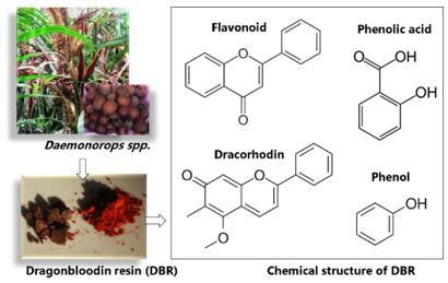
Figure 1. Chemical component of DBR
Daemonorops spp., also known locally as Jernang, is a plant species found in various regions, including Aceh, Jambi, and Kalimantan in Indonesia. The resin has shown pharmacological activities, including antimicrobial, antiviral, anti-inflammatory, and antitumor properties [7-9]. Additionally, the resin extract has been found to contain secondary metabolites such as flavonoids and phenolic compounds [4, 7, 10]. The shape of the tree, fruit, resin, and the chemical structure of the DBR component can be seen in Figure 1 [11].
Phenolic compounds are naturally occurring substances found in plants that have antioxidant and pro-oxidative properties [12]. Phenolic compounds are classified into various groups based on their structural elements and the number of aromatic rings [13, 14]. These groups include simple phenols, phenolic acids, flavonoids, xanthones, stilbenes, lignans, and more. They are characterized by the presence of a hydroxyl (-OH) group attached to an aromatic ring [15]. Flavonoids are a class of naturally occurring compounds that are widely distributed in plants. They are characterized by a flavone structure, which consists of two aromatic rings linked by a three-carbon chain containing an oxygen atom [16]. Phenolic acids are a class of compounds that contain a phenolic ring with one or more carboxylic acid groups (-COOH) attached to it [17]. Meanwhile, Dracorhodin is a red pigment, it is a type of anthocyanin, which is a subclass of flavonoids [18].
These groups have the potential to increase membrane hydrophilicity through reactions with hydrophobic polymers [19]. In chemical modification, the polymer membrane is altered through the formation of new chemical bonds. Membrane modification is primarily related to the introduction of functional groups into the membrane, such as carboxylates (-COOH), amines (-N), hydroxyls (-OH), thiols (-SH), epoxides, and aldehydes [20, 21]. This modification can be achieved by adding additives to the membrane matrix. The hydrophilicity of the polymer membrane will increase, leading to a decrease in surface roughness and an increase in charge properties [22, 23]. When DBR is combined with a membrane dope solution as an additive, some of its components can remain in the polymer matrix and induce hydroxyl and polar compounds to increase hydrophilicity. As a result, it can increase permeability and anti-fouling properties. Hydrophilic surfaces readily induce the formation of a hydration layer at the membrane-water interface. The hydration layer repels foulants, which are usually hydrophobic on the membrane surface [24, 25].
As described above, DBR may have potential as a membrane additive. Therefore, in order to validate this hypothesis, this study analyzed the important characteristics of DBR, including functional groups, morphology, elemental composition, particle charge, and antibacterial activity. Each characteristic is always related to the potential of DBR as a membrane modifier agent. The results can be used as a basic knowledge for increasing membrane hydrophilicity by bio additive.
2.1 Material
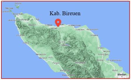
The material used in this research was dragon blood resin from the Daemonorops draco Blume species, obtained from farmers in the Kab. Bireuen, Aceh Province, Indonesia (Figure 2). Based on literature, DBR from Aceh has the highest dracorhodin content [9]. The DBR used is DBR Grade A, which is obtained by dry extraction. The resin was extracted by farmers and was available in powder form. We purchased it and did not purify this material. Prior to characterization, we reduced the particle size with a mortar and pestle, then sieved with 100 mesh. N-methyl-2-pyrrolidone (NMP) and ciprofloxacin as a control solution were also used to antibacterial analysis.
Figure 2. Location where the sample was taken (https://maps.app.goo.gl/9h7DXTui5kJmwfga9)
2.2 Experimental
DBR characterization includes analysis of functional groups, morphology, elemental composition, particle charge, and anti-bacterial activity. Analysis of DBR functional groups using the Fourier Transform Infrared (FTIR, PerkinELmer Inc) instrument. The IR spectrum is measured in the wave number range 650 - 4000 cm-1.
Analysis of the DBR particle charge using the Zeta Potential instrument (Malvern Zetasizer Nano ZSP). A total of 0.05 grams of DBR was dissolved in 25 mL of distilled water to make a colloidal solution. Then a small amount of the solution is entered into the zeta cell. Zeta potential readings were carried out 9 times and the average value was taken. Zeta measurements were carried out at neutral pH.
SEM-EDS device (Phenom ProX, Thermo scientific, Japan) was used to visualize the surface morphology of each membrane and analyze elementals. The DBR antibacterial activity test used two methods, which are total plate count (TPC) and disc diffusion Kirby Bauer method. The TPC method is carried out by observing the number of E. coli bacteria that grow on agar media that has DBR solution spread over its surface. Meanwhile, the Kirby Bauer disc diffusion method is carried out by observing the inhibition zone that forms between discs that have been soaked in DBR solution.
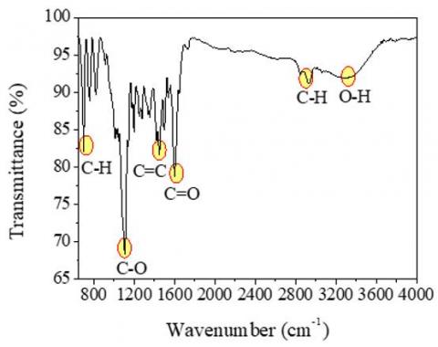
3.1 FTIR
Figure 3 shows FTIR spectra obtained for the identification of DBR chemical bonds. The C-H stretching of the alkene bond can be identified at peaks 697, 755, 825, and 2944 cm-1. Subsequently, peaks in the range of 1050-1310 cm-1 are associated with the C-O stretching in the carboxyl group, specifically esters. Peaks at wave numbers 1452 and 1498 cm-1 correspond to the double bond C=C (aromatic ring), while those at 1603 and 1740 cm-1 indicate C=O groups. Meanwhile, the peak at 3292 cm-1 signifies the O-H group of carboxylic acid.
Figure 3. FTIR spectra of dragon blood resin
The detected chemical bonds in the spectrum suggest the presence of three main elements in the DBR molecular structure: C, H, and O. These findings are consistent with previous reports, where DBR contains several components such as dracorhodin, biflavonoids, polysaccharides, flavans, dicoflavans, abietic acid, and dammaradienol [26]. All of these components have the chemical formula CxHyOx. Similar results have also been reported by studies [27, 28], using DBR obtained from different species. Following these outcomes, DBR as a new bio-additive in membrane matrices can provide carboxyl and phenol groups, both of which are hydrophilic, thereby potentially enhancing the wetting properties of DBR-containing membranes.
3.2 SEM-EDX
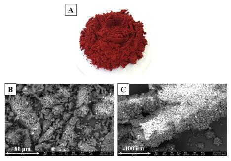
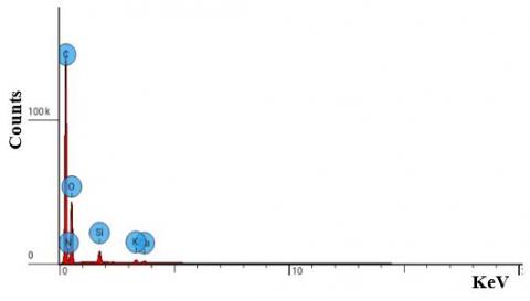
In this study, red resin (DBR) from the genus Daemonorops was utilized (Figure 4a). Although internationally known as 'Dragon Blood Resin', this substance does not originate from actual dragon's blood but is instead extracted from the fruit of rattan through a simple extraction process. SEM analysis results revealed non-uniform particle sizes and shapes (amorphous) (Figure 4b). The observed microscopic structure resembled that of coral, featuring small cavities (Figure 4c). Elemental analysis results (Figure 5) demonstrated several DBR components, including carbon (C = 53.8%), oxygen (O = 34.1%), nitrogen (N = 8.2%), silica (Si = 1.7%), potassium (K = 1.4%), and calcium (Ca = 0.8%). Thus, it can be deduced that the highest composition of DBR originates from carbon and oxygen components, corroborating the FTIR spectrum readings. DBR is composed of hydrophobic segments due to its carbon content, causing it to be sparingly soluble in water. Nevertheless, DBR also possesses hydrophilic segments due to the presence of oxygen, nitrogen, and silica, which are expected to positively contribute to enhanced hydrophilicity of the membrane.
Figure 4. (a) Particles, (b-c) Surface morphology of DBR
Figure 5. EDS spectrum of DBR
3.3 Zeta potential
Zeta potential is an analysis employed for determining the surface charge of particles in colloidal solutions. Particles with a positive zeta potential will interact with negatively charged surfaces, and vice versa. Zeta potential values typically range from +100 to -100 mV. The magnitude of the zeta potential provides insights into particle stability, with higher values indicating an increased potential for electrostatic repulsion. Particles with zeta potential values ranging from > +25 mV to < -25 mV usually exhibit high levels of stability [29, 30]. The zeta potential analysis results for DBR are presented in Table 1, indicating an average value of -44.42 mV. The negative value suggests that DBR possesses antifouling properties, as organic pollutants in water commonly carry a negative charge.
Table 1. Zeta potential result of DBR
|
Run Time |
T (℃) |
ZP (mV) |
Mob (µmcm/Vs) |
Cond (mS/cm) |
|
1 |
25,0 |
-42,6 |
-3,343 |
0,0139 |
|
2 |
25,0 |
-41,8 |
-3,273 |
0,0249 |
|
3 |
25,0 |
-38,3 |
-3,003 |
0,0140 |
|
4 |
25,0 |
-47,3 |
-3,711 |
0,0174 |
|
5 |
25,0 |
-49,1 |
-3,852 |
0,0142 |
|
6 |
25,0 |
-43,5 |
-3,409 |
0,0241 |
|
7 |
25,0 |
-47,0 |
-3,684 |
0,0153 |
|
8 |
25,0 |
-42,5 |
-3,328 |
0,0173 |
|
9 |
25,0 |
-47,7 |
-3,741 |
0,0152 |
|
Average ZP |
-44.42 mV |
|||
3.4 Antibacterial analysis
3.4.1 Total Plate Count (TPC)
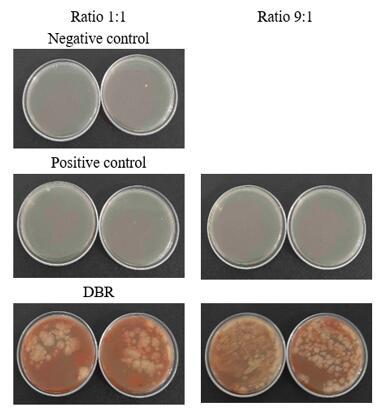
The TPC method involved mixing a solution of DBR with a solution of E. coli, and the mixture was then cultured on agar media. For this test, a 99.5% NMP solution was used as a negative control, and a ciprofloxacin solution (at a certain concentration) was used as a positive control. Treatment using a positive control aims to compare the pattern of inhibition and the ability of the antibacterial activity of the sample to inhibit the test bacteria. The choice of using NMP as a negative control was since DBR is low solubility in aqueous media and requires an organic solvent for dissolution. The DBR sample was prepared by dissolving 1 gram of DBR powder in 50 mL of NMP. Different ratios between DBR and E. coli were applied to observe the concentration-dependent effect of DBR on inhibiting E. coli growth. The tested DBR solutions included ratios of 1:1 and 9:1 (sample: E. coli), as well as ciprofloxacin solutions. The incubation temperature was set at 37℃ for 24 hours. The results of the antibacterial activity test using the TPC method are quantitatively summarized in Table 2 and qualitatively in Figure 6.
Figure 6. Illustrates the growth visualization of E. coli bacteria in each sample
Based on the results presented in Table 2 and Figure 6, it is evident that the 99.5% NMP solution exhibits a remarkably strong antibacterial activity, even rivaling ciprofloxacin, which serves as the positive control. Related studies on the use of NMP as an antimicrobial gel have shown that NMP possesses a good antibacterial activity in inhibiting the growth of S. aureus, E. coli, and C. albicans. The antibacterial property of NMP improves with increasing concentration, except when it is below 20%. This is attributed to NMP's ability to dissolve lipids in cell membranes and induce membrane damage in microorganisms [31, 32]. An interesting finding arises from the antibacterial activity test of DBR, as indicated in Table 2, where no E. coli bacteria grew under both ratios. However, qualitatively, Figure 6 shows bacterial growth on the agar medium. To confirm the type of bacteria that grew, microscopic observations were conducted using light microscopy (Figure 7).
Table 2. The data of the total bacterial plate count test results in the DBR samples
|
No. |
Sample |
E. coli (CFU/mL) |
|||
|
Ratio 1:1, V= 0.5 |
Ratio 9:1, V= 0.5 |
||||
|
Run 1 |
Run 2 |
Run 1 |
Run 2 |
||
|
1 |
Control - |
2 |
1 |
- |
|
|
2 |
Control + |
2 |
1 |
0 |
0 |
|
3 |
DBR |
0 |
0 |
0 |
0 |
Figure 7. Morphology of bacteria that grew in the DBR sample
Based on the analysis of morphological features, it has been determined that the type of bacteria that grew belongs to the Bacillus group. Bacillus is a genus of rod-shaped bacteria that are Gram-positive and commonly found in soil and water. Several types of Bacillus bacteria are undesirable, such as B. cereus, which can cause food spoilage in canned products and lead to rapid food poisoning. However, there are also beneficial Bacillus species like B. subtilis, which is used in antibiotic production. It is suspected that the presence of Bacillus on the agar medium, where the DBR and E. coli solutions were spread, is due to common laboratory culture contaminants (which also affected Louis Pasteur's experiments). These bacteria can form spores that remain inactive under adverse environmental conditions. These endospores can survive for extended periods, being resistant to heat, chemicals, and sunlight, thus having a high likelihood of prolonged survival [33-35]. Based on these findings, it is still uncertain whether the appearance of Bacillus is a result of specific compounds present in the DBR.
3.4.2 Disk diffusion
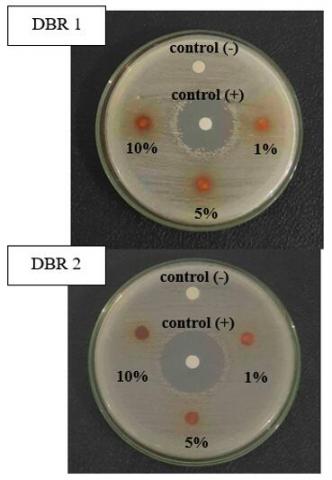
In addition to the TPC testing, the disc diffusion method was also employed to support the argumentation. The method used was the Kirby Bauer disc diffusion method using E. coli ATCC 25922 with a McFarland standard turbidity of 0.5. The analysis was conducted on two samples with different storage time. For DBR 1, NMP was used as the solvent, while for DBR 2, an aqua-bides was employed. The incubation temperature was set at 37℃ for 24 hours. The quantitative testing results are summarized in Table 3 for DBR 1 and Table 4 for DBR 2. The visualization of inhibition zones for both samples can be seen in Figure 8.
Table 3. Inhibition zones in DBR first sample
|
No. |
Sample |
Concentration (%) |
Inhibition Zones (mm) |
||
|
Run 1 |
Run 2 |
Run 3 |
|||
|
1 |
Control - (NMP) |
- |
6.48 |
6.55 |
6.69 |
|
2 |
Control + |
0.1 |
24.70 |
24.41 |
24.30 |
|
3 |
DBR |
1 |
0 |
0 |
0 |
|
5 |
0 |
0 |
0 |
||
|
10 |
0 |
0 |
0 |
||
Table 4. Inhibition zones in DBR second sample
|
No. |
Sample |
Concentration (%) |
Inhibition Zones (mm) |
||
|
Run 1 |
Run 2 |
Run 3 |
|||
|
1 |
Control -(Aqua-bides) |
- |
0 |
0 |
0 |
|
2 |
Control + |
0.1 |
28.60 |
28.51 |
28.65 |
|
3 |
DBR |
10 |
0 |
0 |
0 |
|
25 |
0 |
0 |
0 |
||
|
50 |
0 |
0 |
0 |
||
Figure 8. Inhibition zones of DBR
In the antibacterial activity testing, the formation of clear inhibition zones around the discs immersed in the samples was observed. The results confirmed that NMP possesses antibacterial properties, while the DBR additive does not. No inhibition zones were formed in both DBR samples. Initially, during the testing with DBR 1, the assumption was that the lack of antibacterial activity might be due to the extended storage time, which could indicate expired material. However, this argument was refuted by the second testing using fresh DBR samples. In the second testing, an aqueous solution was used as the solvent to avoid the antibacterial influence of NMP. Nonetheless, the results obtained showed no difference; inhibition zones did not form even when the DBR concentration was increased to 50% (w/w).
DBR consists of various chemical compounds, one of which is phenol and dracorhodin. This compound is responsible for enhancing the antibacterial properties of DBR. The most reasonable possibility for the lack of antibacterial activity in DBR is that the two active compounds are inhibited due to the non-purification. Although "Grade A" DBR is used to obtain antibacterial activity, DBR still requires post-treatment to remove impurities in it. Khairan et al. [36] reported that DBR could inhibit bacterial growth after maceration in n-hexane solvent for 5 days. Therefore, these results indicate that DBR powder does not have confirmed antibacterial activity. Consequently, DBR has no potential as a membrane modifier to enhance anti-biofouling properties. However, the possibility of such potency still exists if DBR is subjected to post-treatment such as maceration to remove impurities.
In this research, DBR has been successfully characterized and reviewed for its potential as a membrane additive. The results show that this additive contains carboxyl and phenol groups, both of which are hydrophilic, and has an amorphous morphology. Supported by zeta potential analysis which has a negative value, namely -44.52 mV, where DBR has the potential to provide electrostatic repulsion with foulants which generally have a negative charge. These two characteristics show the prospect of DBR as a membrane additive with the aim of increasing hydrophilicity as well as anti-fouling properties. The results of the study suggest that DBR is a promising membrane additive for antifouling purposes. However, this potential is limited to the control of organic foulants, such as humic acid, and inorganic foulants, such as heavy metals. Due to the lack of antibacterial activity in DBR resin powder, its addition proves insufficient to significantly inhibit bacterial growth in microorganism-containing foulants. Consequently, DBR requires post-treatment for effective antibiofouling applications. Further research is required to better understand the effect of DBR addition on hydrophobic membrane modification.
This research was funded by The Indonesian Ministry of Education, Culture, Research, and Technology as part of Doctoral Dissertation Research (PDD) 2023. The study was carried out at National Research Center on Membrane Technology (MEM-TEK), Istanbul Technical University, Turkey; and Research Center for Advanced Membrane and Film Technology, KOBE University, Japan.
[1] Gupta, D., Bleakley, B., Gupta, R.K. (2008). Dragon's blood: botany, chemistry and therapeutic uses. Journal of ethnopharmacology, 115(3): 361-380. https://doi.org/10.1016/j.jep.2007.10.018
[2] Baumer, U., Dietemann, P. (2010). Identification and differentiation of dragon's blood in works of art using gas chromatography/mass spectrometry. Analytical and bioanalytical chemistry, 397: 1363-1376. https://doi.org/10.1007/s00216-010-3620-0
[3] Saxena, G., Mittal, A., Siddiqui, A.W. (2019). Evaluation of acute and subchronic toxicity of dragon blood resin extract. Journal of Drug Delivery and Therapeutics, 9(2): 362-366. https://doi.org/10.22270/jddt.v9i2.2435
[4] Fan, J.Y., Yi, T., Sze-To, C.M., et al. (2014). A systematic review of the botanical, phytochemical and pharmacological profile of Dracaena cochinchinensis, a plant source of the ethnomedicine “dragon’s blood”. Molecules, 19(7): 10650-10669. https://doi.org/10.3390/molecules190710650
[5] Liu, Y., Zhao, X., Yao, R., Li, C., Zhang, Z., Xu, Y., Wei, J.H. (2021). Dragon’s blood from dracaena worldwide: Species, traditional uses, phytochemistry and pharmacology. The American Journal of Chinese Medicine, 49(6): 1315-1367. https://doi.org/10.1142/S0192415X21500634
[6] Al-Awthan, Y.S., Bahattab, O.S. (2021). Phytochemistry and pharmacological activities of Dracaena cinnabari resin. BioMed Research International, 2021: 1-7. https://doi.org/10.1155/2021/8561696
[7] Jura-Morawiec, J., Tulik, M. (2016). Dragon’s blood secretion and its ecological significance. Chemoecology, 26: 101-105. https://doi.org/10.1007/s00049-016-0212-2
[8] Zheng, Q., Xu, M., Yang, C., Wang, D., Li, H., Zhu, H., Zhang, Y. (2012). A new red pigment from Chinese Dragon's blood, the red resin of Dracaena cochinchinensis. Bulletin of the Korean Chemical Society, 33(12): 4204-4206. https://doi.org/10.5012/BKCS.2012.33.12.4204
[9] Waluyo, T.K., Wibowo, S. (2018). Dracorhodin: A potential marker compound for detecting the presence of dragon’s blood resin from Daemonorops originated from Indonesia. Biodiversitas Journal of Biological Diversity, 19(5): 1665-1671. https://doi.org/10.13057/biodiv/d190510
[10] Escobar, J.D., Prieto, C., Pardo-Figuerez, M., Lagaron, J.M. (2018). Dragon’s blood sap: storage stability and antioxidant activity. Molecules, 23(10): 2641. https://doi.org/10.3390/molecules23102641
[11] Gupta, D., Bleakley, B., Gupta, R.K. (2008). Dragon's blood: botany, chemistry and therapeutic uses. Journal of ethnopharmacology, 115(3): 361-380. https://doi.org/10.1016/j.jep.2007.10.018
[12] Tatipamula, V.B., Kukavica, B. (2021). Phenolic compounds as antidiabetic, anti‐inflammatory, and anticancer agents and improvement of their bioavailability by liposomes. Cell biochemistry and function, 39(8): 926-944. https://doi.org/10.1002/cbf.3667
[13] Al Mamari, H.H. (2021). Phenolic compounds: Classification, chemistry, and updated techniques of analysis and synthesis. Phenolic Compounds: Chemistry, Synthesis, Diversity, Non-Conventional Industrial, Pharmaceutical and Therapeutic Applications, 73-94. https://doi.org/10.5772/intechopen.98958
[14] Zeb, A., Zeb. (2021). Phenolic Antioxidants in Foods: Chemistry, Biochemistry and Analysis, Berlin/Heidelberg, Germany: Springer, pp. 385-411. https://doi.org/10.1007/978-3-030-74768-8_2
[15] Vuolo, M.M., Lima, V.S., MARÓSTICA, M. (2019). Chapter 2-Phenolic Compounds: Structure, Classification, and Antioxidant Power, Editor (s): Maira Rubi Segura Campos. Bioactive Compounds, Woodhead Publishing, pp. 33-50. https://doi.org/10.1016/B978-0-12-814774-0.00002-5
[16] Jayashree, B.S., Venkatachalam, H., Mallik, S.B. (2019). Flavones and their analogues as bioactive compounds–an overview. Mini-Reviews in Organic Chemistry, 16(4): 377-391. https://doi.org/10.2174/1570193X15666180418154510
[17] Cordeiro, M.L.D.S., Martins, V.G.D.Q.A., Silva, A.P. D., Rocha, H.A.O., Rachetti, V.D.P.S., Scortecci, K.C. (2022). Phenolic acids as antidepressant agents. Nutrients, 14(20): 4309. https://doi.org/10.3390/nu14204309
[18] Lu, Z., Lu, C., Li, C., Jiao, Y., Li, Y., Zhang, G. (2019). Dracorhodin perchlorate induces apoptosis and G2/M cell cycle arrest in human esophageal squamous cell carcinoma through inhibition of the JAK2/STAT3 and AKT/FOXO3a pathways. Molecular medicine reports, 20(3): 2091-2100. https://doi.org/10.3892/mmr.2019.10474
[19] Upadhyaya, L., Qian, X., Wickramasinghe, S.R. (2018). Chemical modification of membrane surface—overview. Current opinion in chemical engineering, 20: 13-18. https://doi.org/10.1016/j.coche.2018.01.002
[20] Alenazi, N.A., Hussein, M.A., Alamry, K.A., Asiri, A.M. (2017). Modified polyether-sulfone membrane: A mini review. Designed monomers and polymers, 20(1): 532-546. https://doi.org/10.1080/15685551.2017.1398208
[21] Otitoju, T.A., Ahmad, A.L., Ooi, B.S. (2018). Recent advances in hydrophilic modification and performance of polyethersulfone (PES) membrane via additive blending. RSC advances, 8(40): 22710-22728. https://doi.org/10.1039/C8RA03296C
[22] Ambarita, A.C., Arahman, N., Bilad, M.R., et al. (2023). Polyethersulfone membrane coated with Dragon blood resin: Effect of input parameters and optimization using response surface methodology. South African Journal of Chemical Engineering, 45(1): 30-41. https://doi.org/10.1016/j.sajce.2023.04.005
[23] Chen, J., Yang, J., Ma, L., Li, J., Shahzad, N., Kim, C. K. (2020). Structure-antioxidant activity relationship of methoxy, phenolic hydroxyl, and carboxylic acid groups of phenolic acids. Scientific reports, 10(1): 2611. https://doi.org/10.1038/s41598-020-59451-z
[24] Ambarita, A.C., Mulyati, S., Arahman, N., Bilad, M.R. (2023). Characterization and performances of hydrophobic polymer membrane modified with dragon’s blood resin. In AIP Conference Proceedings, 2613(1). https://doi.org/10.1063/5.0120373
[25] Hosseini, S.M., Banijamali, M.S., Farahani, S.K., Bandehali, S. (2022). Enhancing antifouling and separation characteristics of carbon nanofiber embedded poly ether sulfone nanofiltration membrane. Korean Journal of Chemical Engineering, 39(9): 2491-2498. https://doi.org/10.1007/s11814-022-1088-1
[26] Edward, H.G., de Oliveira, L.F.C., Quye, A. (2001). Raman spectroscopy of coloured resins used in antiquity: Dragon's blood and related substances. Spectrochimica Acta Part A: Molecular and Biomolecular Spectroscopy, 57(14): 2831-2842. https://doi.org/10.1016/S1386-1425(01)00602-3
[27] Oliveira, R.N., Mancini, M.C., Oliveira, F.C.S.D., Passos, T.M., Quilty, B., Thiré, R.M.D.S.M., McGuinness, G.B. (2016). FTIR analysis and quantification of phenols and flavonoids of five commercially available plants extracts used in wound healing. Matéria (Rio de Janeiro), 21: 767-779. https://doi.org/10.1590/S1517-707620160003.0072
[28] Maqtari, M.A. (2015). Lupane-type triterpenoids derivatives from resin of the Socotra dragon tree Dracaena cinnabari Balf. Jordan Journal of Chemistry, 10(2): 108-118. https://doi.org/10.12816/0026451
[29] Barhoum, A., García-Betancourt, M.L., Rahier, H., Van Assche, G. (2018). Physicochemical characterization of nanomaterials: Polymorph, composition, wettability, and thermal stability. In: Emerging Applications of Nanoparticles and Architecture Nanostructures, Elsevier, pp. 255-278. https://doi.org/10.1016/B978-0-323-51254-1.00009-9
[30] Bajpai, P. (2018). Chapter 4 - Additives for Papermaking. Biermann’s Handbook of Pulp and Paper, Elsevier, pp. 77-94. https://doi.org/10.1016/B978-0-12-814238-7.00004-0
[31] Phaechamud, T., Mahadlek, J., Charoenteeraboon, J., Choopun, S. (2012). Characterization and antimicrobial activity of N-methyl-2-pyrrolidone-loaded ethylene oxide-propylene oxide block copolymer thermosensitive gel. Indian Journal of Pharmaceutical Sciences, 74(6): 498. https://doi.org/10.4103/0250-474X.110574
[32] Kim, M.A., Neelakantan, P., Min, K.S. (2022). Effect of N-2-methyl-pyrrolidone on Enterococcus faecalis biofilms. Dental Materials Journal, 41(5): 774-779. https://doi.org/10.4012/dmj.2022-012
[33] Su, Y., Liu, C., Fang, H., Zhang, D. (2020). Bacillus subtilis: a universal cell factory for industry, agriculture, biomaterials and medicine. Microbial cell factories, 19(1): 1-12. https://doi.org/10.1186/s12934-020-01436-8
[34] Tu, Z., Setlow, P., Brul, S., Kramer, G. (2021). Molecular physiological characterization of a high heat resistant spore forming Bacillus subtilis food isolate. Microorganisms, 9(3): 667. https://doi.org/10.3390/microorganisms9030667
[35] Owusu-Darko, R., Allam, M., Ismail, A., Ferreira, C.A., Oliveira, S.D.D., Buys, E.M. (2020). Comparative genome analysis of Bacillus sporothermodurans with its closest phylogenetic neighbor, Bacillus oleronius, and Bacillus cereus and Bacillus subtilis groups. Microorganisms, 8(8): 1185. https://doi.org/10.3390/microorganisms8081185
[36] Khairan, K., Arini, M., Idroes, R., Awang, K., Jacob, C. (2023). Antibacterial activity of n-hexane dragon’s blood resin extract (Daemonorops draco wild Blume) from Bener Meriah, Aceh province, Indonesia. Malacca Pharmaceutics, 1(1): 22-29. https://doi.org/10.60084/mp.v1i1.29