Aseel Majid Habeeb*![]() | Nihad Abdul-Ameer Salih
| Nihad Abdul-Ameer Salih![]()
© 2024 The authors. This article is published by IIETA and is licensed under the CC BY 4.0 license (http://creativecommons.org/licenses/by/4.0/).
OPEN ACCESS
Hydroxyapatite, a bio-ceramic material widely utilized in bioengineering, holds significant promise for bone implant applications due to its biocompatibility and non-toxic nature. This study focuses on the organic synthesis of hydroxyapatite with tailored nanoscale properties suitable for integration into functionally graded materials like NiTi/HA, intended for bone implants. Porous NiTi possesses desirable mechanical properties for such applications; however, its limited bioactivity poses a challenge for therapeutic use. Composite structures comprising porous NiTi and hydroxyapatite (HA) offer a viable solution to promote bone ingrowth and implant integration with surrounding tissue. Eggshells serve as the raw material in this research, subjected to calcination at 1000℃ for three hours to yield calcium oxide. Subsequent crushing and mixing with phosphoric acid, followed by milling using a planetary ball mill for twenty-two hours at 45 rpm, produces a homogeneous hydroxyapatite powder (HA). Comprehensive characterization using particle analysis, FTIR, SEM, and XRD confirms the desired properties of the synthesized powder. FTIR analysis verifies the presence of fundamental HA components, while XRD reveals a structure akin to traditional hydroxyapatite powder, featuring characteristic peaks corresponding to (PO43−), (CO32−), and bending OH−. Particle size analysis indicates a range of (0.301) µm to (4.759) µm, with a mean size of (1.088) µm. The findings of this study highlight that hydroxyapatite powder derived from eggshells exhibits favorable particle size, bioactivity, and porous nature, rendering it well-suited for incorporation into bone implant materials.
nanoscale hydroxyapatite, eggshells, calcination, biomedical, calcium, natural, phosphoric acid, bio-waste
Bone grafts are the second most common tissue transplant procedure performed globally, after blood transfusions [1]. Alveolar bone abnormalities can arise for a variety of reasons, but the most frequent ones include osseous deficiency, tumor excision, periodontal disease-induced alveolar bone loss, and subsequent tooth loss [2]. The main reason rehabilitation is required for bone defects are to stop excessive alveolar bone resorption, which jeopardizes the quantity, quality, and shape of the bone. This keeps the insertion of dental implants from failing, preserves the typical anatomic outline, eliminates empty space, offers an aesthetically pleasing restoration, and makes it easier to encapsulate and administer drugs [3, 4]. Mammalian bone grafts, or xenografts, have been utilized for over thirty years and are still in use today. They have many benefits, including excellent biocompatibility, osteoconductive properties, great availability (both in size and quantity), and cheap cost [5]. Up until now, the only materials that could meet the necessary requirements for improved bone substitution ability—such as biocompatibility, osteoconductivity, and osteoinductivity—were autografts and, to a lesser extent, allografts [6-10]. As a result, the natural supplies of apatite for biological synthesis have grown yearly, starting with mammalian bone sources and continuing with fish bones and scales [6-8], egg-shells [9, 10], and marine organism exoskeletons (snails, starfish, coral, and seashells [11, 12]. Alloplastic bone substitutes come in a variety of compositions, sizes, shapes, textures, synthesis techniques, biocompatibility, bioresorbability, high availability, and cost-effectiveness. [13, 14]. Even with these advantages, improvements are still needed to improve their bioactivity, bio tolerance, or absorption into the physiological milieu [15, 16]. Due to the carbonated apatite that is produced having a composition and structure identical to human bone, hydroxyapatite synthesis from hen egg shell sources is now one of the most promising methods [17, 18].
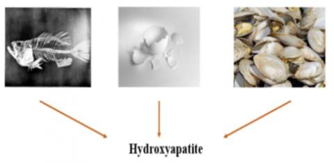
The inorganic mineral hydroxyapatite, often known as HAP [Ca10 (PO4)6 (OH)2], makes up about 70% of bone. Natural bone contains HAp crystals that are in the Nano range in size. Calcium and phosphorus make up most of the basic components of hydroxyapatite, with a calcium-to-phosphate proportion of 1.667. In this process of chemical synthesis, hydrogen ions are removed at extreme temperatures. However, a variety of methods were utilized to examine how to create these nanocrystals. More research is still needed to fully understand how to regulate the crystallinity, shape, and size of these substances [19, 20]. Because of its excellent biocompatibility with soft tissues such as gums, muscles, and skin, hydroxyapatite (HAP) is a great choice for orthopaedic and dental implantation and the components of implantation. Widespread applications for synthetic HAp in the repair of hard tissues include bone healing, bone augmentation, covering implantation, and functioning as fillers in teeth or bone. Normal HA ceramics' poor mechanical strength, on the other hand, often limits their employment to light load-bearing applications. To precisely describe the small-scale features of HAp, recent developments in nanotechnology and nanoscience have rekindled studies of Nano-scale HAp formation [21, 22]. (HA) could be produced from biogenic materials as shown in Figure 1, like fish bones [23], eggshells [24, 25], seashells [10], and coral by the use of several chemical synthesis processes. These are based on reactions that take place in a solid state [26].
Figure 1. HAP natural sources [27]
Academics are very interested in using eggshells to create products with a large added value, like Nano-crystalline hydroxyapatite (HA). Eggshells are made of CaCO3 and other trace elements, like Si, Mg, and others, that are necessary for physiology. HA must have the proper nanoscale properties to be useful in a wide range of biological situations. These characteristics include, among others, their mesoporous nature, surface area, shape, and crystallinity. The rapid production of precursor materials with suitable nanoscale properties for tissue engineering scaffolds, drug/protein delivery carriers, creating bone fillers, and other similar applications appears to be made possible by the formulation of eggshell-derived HA using a variety of organic modifiers in a microwave reactor designed specifically for the task [28]. Natural hydroxyapatite exhibits a notable absence of impurities, possesses a superior level of crystalline structure, and is characterized by its eco-friendly nature [29, 30]. A prior investigation indicated that synthetic hydroxyapatite exhibits significantly lower biodegradability compared to natural hydroxyapatite and calcium phosphate [31]. As of 2017, the majority of the publications listed in the Scopus database described eggshell as a source of hydroxyapatite. Calcium oxide, or CaO, was produced in a number of investigations using high temperature fire calcination [32-36]. To create pure calcium phosphate, the CaO was subsequently treated with diammonium hydrogen phosphate [32, 34, 36]. Other studies employed phosphoric acid sonication [33]. In addition to calcination, calcium chloride solution was also made using the hydrothermal method [37, 38] as a calcium precursor. It was demonstrated that the extraction time could be reduced by using the hybrid method of calcination and microwave-assisted hydrothermal processing [5]. Lala and associates [32] used disodium hydrogen phosphate and calcination to remove hydroxyapatite from eggshells. Their research revealed promising osteogenic potential. Additionally, crystalline hydroxyapatite increased cell viability, indicating that it was more biocompatible than the control sample. Arslan et al. [34] carefully examined the impact of hydroxyapatite derived from an eggshell scaffold in conjunction with human hair keratin and jellyfish collagen. Eggshells were calcined at 1000 °C for three hours in a box furnace to produce calcium oxide. The next step was to add diammonium hydrogen phosphate to create hydroxyapatite. Considering the unique and remarkable method of using bioceramic or biopolymers in regenerative medicine, the researchers proposed that osteoconductive scaffold using human hair keratin, jellyfish collagen, and eggshell-derived Nano hydroxyapatite was a new and cost-effective approach for scaffold fabrication Sultana and colleagues [35] used a novel UV-mediated solid state method to synthesize hydroxyapatite from eggshell without subjecting it to heat treatment. They proposed that the UV-irradiation method, followed by ball milling, can be used to create hydroxyapatite at room temperature. They also noticed that the cell viability test revealed no appreciable cytotoxicity. Cell bioactivity in a simulated body fluid soaking test was within permissible bounds.
Similar to muscles, the composition of bone can be arranged in a pyramid, with hydroxyapatite (HA) crystals at the base of the pyramid. The type and location of bone in the body determine its different composition. Such an arrangement of composition can be lamellar or layer-by-layer [39]. In the present work, we synthesized HA Nano and Micro powders, starting from chicken eggshells (biogenic calcite), Then confirm its formula by chemical and spectroscopic analysis and compare it with scientific references, and then use it in another research to benefit from it medically in preparing a functionally graded material NiTi/HA that can be used in bone implants.
Egg shells and phosphoric acid were used as raw materials. The following steps were taken to create HA powder from egg shells:
The experimental process for preparing HA from egg shells using the calcination method is depicted in Figure 2 below.
The equipment and device used to prepare HA powder from egg shells using the calcination method are shown in Figure 3.
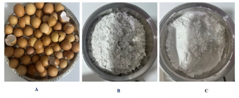
Figure 2. Procedure for making HA powder from eggshells: (A) Egg shells cleaned; (B) Broken and calcinated egg shells; (C) Crushed and milled egg shells
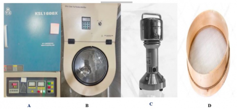
Figure 3. The tool used to make HAP powder from eggshells: (A) furnace, (B) planetary ball, (C) home steel mill, and (D)sieve
3.1 X-ray diffraction
XRD is employed to describe the quantitative and qualitative characteristics of solid compounds as well as to examine the crystallization of compounds and their phase purity .
The relationship that used is:
nλ=2d sin θ
where:
n: is a positive integer.
$\lambda$ : is the incident $\mathrm{X}$-ray beam's wavelength.
d: is the separation of atomic layers in a crystal.Θ:is the Bragg's angle.
Bragg's law is used to interpret X-ray diffraction data [41].
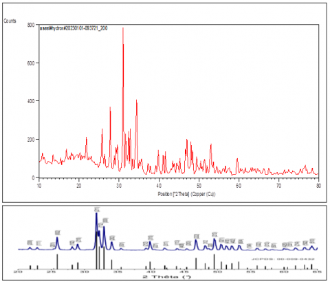
Identification of HAP depends greatly on the location, form, width, and strength of the HAP peak in the XRD spectrum. To match the resulting XRD spectrum to recognized standard pattern analysis, the examination of the HAP, which is made from powdered eggshells, using X-ray diffraction (XRD) is shown in Figure 4 for the 10o to 80o diffracted angle range. When the peaks are compared to those on (JCPDS) card No., it demonstrates high purity of the hydroxyapatite phase (09-0432). These outcomes demonstrated the effectiveness of raw eggshells in producing pure HAP powder.
Figure 4. XRD spectrum for the resulted HAP of eggshells and of pure hydroxyapatite (JCPDS no. 09-0432)
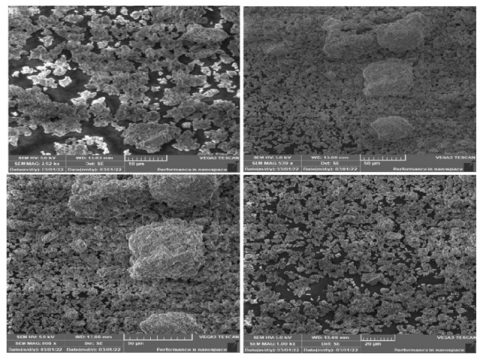
3.2 Scanning Electron Microscope (SEM)
The morphology of hydroxyapatite was studied using a scanning electron microscope (SEM). Figure 5 shows SEM images of Hydroxyapatite particles that formed in agglomerates. The images also show that the synthesised hydroxyapatite is porous in nature. This porous characteristic is desired and can be advantageous when utilised in implants since it makes it easier for the implant and the biological environment to interact.
Figure 5. SEM micrographs of HAP of eggshells
3.3 Analysis of FTIR spectroscopy
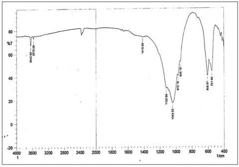
Infrared spectroscopy using Fourier transform can be used to identify the amide, phosphate, and carbonate sets that make up the final powder, in addition to confirming the production of HAP. According to Fourier-transform infrared spectroscopy standard analysis, transmission mode, the carbonate set is present at about (1410-1450) cm-1 and (875) cm-1, and the hydroxide group is present at approximately (3500-3200) cm-1. For the phosphate set, (10) (1049-1090) cm-1, 1950-2200 cm-1, (962) cm-1, and (560) cm-1 are used . As shown in Table 1, the energy beams at (3572.29) cm-1 and (3643.65) cm-1 depict OH-, (1415.80) cm-1 represents the amide set of CO3, and (1043.52) cm-1, (972.16) cm-1, (605.67) cm-1, and (551.66) cm-1 depict the beams for the PO4 set. Figure 6 depicts the constituents of the powder produced by the thermal calcining of egg. FT-IR analysis and comparison with reference spectra showed that they were the active sets for HAP powders.
Figure 6. HAP from eggshells FT-IR spectrum
Table 1. The significant FT-IR stretching of HAP frequencies from eggshell
|
Bands of Infrared Absorption (cm−1) |
Description |
|
1043.52 |
(PΟ43−) |
|
972.16 |
(PΟ43−) |
|
605.67 |
(PΟ43−) |
|
551.66 |
(PΟ43−) |
|
1415.80 |
(CΟ32−) |
|
3643.65 |
OH Bending |
|
3572.29 |
OH Bending |
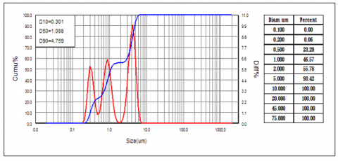
3.4 Analysis of particle size
The particle distribution of micro and Nano-HAP is shown in Figure 7. Due to the high agglomeration caused by the granules' large surface area, The screening medium consisted of distilled water without any dispersants. The particles are between (0.301) µm and (4.759) µm in size, with a size that is on average (1.088) µm.
Figure 7. Analysis of the particle size of HAP from eggshells
In this study, hydroxyapatite was prepared from egg shells and its properties were studied, X-Ray diffraction(XRD) technique was used to investigate the formation of HA powder, By analyzing the XRD pattern and comparing it with the standard hydroxyapatite (HA), it was determined that the raw eggshells were successful in producing pure HAP powder, The FT-IR analysis validates that the resultant powder derived from eggshells comprises identical compounds that are detectable in pure hydroxyapatite (HAP). Through SEM analysis, it was demonstrated that the produced material exhibited a discernible and extremely high porosity. This may be linked to enhanced adherence of biomolecules and possible elevation of osteoconductivity. The particle size was also established, with a range of (0.301 to 4.759) µm and (1.088) µm, Hydroxyapatite, with these micro and nano molecular sizes, has high efficiency in its biomedical applications, especially bone tissue engineering applications. Nanoscale properties enhance biological responses. From the test results, the following conclusions can be drown:
Recommendations for future work:
The authors would like to thank the Department of Ceramics, College of Materials Engineering, University of Babylon, for supporting this work.
[1] Mencía Castaño, I., Curtin, C.M., Duffy, G.P., O’Brien, F.J. (2016). Next generation bone tissue engineering: Non-viral miR-133a inhibition using collagen-nanohydroxyapatite scaffolds rapidly enhances osteogenesis. Scientific Reports, 6(1): 27941. https://doi.org/10.1038/srep27941
[2] Sohn, H.S., Oh, J.K. (2019). Review of bone graft and bone substitutes with an emphasis on fracture surgeries. Biomaterials Research, 23(1): 9. https://doi.org/10.1186/s40824-019-0157-y
[3] Mello, B.F., de Carvalho Formiga, M., de Souza da Silva, L.F., dos Santos Coura, G., Shibli, J.A. (2020). Horizontal ridge augmentation using a xenograft bone substitute for implant-supported fixed rehabilitation: A case report with four years of follow-up. Case Reports in Dentistry, 2020: 6723936. https://doi.org/10.1155/2020/6723936
[4] Ionescu, O.A.N.A., Ciocilteu, M., Manda, V., Neacsu, I., Ficai, A., Amzoiu, E., Stiolica, A.T., Croitoru, O., Neamtu, J. (2019). Bone-graft delivery systems of type PLGA-gentamicin and collagen-hydroxyapatite-gentamicine. Materiale Plastice, 56(3): 534-537. https://doi.org/10.37358/MP.19.3.5224
[5] Dumitrescu, C.R., Neacsu, I.A., Surdu, V.A., Nicoara, A.I., Iordache, F., Trusca, R., Ciocan, L.T., Ficai, A., Andronescu, E. (2021). Nano-hydroxyapatite vs. Xenografts: Synthesis, characterization, and in vitro behavior. Nanomaterials, 11(9): 2289. https://doi.org/10.3390/nano11092289
[6] Sadat-Shojai, M., Khorasani, M.T., Dinpanah-Khoshdargi, E., Jamshidi, A. (2013). Synthesis methods for nanosized hydroxyapatite with diverse structures. Acta Biomaterialia, 9(8): 7591-7621. https://doi.org/10.1016/j.actbio.2013.04.012
[7] Sunil, B.R., Jagannatham, M. (2016). Producing hydroxyapatite from fish bones by heat treatment. Materials Letters, 185: 411-414. https://doi.org/10.1016/j.matlet.2016.09.039
[8] Abdulkadhim, A.A., Abdulameer, N. (2021). Experimental and numerical study to prepare hydroxyapatite powder from fish bones. Transactions on Electrical and Electronic Materials, 22(4): 481-488. https://doi.org/10.1007/s42341-020-00254-4
[9] Goh, K.W., Wong, Y.H., Ramesh, S., Chandran, H., Krishnasamy, S., Sidhu, A., Teng, W.D. (2021). Effect of pH on the properties of eggshell-derived hydroxyapatite bioceramic synthesized by wet chemical method assisted by microwave irradiation. Ceramics International, 47(7): 8879-8887. https://doi.org/10.1016/j.ceramint.2020.12.009
[10] Sabry, A.A.A., Salih, N.A. (2020). Synthetic properties of hydroxyapatite powder prepared from natural eggs shell. Journal of Green Engineering, 10(7): 3498-3507.
[11] Cestari, F., Agostinacchio, F., Galotta, A., Chemello, G., Motta, A., M. Sglavo, V. (2021). Nano-hydroxyapatite derived from biogenic and bioinspired calcium carbonates: Synthesis and in vitro bioactivity. Nanomaterials, 11(2): 264. https://doi.org/10.3390/nano11020264
[12] Núñez, D., Elgueta, E., Varaprasad, K., Oyarzún, P. (2018). Hydroxyapatite nanocrystals synthesized from calcium rich bio-wastes. Materials Letters, 230: 64-68. https://doi.org/10.1016/j.matlet.2018.07.077
[13] Prasadh, S., Wong, R.C.W. (2018). Unraveling the mechanical strength of biomaterials used as a bone scaffold in oral and maxillofacial defects. Oral Science International, 15(2): 48-55. https://doi.org/10.1016/S1348-8643(18)30005-3
[14] Yamada, M., Egusa, H. (2018). Current bone substitutes for implant dentistry. Journal of Prosthodontic Research, 62(2): 152-161. https://doi.org/10.1016/j.jpor.2017.08.010
[15] Selders, G.S., Fetz, A.E., Radic, M.Z., Bowlin, G.L. (2017). An overview of the role of neutrophils in innate immunity, inflammation and host-biomaterial integration. Regenerative Biomaterials, 4(1): 55-68. https://doi.org/10.1093/rb/rbw041
[16] Dewi, A.H., Ana, I.D. (2018). The use of hydroxyapatite bone substitute grafting for alveolar ridge preservation, sinus augmentation, and periodontal bone defect: A systematic review. Heliyon, 4(10): e00884. https://doi.org/ 10.1016/j.heliyon.2018.e00884
[17] Erdem, U., Dogan, M., Metin, A.U., Baglar, S., Turkoz, M.B., Turk, M., Nezir, S. (2020). Hydroxyapatite-based nanoparticles as a coating material for the dentine surface: An antibacterial and toxicological effect. Ceramics International, 46(1): 270-280. https://doi.org/10.1016/j.ceramint.2019.08.260
[18] Agbeboh, N.I., Oladele, I.O., Daramola, O.O., Adediran, A.A., Olasukanmi, O.O., Tanimola, M.O. (2020). Environmentally sustainable processes for the synthesis of hydroxyapatite. Heliyon, 6(4): e03765. https://doi.org/10.1016/j.heliyon.2020.e03765
[19] Akagi, H., Ochi, H., Soeta, S., Kanno, N., Yoshihara, M., Okazaki, K., Yogo, T., Harada, Y., Amasaki, H., Hara, Y. (2015). A comparison of the process of remodeling of hydroxyapatite/poly-D/L-lactide and beta-tricalcium phosphate in a loading site. BioMed Research International, 2015: 730105. https://doi.org/10.1155/2015/730105
[20] Ganachari, S.V., Bevinakatti, A.A., Yaradoddi, J.S., Banapurmath, N.R., Hunashyal, A.M., Shettar, A.S. (2016). Rapid synthesis, characterization, and studies of hydroxyapatite nanoparticles. Adv Mater Sci Res., 1(1): 9-13.
[21] Zhou, H., Lee, J. (2011). Nanoscale hydroxyapatite particles for bone tissue engineering. Acta Biomaterialia, 7(7): 2769-2781. https://doi.org/10.1016/j.actbio.2011.03.019
[22] Rusu, L.C., Nedelcu, I.A., Albu, M.G., Sonmez, M., Voicu, G., Radulescu, M., Ficai, D., Ficai, A., Negrutiu, M., Sinescu, C. (2015). Tetracycline loaded collagen/hydroxyapatite composite materials for biomedical applications. Journal of Nanomaterials, 2015: 3. https://doi.org/10.1155/2015/361969
[23] Ripamonti, U., Crooks, J., Khoali, L., Roden, L. (2009). The induction of bone formation by coral-derived calcium carbonate/hydroxyapatite constructs. Biomaterials, 30(7): 1428-1439. https://doi.org/10.1016/j.biomaterials.2008.10.065
[24] Vecchio, K.S., Zhang, X., Massie, J.B., Wang, M., Kim, C.W. (2007). Conversion of bulk seashells to biocompatible hydroxyapatite for bone implants. Acta Biomaterialia, 3(6): 910-918. https://doi.org/10.1016/j.actbio.2007.06.003
[25] Balázsi, C., Wéber, F., Kövér, Z., Horváth, E., Németh, C. (2007). Preparation of calcium–phosphate bioceramics from natural resources. Journal of the European Ceramic Society, 27(2-3): 1601-1606. https://doi.org/10.1016/j.jeurceramsoc.2006.04.016
[26] Yang, X., Wang, Z. (1998). Synthesis of biphasic ceramics of hydroxyapatite and β-tricalcium phosphate with controlled phase content and porosity. Journal of Materials Chemistry, 8(10): 2233-2237. https://doi.org/10.1039/A802067A
[27] Radulescu, D.E., Neacsu, I.A., Grumezescu, A.M., Andronescu, E. (2022). Novel trends into the development of natural hydroxyapatite-based polymeric composites for bone tissue engineering. Polymers, 14(5): 899. https://doi.org/10.3390/polym14050899
[28] Muthu, D., Kumar, G.S., Gowri, M., Prasath, M., Viswabaskaran, V., Kattimani, V.S., Girija, E.K. (2022). Rapid synthesis of eggshell derived hydroxyapatite with nanoscale characteristics for biomedical applications. Ceramics International, 48(1): 1326-1339. https://doi.org/10.1016/j.ceramint.2021.09.217
[29] Huang, Y.C., Hsiao, P.C., Chai, H.J. (2011). Hydroxyapatite extracted from fish scale: Effects on MG63 osteoblast-like cells. Ceramics international, 37(6): 1825-1831. https://doi.org/10.1016/j.ceramint.2011.01.018
[30] Herliansyah, M.K., Nasution, D.A., Bin Abdul Shukor, M.H., Ide-Ektessabi, A., Wildan, M.W., Tontowi, A.E. (2007). Preparation and characterization of natural hydroxyapatite: A comparative study of bovine bone hydroxyapatite and hydroxyapatite from calcite. Materials Science Forum, 561: 1441-1444. https://doi.org/10.4028/www.scientific.net/MSF.561-565.1441
[31] Sathiyavimal, S., Vasantharaj, S., LewisOscar, F., Selvaraj, R., Brindhadevi, K., Pugazhendhi, A. (2020). Natural organic and inorganic–hydroxyapatite biopolymer composite for biomedical applications. Progress in Organic Coatings, 147: 105858. https://doi.org/10.1016/j.porgcoat.2020.105858
[32] Lala, S.D., Barua, E., Deb, P., Deoghare, A.B. (2021). Physico-chemical and biological behaviour of eggshell bio-waste derived nano-hydroxyapatite matured at different aging time. Materials Today Communications, 27: 102443. https://doi.org/10.1016/j.mtcomm.2021.102443
[33] Patel, D.K., Jin, B., Dutta, S.D., Lim, K.T. (2020). Osteogenic potential of human mesenchymal stem cells on eggshells-derived hydroxyapatite nanoparticles for tissue engineering. Journal of Biomedical Materials Research Part B: Applied Biomaterials, 108(5): 1953-1960. https://doi.org/10.1002/jbm.b.34536
[34] Arslan, Y.E., Arslan, T.S., Derkus, B., Emregul, E., Emregul, K.C. (2017). Fabrication of human hair keratin/jellyfish collagen/eggshell-derived hydroxyapatite osteoinductive biocomposite scaffolds for bone tissue engineering: From waste to regenerative medicine products. Colloids and Surfaces B: Biointerfaces, 154: 160-170. https://doi.org/10.1016/j.colsurfb.2017.03.034
[35] Sultana, S., Hossain, M.S., Mahmud, M., Mobarak, M.B., Kabir, M.H., Sharmin, N., Ahmed, S. (2021). UV-assisted synthesis of hydroxyapatite from eggshells at ambient temperature: Cytotoxicity, drug delivery and bioactivity. RSC Advances, 11(6): 3686-3694. https://doi.org/10.1039/D0RA09673C
[36] Alhasan, H.S., Alahmadi, N., Yasin, S.A., Khalaf, M.Y., Ali, G.A. (2022). Low-cost and eco-friendly hydroxyapatite nanoparticles derived from eggshell waste for cephalexin removal. Separations, 9(1): 10. https://doi.org/10.3390/separations9010010
[37] Liu, J., Yao, R., Guo, J., Gao, T., He, J., Meng, G., Wu, F. (2021). The regulating effect of trace elements Si, Zn and Sr on mineralization of gelatin-hydroxyapatite electrospun fiber. Colloids and Surfaces B: Biointerfaces, 204: 111822. https://doi.org/10.1016/j.colsurfb.2021.111822
[38] Nga, N.K., Chau, N.T.T., Viet, P.H. (2018). Facile synthesis of hydroxyapatite nanoparticles mimicking biological apatite from eggshells for bone-tissue engineering. Colloids and Surfaces B: Biointerfaces, 172: 769-778. https://doi.org/10.1016/j.colsurfb.2018.09.039
[39] Munir, M.U., Salman, S., Ihsan, A., Elsaman, T. (2022). Synthesis, characterization, functionalization and bio-applications of hydroxyapatite nanomaterials: An overview. International Journal of Nanomedicine, 17: 1903-1925. https://doi.org/10.2147/IJN.S360670
[40] Venkatesan, J., Lowe, B., Manivasagan, P., Kang, K.H., Chalisserry, E.P., Anil, S., Kim, D.G., Kim, S.K. (2015). Isolation and characterization of nano-hydroxyapatite from salmon fish bone. Materials, 8(8): 5426-5439. https://doi.org/10.3390/ma8085253
[41] Cullity, B.D. (1956). Elements of X-ray Diffraction. Addison-Wesley Publishing.
[42] Yoganand, C.P., Selvarajan, V., Wu, J., Xue, D. (2008). Processing of bovine hydroxyapatite (HA) powders and synthesis of calcium phosphate silicate glass ceramics using DC thermal plasma torch. Vacuum, 83(2): 319-325. https://doi.org/10.1016/j.vacuum.2008.06.003